Bayram Çolak1(İD), Faruk Aksoy2(İD), Selman Yavuz3(İD), Mehmet Emin Demircili4(İD) 1 Department of General Surgery, Selcuk University School of Medicine, Konya, Turkey
2 Department of General Surgery, Necmettin Erbakan University School of Medicine, Konya, Turkey
3 Department of Metallurgy and Materials Engineering, Selcuk University School of Engineering, Konya, Turkey 4 Department of Medical Microbiyology, Necmettin Erbakan University School of Medicine, Konya, Turkey
Investigating the effect of gold nanoparticles on
hydatid cyst protoscolices under low-power green laser
irradiation
Cite this article as: Çolak B, Aksoy F, Yavuz S, Demircili ME.
Investigating the effect of gold nanoparticles on hydatid cyst protoscolices under low-power green laser irradiation. Turk J Surg 2019; 35 (4): 314-320. Corresponding Author Bayram Çolak E-mail: [email protected] Received: 15.11.2018 Accepted: 27.03.2019
Available Online Date: 16.12.2019
©Copyright 2019 by Turkish Surgical Society Available online at www.turkjsurg.com
DOI: 10.5578/turkjsurg.4354
ABSTRACT
Objective: Various scolicidal agents are applied for the destruction of protoscolices in cysts media. Undesirable complications of the scolicidal agents
limit the techniques to treat the cyst disease. Therefore, new non-toxic scolicidal agents are needed. Upon laser light irradiation, the photothermal gold nanoparticles (AuNPs) convert the absorbed laser light into heat through photothermal effect which kills the surrounding protoscolices by rising the temperature of the cysts media. In this study, we introduced biocompatible AuNPs as a non-toxic scolicidal agent to cure liver hydatid cysts.
Material and Methods: The protoscoleces were collected from the livers of naturally infected sheeps. In each experimental group, 1.5 mL suspensions
of hydatid liquid containing protoscolices were added to test tubes. The test tubes were divided into five groups. Control, AuNPs only, Green laser only, High-dose AuNPs + laser and Low-dose AuNPs + laser groups. Two concentrations (0.4 and 0.8 mL) of AuNPs and three laser powers (30, 50, 150 mW) were applied for 30, 60 and 120 minutes to the groups. Then the ciysts liquid assessed under a light microscope and determined the viability of protoscoleces.
Results: Protoscolices in high-dose AuNPs group were destructed up to 89.30% deaths under 150 mW laser power for 120 minutes. However, negligible
cell deaths were observed in cases where only AuNPs added or only laser irradiated groups. Increasing the dose of AuNPs or laser power or duration of aplication increased the protoscolosidal death rate.
Conclusion: In the study, we have successfully demonstrated that the AuNPs are an effective therapeutic and scolicidal agent to cure hydatid cyst
disease under laser irradiation.
Keywords: Gold, nanoparticle, laser, hydatid cyst
IntRODuCtIOn
Cystic echinococcosis is a zoonosis disease characterized by the formation of cysts in the internal organs of most humans caused by the larvae of a tape worm called
Echinococcus granulosus (EG) (1). In humans, this infection involves cyst evolution
in the lungs, liver and other organs (2,3). In some cases, hydatid cysts are fatal. This disease occurs in many countries worldwide and poses major challenges to human health and economy (4).
There is no ideal non-invasive treatment to cure hydatid cyst disease. Current treat-ment methods for this disease in the liver are surgery, percutaneous aspiration, and drug treatment (mebendazole and albendazole) (5,6). Among these, Punc-ture-Aspiration-Injection-Reaspiration (PAIR) method, as a percutaneous aspiration treatment, has recently gained much attention as a superior method to treat the hydatid cyst disease, especially gharbi type 1,2 cysts (7). Various scolicidal agents including albendazole, 95% alcohol, hypertonic saline, hydrogen peroxide, silver nitrate, cetrimide and ethyl alcoholare used in PAIR method for the sterilization of the cyst contents (7). However, scolicidal agents lead to many complications and the most important adverse effects of these agents are sclerosis and chemical chol-angitis (8-10). The undesirable complications of the scolicidal agents limit the PAIR technique to be a non-invasive method to treat the cyst disease (11). Therefore, new non-invasive scolicidal agents are desperately needed to cure hydatid cysts. In this study, we introduced biocompatible photothermal gold nanoparticles (AuNPs) as a non-invasive scolicidal agent to treat liver hydatid cysts via PAIR
meth-od (12). Upon laser light irradiation, the AuNPs absorb the light and convert into heat through photothermal effect which de-structs the surrounding hydatid cystsby rising the temperature of the cysts media. We evaluated the scolicidal effect of AuNPs under different laser powers against protoscolices of hydatid cysts in this study.
MAtERIAl and MEthODs
Ethics approval for this study was granted by the ethical com-mittee. The protoscoleces of E. granulosus were collected from the livers of naturally infected sheep and goats. In each experi-mental group, 1.5 mL suspensions of hydatid liquid containing protoscolices were added to 42 test tubes in total. All the exper-imental test tubes were randomly assigned to five groups. Each study was repeated five times.
synthesis of Gold nanoparticles (AunPs)
The gold nanoparticles were produced by Turkevitch method (13). In typical synthesis, 1 mL of 12.7 mM aqueous chloroauric acid (HAuCl4) solution was added to 49 mL of deionized water. The mixture was heated till boiling while stirring. After 5 min-utes, 0.94 mL of 38.8 M trisodium citrate solution was added to the boiling mixture. Within 2-3 minutes, the color of the mix-ture turned to red. The mixmix-ture was stirred for 15 minutes and cooled to room temperature. The samples were centrifuged and washed several times with distilled water to generate AuNPs.
Collection of Protoscolices
The hydatid fluid was aspirated by a syringe and aseptically transferred in to a flask. This was centrifuged at 2500 rpm for 7 minutes by using centrifuge (Nuve NF 1200R multi-purpose cen-trifuge, Istanbul, Turkey) . The supernatant was discarded and the protoscoleces precipitates were washed two times with PBS (pH 7.2) solution. The number of protoscolices per ml was adjusted as 2 × 10³ protoscolices in 0.9% NaCl solution with at least 90%
viability rate. The viability of the protoscolices was confirmed by their flame cell motility and impermeability to 0.1% eosin solu-tion under a light microscope. The cysts which did not have any protoscolices or sufficient number of live protoscolices were not included in the study.
Experiment Groups
Two concentrations (0.4 and 0.8 mL) of AuNPs and three laser powers (30, 50, 150 mW) were applied for 30, 60 and 120 min-utes. 1.5 mL of each hydatid liquid was placed in test tubes and these hydatid liquids were gently mixed. The test tubes were di-vided into five groups. CNI 532 nm green laser (Changchun New Industries Optoelectronics Technology Co., Ltd. Jilin, China) was used as the laser source.
Groups: There were 42 test tubes in total. Each test was
repeat-ed five times. 42 test tubes were usrepeat-ed for each repeat
Group C: (control group) There were 12 tubes in this group. This
group is the test tubes containing hydatid liquid. No laser was applied and no AuNPs was added.
Group A: (AuNPs group only) There were 3 tubes in this group.
AuNPs were added to test tubes containing hydatid liquid. No la-ser was applied. (AuNPs-30 min, AuNPs-60 min, AuNPs-120 min).
Group G: (Green laser group only) There were 9 tubes in this group.
Test tubes containing hydatid liquid were only exposed to laser. AuNPs were not added to the test tubes. These test tubes were exposed to 30 mW (30 mW-30 min, 30 mW-60 min, 30 mW-120 min), 50 mW (50 mW-30 min, 50 mW-60 min, 50 mW-120 min) and 150 mW (150 mW-30 min, 150 mW-60 min, 150 mW-120 min) green lasers.
Group H: (High-dose AuNPs group) There were 9 tubes in this
group. A high dose of AuNPs (0.8 mL) was added to the test tubes containing hydatid liquid. Then, these tubes were exposed to 30 mW (30 mW-30 min, 30 mW-60 min, 30 mW-120 min), 50 mW (50 mW-30 min, 50 mW-60 min, 50 mW-120 min) and 150 mW (150 mW-30 min, 150 mW-60 min, 150 mW-120 min) green lasers.
Group L: (Low-dose AuNPs group) There were 9 tubes in this
group. A low dose of AuNPs (0.4 mL) was added to hydatid liquid containing test tubes. Then, these tubes were exposed to 30 mW (30 mW-30 min, 30 mW-60 min, 30 mW-120 min), 50 mW (50 mW-30 min, 50 mW-60 min, 50 mW-120 min) and 150 mW (150 mW-30 min, 150 mW-60 min, 150 mW-120 min) green lasers.
Processes of Exposed of Groups
Group A: 0.8 mL of AuNPs were added to all the test tubes
con-taining 1.5 mL cyst fluid. Laser was not applied. These tubes were kept for 30, 60 and 120 minutes. At the end of the period, 1.5 mL of 1% Eosin Y was added to each test tubes and then the test tubes were kept in the incubator (37°C) for five minutes. After dyeing process completion, equal amounts of cells were taken from each test tube and then assessed under a light microscope.
Group G: AuNPs were not added to any of the test tubes
con-taining 1.5 mL cyst fluid. A power of 30 mW, 50 mW or 150 mW green laser was applied for 30, 60, 120 minutes. At the end of the period, the test tubes were taken and 1.5 mL of 1% Eosin Y was added to each of the test tubes. After keeping these test tubes in the incubator (37°C) for five minutes, equal amounts of cells were taken from each test tube. These cell samples were assessed under a light microscope.
Group L: A low dose (0.4 mL) of AuNPs was added to all the test
tubes having 1.5 mL of cyst fluid. A power of 30 mW, 50 mW or 150 mW green laser was applied for 30, 60, 120 minutes. At the end of the period, 1.5 mL of 1% Eosin Y was added to each test tubes and these test tubes were kept in the incubator (37°C) for five minutes. The dyeing process was completed. Equal amounts
of cells were taken from each test tube and these cell samples were assessed under a light microscope.
Group H: A high dose (0.8 mL) of AuNPs was added to all the test
tubes containing 1.5 mL of cyst fluid. Laser power of 30 mW, 50 mW or 150 mW was applied for 30, 60 and 120 minutes. At the end of the period, the test tubes were taken and 1.5 mL of 1% Eosin Y was added to each test tubes. Then these were kept in the incubator (37°C) for five minutes for dyeing process. Equal amounts of cells were taken from each test tube and the sam-ples were assessed under a light microscope.
Viability test
Eosin exclusion test was used to determine the viability of protos-coleces (14). After exposure to the eosin 0.1% (1 g of eosin pow-der in 1000 mL distilled water), the alive and dead protoscoleces were analyzed. Alive protoscoleces remain colorless and show characteristic muscular movements and flame cell activity, while dead protoscoleces absorb eosin and have a red color.
statistical Analysis
Mortality rate of protoscolices (%) = the number of dead pro-toscolices/total number of protoscolices x 100% (15).
All the experiments were performed in quintuplicate. Data col-lection was performed using Microsoft Excel 2007 (Microsoft, Remond, WA, USA), and statistical analysis was undertaken us-ing Statistical Package for the Social Sciences 16.0 (SPSS Inc., Chicago, IL, USA) with the analysis of variance (ANOVA). More-over, Tukey’s Honest Significant Difference (HSD) test was used for categorical variables. Continuous variables were reported as the mean ± standard deviation. A value of p< 0.01 indicated significant differences between groups.
REsults
Results of Ex-Vivo Microbiological Examinations
Protoscolex death was only observed in the groups with AuNPs containing protoscolices in hydatid cyst liquid under laser irra-diation (group L and group H) (Table 1). In these groups, the amount of cell deaths depends on the duration of laser
irradi-ation and the laser power. Whenever the durirradi-ation of the laser irradiation or laser power increases, the amount of cell deaths rises. For instance, protoscolices in group H were destructed up to 89.30 % deaths under 150 mW laser power for 120 minutes. However, negligible cell deaths were observed in cases where only AuNPs added or only laser irradiated samples which are group G and group A.
In conclusion, a significant increase for the number of protosco-lices deaths in hydatid cyst liquid is observed whenever longer laser irradiation, higher laser power or higher amount of AuNPs is applied.
statistical Analysis Results
Groups were compared according to their AuNPs doses and laser irradiation (low-dose AuNPs with laser, high-dose AuNPs with laser, only laser, and only AuNPs). The highest mean was associated with the high-dose AuNPs group (Group H), then the low-dose AuNP group (Group L) followed by only laser (Group G) and then only AuNP group (Group A) (p< 0.01).
In addition, the groups were also compared according to their processing times (30 min, 60 min, 120 min). The highest mean was associated with the 120 min process groups, followed by the 60 min process groups, and then the 30 min process groups (p< 0.01).
According to the power of the laser applied (30 mW, 50 mW, 150 mW), the groups were compared. The highest mean was associated with the 150 mW group, followed by the 50 mW group, and then 30 mW group.
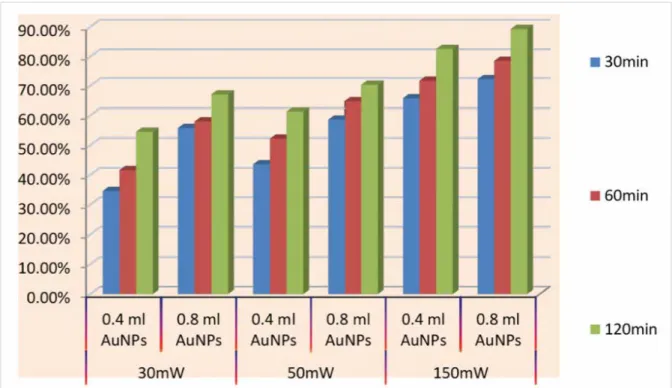
When the AuNP dose, process time and laser power were taken into account, the highest average protoscolices deaths in hy-datid cyst liquid was associated with the group H having high-dose AuNPs which exposed to 150 mW laser power for 120 min-utes irradiation (p< 0.01) as shown in Figure 1. In contrast, the lowest average protoscolices deaths was associated with the group G or group A which are having AuNPs without any laser exposure or only laser irradiation without any AuNPs addition, respectively (p< 0.01) (Figure 2).
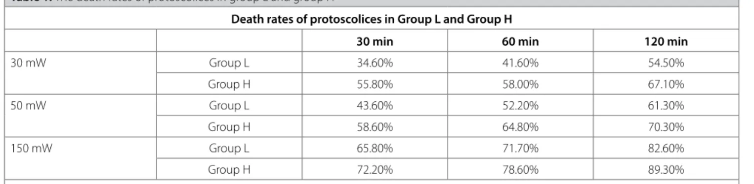
table 1. The death rates of protoscolices in group L and group H
Death rates of protoscolices in Group l and Group h
30 min 60 min 120 min
30 mW Group L 34.60% 41.60% 54.50% Group H 55.80% 58.00% 67.10% 50 mW Group L 43.60% 52.20% 61.30% Group H 58.60% 64.80% 70.30% 150 mW Group L 65.80% 71.70% 82.60% Group H 72.20% 78.60% 89.30%
DIsCussIOn
Hydatid cyst disease is an important and common public health problem worldwide. In the endemic areas of countries and regions such as Peru, Argentina, East Africa, Middle Asia and China, the disease affects 50 out of 100,000 people each year while its prevalence is between 5% and 10% (1,2).
The present optional therapies for hydatid cyst disease are prob-lematic in many ways. Surgical methods are all invasive and come with the risk of recurrence and the rupture and spillage of the contents (14). Where chemotherapy is concerned, albendazole
and mebendazole are the preferred drugs. However, their con-centrations in the hydatid cyst are not sufficient to cure the dis-ease because the cyst’s wall is too thick (16). Clinical studies have shown that the medication used for treatment could not always kill the protoscoleces (17). Percutaneous aspiration, injection and reaspiration (PAIR) is a percutaneous technique which is a signif-icant and versatile alternative for the conventional methods, es-pecially in the treatment of early-stage cysts (18).
There is a desperate need for a scolicidal agent to be used in PAIR method to effectively cure hydatid cyst protoscoleces.
Figure 1. The death rates of protoscolices treated with laser (30 mW, 50 mW, 150 mW) and AuNPs (0.4 mL, 0.8 mL) in anisochrony (30
min, 60 min, 120 min).
Figure 2. (Left) the death rates of protoscolices treated with laser solely (30 mW, 50 mW, 150 mW) for different durations (30 min,
60 min, 120 min); (right) the death rates of protoscolices treated with AuNPs only (0.4 mL, 0.8 mL) for different durations (30 min, 60 min, 120 min).
According to the literature, a large number of scolicidal agents are used in PAIR, such as; Albendazole, 95% alcohol, hyperton-ic saline, hydrogen peroxide, silver nitrate, cetrimide and ethyl alcohol. However, many of these agents have adverse effects, therefore, limit their usage in the treatment of hydatid cyst dis-ease (19). Formalin was the first scolicidal agent used, howev-er, it is not used today due to its toxic effect (20). Radiologists prefer using another scolicidal agent, ethyl alcohol, which is in-flammable and volatile (21). This characteristic of ethyl alcohol has led to the restriction of its use in surgery. Also, ethyl alco-holcan damage the bile duct’s epithelium, leading to sclerosing cholangitis (22). That is why, it is not used to treat hydatid cysts which have bile duct communication. In addition, due to low scolicidal efficiency and the resultant complications, the usage of hydrogen peroxide as a scolicidal agentin the treatment of hydatid disease is very limited (23). A disinfectant chemical, povidone-iodine (PVP-I), is also used as a scolicidal agent. The complications of PVP-I are PVP (polyvinlpyrrolidone) storage disease, renal shutdown and sclerosing serositis (23). Nowadays, most effective scolicidal agent frequently used is hypertonic sa-line. Hypertonic saline is nearly 100% effective on protoscolic-es (24); however, it could lead to hypernatremia, neurological side effects and intracranial bleeding (25). Furthermore, the use of hypertonic saline should be avoided in treatments ofcysts which open to the bile duct due to very high possibility to have chronic sclerosing cholangitis disease (20). Another effective scolicidal agent is cetrimide-chlorhexidine (Savlon) and it hasa scolicidal effect even a minimal usage of 0.1%. However, like hypertonic saline, cetrimide-chlorhexidine should also not be used to treat cysts associated with the bile duct (26). Although effective cure rate of scolicidal agents are present for the treat-ment of hydatid cyst protoscoleces, these agents possess very harmful adverse effects including chronic sclerosing cholangitis (12,14). Even if the surgeons are not willing to use these toxic agents, the usage of these agents is unfortunately a common practice in clinic due to deficiency of ideal non-toxic scolicidal agent (27). Therefore, there is an extreme need for a non-inva-sive agent which has a significant scolicidal effect to cure the hydatid cyst protoscoleces without any harmful effects and complications.
Anderson and Loveless have reported a successful destruction of protoscoleces, namely, E. granulosus protoscolices, by varying temperature in cyst media (28). In their study, different degrees of temperature were applied to protoscolices in cyst fluid from the lungs and livers of infected sheep. The durations of the pro-toscolices deaths were 16 days at 20°C, 8 days at 30°C, 4 days at 40°C, and 2 hours at 50°C. According to this study, high tem-peratures above 40°C effectively kill the protoscolices which is also reported in various studies in literature (29,30). As a result, a scolicidal agent which releases heat to the surroundings mean-ing increase the temperature of the protoscolices media above
40°C could be used as a new scolicidal agent to cure the pro-toscolices in cyst fluid.
In this study, non-toxic AuNPs was introduced and successfully applied as a new scolicidal agent to cure the hydatid cysts pro-toscolices in the liver. Under laser irradiation, the AuNPs is a heat generator which increase the temperature of the surroundings, protoscolices in this case, and eventually kill the protoscolices. Under different laser powers and irradiation durations, we eval-uated the scolicidal effect of AuNPs against protoscolices of hy-datid cysts on ex-vivo model.
Gold nanoparticles are considered as harmless, stable and biocompatible materials which are frequently used in medi-cal research (15). Various reports in literature have shown that AuNPs do not possess anycytotoxic and genotoxic effects, and any known systematic or local side effects (31,32). Besides the inertness and non-toxic character, AuNPs have a unique prop-erty which is called “photothermal effect”. Spherical AuNPs can transform the absorbed green laser light energy into heat ener-gy through photothermal effect (33,34). The heat will dissipate into the surrounding media and this localized heating by using AuNPs can cause thermal cellular destruction (35,36). We ben-efit this destruction process in the treatment of protoscolices in liver hydatid cysts and kill this protoscolices via the tempera-ture increase after localized heating by AuNPs. That is to say, this study facilitated the application of laser to AuNPs in order to rise the temperature of the cyst fluid which eventually cause the deaths of all the protoscolices.
In the present study, besides a control group (Group C), we used 4 different groups such as; only AuNPs added samples (Group A), only laser irradiation applied samples (Group G), low-dose AuNPs added and laser irradiated groups (Group L), high-dose AuNPs added and laser irradiated groups (Group H). We applied three different laser powers of 30 mW, 50 mW, and 150 mW for different durations (30 min, 60 min, 120 min). Due to the usage of low-power and harmless laser powers, we irradiat-ed the cyst liquid containing protoscolices for longer periods in order to result a successful mortality. The experimental results show that scolicidal activity of AuNPswas increased by raising the laser power with higher concentrations of AuNP dose and extending the duration of irradiation process. These result a rise in temperature of the hydatid cysts media which kills protosco-licesin significant incidences of mortality. However, when only irradiation of laser or only inclusion of AuNPs to hydatid cysts liquid, there was only negligible deaths of protoscolices which is within the range of acceptable deviation.
COnClusIOn
We have successfully demonstrated the usage of AuNPs as a therapeutic and scolicidal agent to cure hydatid cyst disease un-der laser irradiation. The main advantages of AuNPs compared
with the other scolicidal agents are non-invasive and biocom-patible scolicidal agent. Our results show that higher laser pow-ers and AuNPs amounts with longer irradiation durations result effective destruction of protoscolices in liver hydatid cysts due to a sharp increase in temperature. AuNPs possess a scolicidal effect and could use in PAIR applications. Hence, this approach could produce a better treatment for various hydatid cyst diseas-es, even in the treatment of cysts associated with the bile duct.
Ethics Committee Approval: Ethics approval for this study was granted by
the ethical committee.
Informed Consent: Written informed consent was obtained from patients
who participated in this study.
Peer-review: Externally peer-reviewed.
Author Contributions: Concept - B.Ç.; Design - B.Ç., F.A.; Supervision - F.A.,
S.Y.; Resource - B.Ç., M.E.D.; Materials - B.Ç.; Data Collection and/or Processing - F.A., B.Ç.; Analysis and/or Interpretation - B.Ç., M.E.D.; Literature Search - S.Y., B.Ç.; Writing Manuscript - B.Ç., F.A.; Critical Reviews - F.A.
Conflict of Interest: The authors have no conflicts of interest to declare. Financial Disclosure: The authors declared that this study has received no
financial support.
REFEREnCEs
1. Craig PS, McManus DP, Lightowlers MW, Chabalgoity JA, Garcia HH, Gavidia CM, et al. Prevention and control of cystic echinococcosis. Lancet Infect Dis 2007;7:385-94.[CrossRef]
2. Budke CM, Deplazes P, Torgerson PR. Global socioeconomic impact of cystic echinococcosis. Emerg Infect Dis 2006;12(2):296–303.[CrossRef]
3. Manfredi MT, DiCerbo AR, Zanzani S, Moriggia A, Fattori D, SiboniA, et al. Prevalence of echinococcosis in humans, live stock and dogs in northern Italy. Parasitol Res 2011;48:59–66.[CrossRef]
4. Ralph T, Bryan MD, Schantz VMD, et al. Parasitic diseases branch, di-vision of parasitic diseases, center for infectious diseases, Centers for Disease Control, Public Health Service, US Department of Health and Human Services. Atlanta, 1989;30333.
5. Moazeni M, Larki S. In vitro effectiveness of acidic and alkline solutions on scolices of hydatid cyst. Parasitol Res 2010;106(4):853-6.[CrossRef]
6. Adas G, Arikan S, Kemik O, Oner A, Sahip N, Karatepe O. Use of alben-dazole sulfoxide, albenalben-dazole sulfone, and combined solutions as scolicidal agents on hydatid cysts (in vitro study). World J Gastroen-terol 2009;15(1):112-6.[CrossRef]
7. Caglar R, Yuzbasioglu MF, Bulbuloglu E, Gul M, Ezberci F, Kale IT. In vitro effectiveness of different chemical agents on scolices of hydatid cyst. J Invest Surg 2008;21:71-5.[CrossRef]
8. Coşkun İ, İrfanoğlu ME, Uzunköy A, et al. Skolisidal maddelerin safra yollarına etkileri. Çağdaş Cerrahi 1992;75:946-50.
9. Arikoglu H. Histologic evaluation of the efficacy of different scolicidal agents on the viability of scolices: an in vitro study. Postgraduatethe-sis. Selçuk Üniversity, Konya, Turkey 1996;53.
10. Abbasi A, Shadmehr B, Ghaffarinejat MH, et al. Scolicidal agents can cause sclerosing cholangitis. London J Hepatic Pancreatic Biliary Surg 1990;2:157.
11. Üstünsöz B, Akhan O, Kamiloğlu M.A. Percutaneous treatment of hydatid cyst: long-term results. AJR 1999;172:91-6.[CrossRef]
12. Faraday M. Experimental relations of gold (and other metals) to light. Philosophical Transactions of the Royal Society 1857;147:145-81.
[CrossRef]
13. Turkevitch J, Stevenson PC, Hillier J. Nucleation and growth process in the synthesis of colloidal gold. Discuss Faraday Soc 1951;11:55-75.
[CrossRef]
14. Gottstein B, Eckert J, Woodtli W. Determination of parasite specific immunoglobulin susing the ELISA in patients with echinococcosis tre-ated with mebendazole. Parasitol Res 1984;70(3):385-9.[CrossRef]
15. Zou X, Wang J, Zhao H, Zhang J, Wua W, Ye B. Echinococcus gra-nulosus: protoscolicidal effect of high intensity focused ultrasound. Experimental Parasitology 2009;121:312-6.[CrossRef]
16. Liu AB, Cai H, Ye B, Chen LL, Wang MY, Zhang J, et al. The damages of high intensity focused ultrasound to transplanted hydatid cysts in ab-dominal cavities of rabbits with aids of ultrasound contrast agentand superabsorbent polymer. Parasitol Res 2013;112:1865-75.[CrossRef]
17. Gil-Grande LA, Rodriguez-Cabeiro F, Prieto JG, Sanches-Ruano JJ, Bra-sa C, Aguilar L, et al. Randomised controlled trial of efficacy of alben-dazole in intraabdominal hydatid disease. Lancet 1993;324:1269-72.
[CrossRef]
18. Kabaalioğlu A¸ Alimoglu E, Apaydin A. Percutaneous imaging-guided treatment of hydatid liver cysts: do long-term results make it a first choice? Eur J Radiol 2006;59:65-73.[CrossRef]
19. Sungur İ. Güncel bazı kimyasal maddelerin in vitro skolisidal etkileri-nin araştırılması. C.Ü. Tıp Fak Der 1979;4:317-26.
20. Besim H, Karayalçın K, Hamamcı O. Scolicidal agents in hydatid cyst surgery. HPB Surgery 1998;10:347-51.[CrossRef]
21. Giorgio A, Tarantino L, Francica G, Mariniello N, Aloisio T, Soscia E, et al. Unilocular hydatid liver cyst: treatment with US-guided, double percu-taneous aspiration and alcohol injection. Radiology 1992;184:705-10.
[CrossRef]
22. Castellano G, Moreno-Sanchez D, Gutierrez J, Moreno-Gonzalez E, Colina F, Solis Herruzo JA. Caustic sclerosing cholangitis. Report of four cases and a cumulative review of the literature. Hepato Gastro-enterology 1994;41:458-70.
23. Le Veen HH, Le Veen FR, Le Veen GE. The mythology of povidone-iodin and the development of self-sterilizing plastics. SGO 1993;176:183-9. 24. Erzurumlu K, Hokelek M, Baris S, Sahin M, Birinci A, Amanvermez R,
et al. Effect of albendazole sulfoxide solution on the scolices and the hepatobiliary system. Eur Surg Res 1998;30(6):433-8.[CrossRef]
25. Kayaalp C, Balkan M, Aydin C, Ozgurtas T, Tanyuksel M, KirimliogluV, et al. Hypertonic saline in hydatid disease. World J Surg 2001;25(8):975-9.
[CrossRef]
26. Tozar E, Topcu O, Karayalcin K, Akbay SI, Hengirmen S. The effects of cetrimide–chlorhexidine combination on the hepato-pancreatico-bi-liary system. World J Surg 2005;29:754-8.[CrossRef]
27. Prasad J, Bellamy P, Stubbs RS. Instillation of scolicidal agents into he-patic hydatid cysts: can it longer be justified? New Zealand Medical Journal 1991;104:336-7.
28. Andersen FL, Loveless RM. Survival of protoscolices of Echinococcus granulosus constant temparatures. J Parasitol 1978;64:78-82.[CrossRef]
29. Thanos L, Mylona S, Brontzakis P, Ptohis N, Karaliotas K. A complicated postsurgical echinococcal cyst treated with radiofrequency ablation. Cardiovasc Intervent Radiol 2008;31:215-8.[CrossRef]
30. Zhang J, Ye B, Kong J, Cai H, Zhao Y, Han X, et al. In vitro protoscolici-dal effects of high-intensity focused ultrasound enhanced by a supe-rabsorbent polymer. Parasitol Res 2013;112:385-91.[CrossRef]
31. Connor EE, Mwamuka J, Gole A, Murphy KJ, Wyatt MD. Gold nanopar-ticles are taken up by human cells but do not cause acute cytotoxicity. Small 2005;3:325-7.[CrossRef]
32. Schulz M, Ma-Hock L, Brill S, Strauss V, Treumann S, Groters S, et al. In-vestigation on the genotoxicity of different sizes of gold nanoparticles administered to the lungs of rats. Mutat Res 2012;745(1–2):51-7.
[CrossRef]
33. Skirtach AG, Dejugnat C, Braun D, Susha AS, Rogach AL, Parak WJ, et al. The role of metal nanoparticles in remote release of encapsulated materials. Nano Lett 2005;5:1371-7.[CrossRef]
34. Pissuwan D, Valenzuela SM, Cortie MB. Therapeutic possibilities of plas-monically heated gold nanoparticles. Trends Biotechnol 2006;24:62-7.
[CrossRef]
35. Lee S. Surface Modification of Gold Nanoparticles and Their Applicati-on in Biomolecular Sensing. Master Thes is, Illinois Institue of Techno-logy. Chicago, 2007.
36. Hirsch LR, Stafford RJ, Bankson JA, Sershen SR, Rivera B, Price RE, et al. Nanoshell-mediated near-infrared thermal therapy of tumors under magnetic resonance guidance. Proc Natl Acad Sci USA
2003;100:13549-54.[CrossRef]
Düşük güçte lazer uygulanmış altın nanoparçacıklarının hidatik kist protoskoleksleri
üzerine etkisinin araştırılması
Bayram Çolak1, Faruk Aksoy2, Selman Yavuz3, Mehmet Emin Demircili4 1 Selçuk Üniversitesi Tıp Fakültesi, Genel Cerrahi Anabilim Dalı, Konya, Türkiye
2 Necmettin Erbakan Üniversitesi Tıp Fakültesi, Genel Cerrahi Anabilim Dalı, Konya, Türkiye
3 Selçuk Üniversitesi Mühendislik Fakültesi, Metalurji ve Malzeme Mühendisliği Bölümü, Konya, Türkiye 4 Necmettin Erbakan Üniversitesi Tıp Fakültesi, Tıbbi Mikrobiyoloji Anabilim Dalı, Konya, Türkiye
ÖZET
Giriş ve Amaç: Hidatik kistin içindeki protoskoleksleri öldürmek için çeşitli skolosidal ajanlar kullanılmıştır. Skolosidal ajanların istenmeyen
komp-likasyonları, hidatik kistin tedavi yöntemini sınırlandırmaktadır. Bu nedenle vücuda zararlı olmayan yeni skolosidal ajanlara ihtiyaç duyulmaktadır. Fototermal altın nanopartikülleri (AuNPs) kist sıvısı içindeki protoskolekslerin etrafını sararak lazer ışını altında, ışınların enerjisini absorbe edip ısı enerjisine dönüştürmek suretiyle protoskoleksleri öldürmektedir. Çalışmada, biyo-uyumlu fototermal altın nanopartiküllerini (AuNPs) karaciğer hidatik kistlerini tedavi etmek için toksik olmayan bir skolosidal ajan olarak uyguladık.
Gereç ve Yöntem: Protoskoleksler infekte koyun karaciğerlerinden elde edildi. Her bir çalışma grubu için içinde 1,5 mL kist sıvısı içeren tüpler
oluşturuldu. Tüpler 5 gruba ayrıldı. Kontrol grubu, sadece AuNPs eklenen grup, sadece lazer uygulanan grup, yüksek doz AuNPs eklenip lazer uygulanan grup, düşük doz AuNPs eklenip lazer uygulanan gruplar idi. İki farklı konsantrasyonda (0,4 ve 0,8 mL) AuNPs ve üç farklı güçte lazer (30, 50, 150 mW), 30, 60, 120 dakika sürelerle gruplara uygulandı. Daha sonra kist sıvıları ışık mikroskobu altında incelendi ve protoskolekslerin canlılıkları değerlendirildi.
Bulgular: Yüksek doz AuNPs eklenen ve 150 mW lazerin 120 dakika uygulandığı grupta %89.30 protoskoleks ölümü sağlandı. Fakat sadece AuNPs
eklenen ve sadece lazer uygulanan gruplarda anlamlı ölüm oranları ile karşılaşılmadı. AuNPs dozunun, lazer gücünün veya sürenin arttırılması ile protoskoleks ölümünün de arttığı tespit edildi.
Sonuç: Çalışmada, AuNPs’nin lazer ışını altında, hidatik kist tedavisinde etkili bir skolosidal ajan olabildiği başarılı bir şekilde gösterildi. Anahtar Kelimeler: Altın, nanoparçacık, lazer, hidatik kist
DOİ: 10.5578/turkjsurg.4354
ORİJİNAL ÇALIŞMA-ÖZET Turk J Surg 2019; 35 (4): 314-320