REVEALING THE ROLE OF MED14
IN
POL-II TRANSCRIPTION REGULATION
A THESIS SUBMITTED TO
THE GRADUATE SCHOOL OF ENGINEERING AND SCIENCE
OF BILKENT UNIVERSITY
IN PARTIAL FULFILLMENT OF THE REQUIREMENTS FOR
THE DEGREE OF
MASTER OF SCIENCE
IN
MOLECULAR BIOLOGY AND GENETICS
By Yasemin Barış August 2017
ii
REVEALING THE ROLE OF MED14 IN POL-II TRANSCRIPTION
REGULATION
By Yasemin Barış August 2017
We certify that we have read this thesis and that in our opinion it is fully adequate, in scope and in quality, as a thesis for the degree of Master of Science.
Murat Alper Cevher (Advisor)
Ali Osmay Güre
Sreeparna Banerjee
Approved for the Graduate School of Engineering and Science:
Ezhan Karaşan
Director of the Graduate School of Engineering and Science
iii
ABSTRACT
REVEALING THE ROLE OF MED14 IN POL-II TRANSCRIPTION REGULATION
Yasemin Barış
M.Sc. in Molecular Biology and Genetics Advisor: Murat Alper Cevher
August 2017
Transcription of protein coding genes by RNA polymerase II (Pol II) is a multi-step process each of which requires a series of factors including coactivators. 30-subunit (the subunits are organized into discrete modules – head, middle, tail, and kinase) -2mDa human Mediator complex is the key coactivator in transcription not only for facilitating the establishment of pre initiation complex (PIC) by acting as a bridge between gene-specific activators and Pol II but also and more importantly for regulating the Pol II activity at all pre-initiation, elongation and re initiation steps. Although the diversity in the function of Mediator lies in its flexible conformation and variable subunit organization (for instance, four subunit CDK8 module can both bind to and disassociate from Mediator), detailed mechanistic studies regarding how Mediator interacts with different activators/repressors to regulate transcription, how Mediator facilitates basal transcription through interactions with Pol II and general transcription factors (GTFs) to establish a proper PIC on promoter DNA and how the regulation of subunit exchanges/rearrangements on the Mediator results in architectural and functional outcome are not well characterized. By using Multibac baculovirus expression system, the previous studies have shown that the reconstitution of a functional 15-subunit human core Mediator complex (composed of head and middle modules together with Med14) is sufficient to support basal transcription as well as selective activator-dependent transcription in vitro both with purified factors and with nuclear extracts (in the presence of Med26) as the source of GTFs. This study has uncovered a mechanism by which human core Mediator facilitates transcription by directly interacting with Pol II via its Med14 subunit and recruiting Pol II to target gene promoters.
This is the first time that i unraveled the mechanism of how Pol II binds to Mediator and how it is being recruited to target gene promoters for proper PIC assembly and transcription. This way, i shed light into how protein coding genes are universally regulated via the Mediator complex and in the
iv
future , I will be in a better position to specifically target selected genes by reconstituting the entire Mediator complex and characterizing activator-Mediator-Pol II crosstalk.
Key Words: Human Mediator Complex, Med14, RNA polymerase II, transcription, GTFs
v
ÖZET
POL-II TRANSKRİPSİYON
REGÜLASYONUNDA
MED14 FONKSİYONUNUN
BELİRLENMESİ
Yasemin Barış
Moleküler Biyoloji ve Genetik, Yüksek Lisans Tez Danışmanı : Murat Alper Cevher
Ağustos 2017
Protein kodlayan genlerin RNA polimeraz-II aracığıyla transkripsiyonu, koaktivatörleri de kapsayan bir çok faktörün katılımını gerektiren, çok aşamalı bir süreçtir.30 alt-ünitelik 2mDa insan Mediator kompleksi, Pol II aktivitesini transkripsiyonun tüm aşamalarında kontrol edebilen ve gene-özgü aktivatörlerle transkripsiyon başlama öncesi kompleks arasında bir köprü görevi gören, en önemli koaktivatördür. Mediator kompleksinin fonksiyonel çeşitliliği her ne kadar değişebilen alt-ünite kompozisyonuna ve esnek yapısına bağlı olsa da, Mediator’ın transkripsiyonda görevli proteinlerle olan etkileşimi, transkripsiyon öncesi kompleksi ilgili DNA promotörlerine nasıl getirdiği, ve aynı zamanda farklı kompozisyonlarda bulunmasının sebebi ve yarattığı sonuçlara ilişkin detaylı mekanistik çalışmalar yapılmamıştır. Yakın tarihte Multibac-ekspresyon sistemini kullanarak yapılan çalışmalar, 15 alt –ünitelik fonksiyonel insan Mediator kompleksini (head ve middle modülleriyle birlikte med14 ve med26 altünitelerini kapsayan) in vitro sentezleyebilmiş ve çekirdek olarak adlandırılan bu Mediator kompozisyonun basal transkipsiyonu ve aktivatorlerce sağlanan transkripsiyonu sağlayabildiğini göstermiştir. In vitro transkripsiyon deneyleri, hem saflaştırılarak izole edilen genel transkripsiyon faktörleriyle hem de hali hazırda faktörleri barındıran çekirdek özütleriyle yapılmıştır. (Genel transkripsiyon faktörlerini barındıran özütlerle yapılan deneyde, çekirdek Mediator’ın transkripsiyonu gerçekleştirmesi için med26 alt-ünitesi de bağlanmıştır.)
vi
Bu çalışma Mediator-Pol II interaksiyonun Med14 alt birimiyle sağlandığı ve çekirdek Mediator ‘ın bu interaksiyon sonucu transkripsiyonu gerçekleştirebildiğini ilk kez gösteren özgün bir çalışmadır. Böylelikle ilk kez Pol II ‘nun Mediator kompleksine nasıl bağlandığını ve hedef gen promotorlerine nasıl getirildiğini göstermiş oldum. Mediator kompleksinin protein kodlayan genleri nasıl kontrol ettiğini açıklığa kavuşturarak hali hazırda başladığım tüm Mediator kompleksinin invitro senteziyle birlikte aktivator-Mediator – Pol II interaksiyon mekanizmasını detaylı olarak açıklamayı hedeflemekteyim.
Anahtar Kelimeler: İnsan Mediator Kompleksi, Med14, RNA Polymeraz II, Transkripsiyon, GTFs.
vii
To my grandfather who has always been my
source of strength, encouraged me with endless
hope and has been my well of wisdom.
viii
Acknowledgements
I would like to express my deepest appreciation for my best mentor, my advisor Assist. Prof. Murat Alper Cevher. His precious scientific approach and great guidance in my work have rendered me as a young scientist who is searching for the truth in any time and is not losing the hope even in the worst situation. His patience and endless support to me, the great scientific experiences which he has provided to me will guide me for my future career. I’m also deeply grateful for his trust to me in my work as well as the kindest friendship that I can not imagine to see from any advisors. I am sure about that I am the luckiest person in academia having the best advisor ever.
I would like to thank my dearest family for their care and support in my academic life. My mom and dad, Naime Barış and Vahap Barış, I would like to deeply thank for your endless patience and love to your children. My little brother Eren Barıs, I appreciate the joy you’ve given me during my hard times in Bilkent and my grandma and grandpa Seher Eren and Sadi Eren who nurtured me with great care and thought me the importance of honesty and loyality in any time of life. Last but not least, my uncles Erdoğan Eren and Sadi Eren and my aunts Aysel Eren and Hülya Eren many thanks for your love and friendship and being my best friends forever.
I would like to express my deep gratitude to Boğaziçi University and MBG family that gave me a great passion and enthusiasm for science.
I specifically want to thank to Cem Durmuş, Uğur Kahya, Ayşe Sedef Köseer and Güven Akçay for their friendship and help in Bilkent and my dormitory manager Nimet Kaya for her special care for my health and make me feel like I am at home in Bilkent. I also thank to Emre Bilen for his precious opinions and help in my work and for most enjoyable times that I spent in Ankara. Finally, I am deeply grateful to Doğan Çolakoğlu for his endless support and trust to me in every decision that I have made.
This project was financially supported by European Molecular Biology Organization (EMBO/ 6.8.3.778)
ix TABLE OF CONTENTS
Abstract...iii
Özet...v
Acknowledgement ...viii
Table of Contents ...ix
List of Figures ... .xii
List of Tables ...xiii
Abbreviations ... xiv
Chapter1…Introduction...1
1.1 Eukaryotic Transcription and Identification of Functional Elements in the Transcription Machinery………...1
1.2 Mediator Complex………...4
1.2.1 Early Studies in the Identification of Mediator Complex as a Coactivator ……....4
1.2.2 Structural and Functional studies of Human and Yeast Mediator Complex…….10
1.2.3 Mediator Architecture and Structure-Function Releationship. ………...10
1.2.4 Mediator Complex in the regulation of PIC structure and Function………...13
1.3 Baculovirus Expression System for Recombinant Protein Complex Production………...14
Chapter2…..Materials………...19
2.1 Buffers and Solutions………19
2.1.1 Western Blot, Staining and Nuclear Extract buffers………...19
2.1.2 Buffers for Immobilized Template Recruitment Assay using Streptavidin Dynabeads………....20
x
2.2 Materials………..21
2.2.1 Cell Culture Media, Supplements and Culture Equipment……...21
2.2.2 Antibodies Used in Western Blot and Immunoprecipitation………....22
2.2.3 Kits and other Tools………..23
Chapter 3 …Methods………...24
3.1 Reconstitution and Purification of Partial Human Mediator complexes using Baculovirus Expression System………...24
3.2 The purified protein complexes using Baculovirus Expression System………25
3.3 Purification of Endogenous Polymerase II and Human Mediator Complexes from HeLa Cells Stably Expressing f:rpb9 and f:nut2 Respectively………...27
3.3.1 Cell Culturing………..27
3.3.2 Nuclear Extract Preparation………...27
3.3.3 Pull Down of Endogenous Pol II and Human Mediator Complex………....28
3.4 Silver Staining ………...28
3.5 Protein Quantification………...29
3.6 SDS-PAGE………...…29
3.7 Western Blotting………...30
3.8 Immunoprecipitation for Pol II - Mediator Interaction by Using Purified Full length and truncated f:Med14 proteins………31
3.9 Immunoprecipitation (IP) for Pol II – Mediator Interaction by using Partial Mediator Complexes………..31
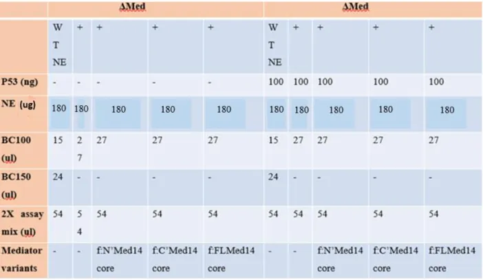
3.10 Immobilized Template Recruitment Assay………....32
xi
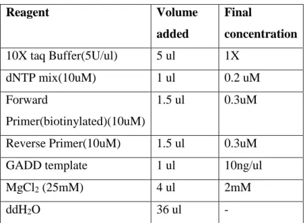
3.10.1 PCR Program for GADD Template………32
3.10.2 Preparation of Dynabeads M-280 streptavidin(DynaI)………..33
3.10.3 Immobilized Template Recruitment Assay Design………33
3.11 Pull Down of Pol II- Core Mediator Complex by Using His-Tag Dynabeads………...35
3.12 Chemical Crosslinking of Pol II – Core Mediator Complex………..35
Chapter 4…Results………...36
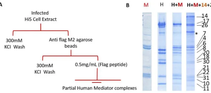
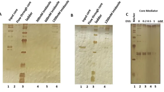
4.1 Purification of Partial Human Mediator Complexes Using Baculovirus Expression System..36
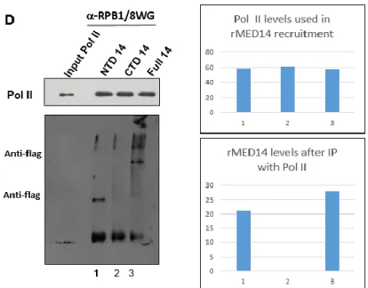
4.2 Immunoprecipitation Reveals a Critical Role for Med14 in the Regulation of Pol II activity in vitro...37
4.3 Med14 directly interacts with Pol II via its N' terminus in the context of Mediator………….39
4.4 Pol II interaction with core Mediator is independent from Med26………...41
4.5 N' Med14 Dependent Pol II recruitment to promoter DNA Revealed by Immobilized Template Recruitment Assay………...43
4.6 Activator Facilitated Recruitment of Pol II to the promoter by N'Med14 core Complex…….44
4.7 Chemical Crosslinking of Pol II- Core Mediator Complex………..46
Chapter 5 …..Discussion……….49
Chapter 6…. Conclusions and Future Perspectives………...54
Bibliography………..………..…57
Appendix…Copyright Permissions ………...………...64
xii
List of Figures:
Figure1.1: The general mechanism of Eukaryotic Transcription……….1 Figure 1.2.1 Mediator Dependent Transcription Activation………....9 Figure 1.2.3 Structure of S.pombe Mediator Head Module………. ……….…..11 Figure 1.3 Multibac Expression system strategy showing the cloning approach of the system which enables high yield production of protein complexes………17 Figure 4.1 Reconstituted Head , Middle ,H+M and core Mediator complexes using Multibac system………..37 Figure 4.2 Med14 directly interacts with Pol II via its amino terminus………...39 Figure 4.3 Immunoprecipitation validates the direct interaction between Pol II- N'Med14 in the context of Mediator………..41 Figure 4.4 Pol II does not directly interact with Med26……….42 Figure 4.5 Immobilized Template Assay shows the requirement of N' Med14 in recruitment of Pol II to the promoter by Mediator complex………..44 Figure 4.6 p53 enhanced recruitment of Pol II to the promoter dependent on N'Med14………...46 Figure 4.7 The optimal DSS concentration was adjusted in chemical crosslinking of
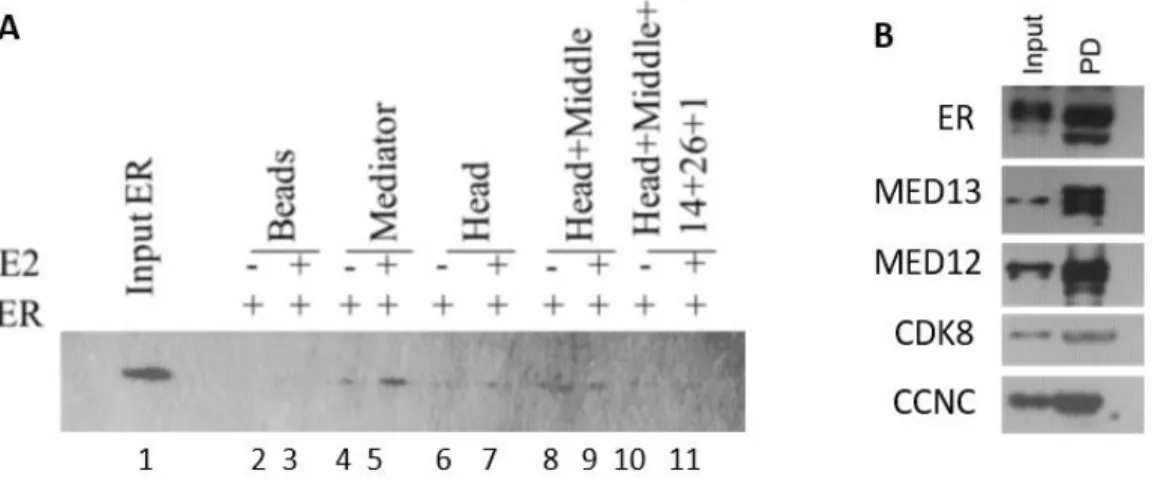
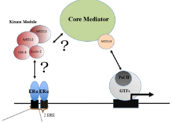
Pol II-core for MS analysis………...48 Figure 5 The proposed mechanism of the regulation of transcription by the Mediator Complex...54 Figure 6.1 Reconstituted Mediator complex and ERα interaction studies………...55 Figure 6.2 Proposed mechanism for ERα- Mediator interaction………...56
xiii
List of Tables:
2.1.1 Western Blot, Staining and Nuclear Extract buffers………19
2.1.2 Buffers for Immobilized Template Recruitment Assay using Streptavidin Dynabeads…..20
2.1.3 His-Tag Dynabeads Pulldown Buffers……….21
2.2.1 Cell Culture Media, Supplements and Culture Equipment………..21
2.2.2Antibodies Used in Western Blot and Immunoprecipitation………22
2.2.3 Kits and other Tools……….23
3.2 The purified protein complexes using Baculovirus Expression System………...25
3.6 Components in SDS-PAGE gel preparation………...29
3.10.1 PCR Program for GADD Template………..32
xiv
Abbreviations:
AcNPV Autographa californica nuclear polyhedrosis virus APS Ammonium persulphate
BAC Artificial bacterial chromosome BSA Bovine serum albumin
CBB Coomessie Brilliant Blue CDK7 Cyclin dependent kinase 7 CDK8 Cyclin dependent kinase 8
CRSP Cofactor Required for Sp1 Activation
CTD Carboxyl terminal domain of RNA polymerase-ll CX-MS Chemical crosslinking coupled to Mass Spectrometry DSS Disuccinimidyl suberate
ERα Estrogen Receptor Alpha FBS Fetal Bovine Serum
GTF General Transcription Factor kDa kilo Dalton
MED Mediator
NAT Negative Regulator of Activated transcription NC Negative cofactor
NELF Negative elongation factor NHR Nuclear Hormone Receptors
xv PC Positive cofactor
PIC Pre-initiation complex Pol- II RNA Polymerase II PBS Phosphate buffered saline
PBST Phosphate buffered saline tween20
SDS-PAGE Sodium Dodecyl sulfate polyacrylamide gel electrophoresis SMCC Human SRB/MED Cofactor Complex
SRB Suppressor of RNA polymerase B TAF TBP associated factor
TBP TATA box binding protein
1
CHAPTER 1
INTRODUCTION
1.1 Eukaryotic Transcription and Identification of Functional Elements in the Transcription Machinery:
Transcription of eukaryotic protein-coding genes by RNA polymerase II (Pol II) is a multi-step process including the events [1] : opening up the chromosome by decondensation of the relevant locus, histone modifications that enable the locus to be recognized by activators /repressors, binding of those activators and coactivators to the enhancers and promoters and finally recruitment of the transcription machinery to the core promoter.
In the native stage of DNA, promoters and related regulatory elements are packaged in nucleosomes, the compact structures wrapped around 146 base pairs(bp) DNA and a histone octamer comprising 2 copies of the core histones, H2A,H2B,H3 and H4.[2-3] Since this condensed shape is not accessible to the proteins that will further recruit the transcription machinery to the site of transcription initiation site, nucleosome itself has a repressive role in transcription.[4] This repressive role could be explained in different mechanisms: First, DNA modifying enzymes, activators and general transcription factors(GTFs) are occluded to bind with the template to enable initiation by Pol II and hence the formation of the preinitiation complex.[4] Second, nucleosome chains could be coiled and folded back upon itself repressing the whole region of transcription. [5] Third, nucleosomes could interact with the proteins in heterochromatin region that will further block the gene expression even in a hereditary manner. [6]
Work on chromatin structure and gene activation mechanisms have revealed the important aspects of proteins named as activators /repressors with the advantage of biochemical studies. Activators couple the transcription to a particular need for any cell type by being specific to each gene or any related gene families.
2
They are activated in response to a physiological stimulus as in the case of nuclear hormone receptors (NHRs) such as estrogen receptor alpha. [7]
Moreover, they could be maintained in an inactive state in the cytoplasm by being associated to an inhibitory protein, then with a particular external signal, they could be released and could enter the nucleus with its fully functional form. [8] When they enter, they bind to their cognate sequence (enhancers) on DNA and execute the recruitment of the transcription machinery to the promoter. As the explanation implies, activators could accomplish those missions by having a distinct DNA binding and activation domains. [9]
Since the initiation of transcription includes two main steps; opening up chromatin followed by recruitment of the factors by activators and the interaction of Pol II with the GTFs, it was noteworthy to speculate how those activators stimulate initiation of transcription with the assistance of transcription machinery. Scientists initially discovered the factors necessary for transcription of Pol II class genes by first using crude cell extracts and then by using intensive chromatographic techniques to purify all such components from the extracts. [10] General transcription factors (TFIIA,TFIIB,TFIIF,TFIID) have been purified and named based on their chromatographic fractions isolated under particular salt concentrations.[10]. After identification of such components, the mechanism by which the activator could promote the transcription initiation through recruiting TFIID to TATA nucleotide sequence and later how this recruitment would act as a scaffold for the pre initiation complex assembly has been proposed.[11] However, later works showed that isolated yeast and metazoan TATA binding protein (TBP) could supplant for TFIID and enhance basal transcription [12] even if its occupation in the extracts are much lower than TFIID itself. Since the works also proved that TBP alone could not satisfy the needs for activator dependent transcription, scientists clarified this functional distinction by discovering the subunits of TFIID (TBP associated factors-TAFs) that were required to bring activators to the transcription machinery and function as coactivators to regulate activator dependent transcription. [13]
3
Biochemical studies regarding to the mechanism of transcription led to the identification of other accessory elements that regulate the transcription in both negative and positive manners. In vitro transcription assays performed with purified factors (namely GTFs) necessitated other factors that are crucial to promote both basal and activator driven transcription.
The biochemical fractionation of mammalian cellular extracts as well as reconstitution of transcription in vitro, elicited another group of coactivators different from TAFs explained above. [14] First, the crude used in the in vitro transcription assays were fractionated and this fraction was named as USA ‘upstream stimulatory activity’. [15] The fraction contained both positive and negative activities that were later on termed as PCs and NCs (positive and negative co-factors), respectively. [14] NC2 component of USA comprised of two subunits as Dr1 and Drap1 was identified as a repressor of TATA promoters. [15] PC2 and PC4 were also identified as coactivators the latter is defined as the enhancer of activator dependent transcription by repressing basal transcription. PC2, on the other hand was later shown to belong to the multi-subunit Mediator coactivator complex that will be explained in detail under the title of Mediator Complex later on. [16]
Figure1.1: The general mechanism of Eukaryotic Transcription [16]
As depicted in figure 1.1, the chromatin is available for transcriptional activators that recruit a series of chromatin coactivators like histone acetylases that modify the nucleosomes at particular
4
histone residues and enable the nucleosome to mobilize through ATP-requring reactions to further provide the accessibility to the DNA. When a particular region on the DNA marked by some acetylation(Ac) or methylation(Me) on their histones, it indicates that those regions will be repressed or expressed under specific sets of regulatory proteins. Thereafter, the activators recruit Mediator complex that will subsequently recruit general transcription factors (TFIIA, TFIIB, TFIID, TFIIE, TFIIF and TFIIH) and Pol II enhancing the formation of PIC. Initiation process will bring a structural change on the Mediator complex, enabling further elongation or pausing of Pol II depending on the needs of the regulatory control and certain physiological conditions. In the elongation process, Pol II is associated with elongation factors including DSIF and P-TEFb and the concominant capping event (7-methyl-guanosine 7MeG) occurs on the newly transcribed (nascent) RNA. Moreover, carboxyl terminal domain (CTD) of RPB1 is phosphorylated at serine2 and serine5 residues, through the actions of TFIIH and PTEF-b. In the case of pausing, Pol II is engaged but not stalled, having the capacity to continue transcription, is approximately +50 nucleotide away from the promoter. As seen, CTD domain is only phosphorylated at serine2 due to blockage of binding of PTEF-b by NELF. [17]
1.2 Mediator Complex:
1.2.1 Early Studies in the Identification of Mediator Complex as a Coactivator
Mediator Complex was initially studied mostly on yeast and the early studies identified the function of Mediator as an activity in the crude fraction reversing the transcription inhibition in yeast nuclear extracts. [18] When those extracts were included in the transcription reaction performed by using GAL4-VP16 hybrid activator consisting of GAL4 DNA binding domain and the activation domain of VP16 [19], the inhibition by T rich element in the reaction was relieved independent from the actions of other factors importantly TFIID and Pol II were sufficient to induce transcription in vitro in the purified system. [18]
5
It was the first time that Mediator come to the transcription field as a coactivator, yet further studies were needed to identify it as a separate entity.
The term RNA polymerase holoenzyme was introduced after the discovery of such coactivator function in the yeast crude fraction. [20] Multi-protein holoenzyme consisted of Pol II, GTFs and other regulatory elements that were previously purified as yeast SRB proteins [21], stoichiometrically. In the holoenzyme model, the complex consisting of Pol II, TFIIB (yeast factor e),TFIIH ( yeast factor h) , TFIIF(yeast factor g) and SRB regulatory proteins (4,5,2,6) can be formed before the binding to the DNA, and in the transcription assays, it was capable of initiating transcription with the addition of TBP and factor a (TFIIF).[21] The holoenzyme was very stable and existed as a single complex on the gel filtration without losing its intact form after many purification steps. It also precipitated against antibodies in the holoenzyme confirming that the complex was indeed a –single separate entity. [20,21] The role of SRB regulatory proteins, later on identified as Mediator subunits, was to somewhat stabilize the interaction between GTFs and pol II or more importantly to regulate activators in the initiation of transcription. [20]
20 subunit-yeast mediator complex was first identified in the course of transcription system performed with basal transcription factors and Pol II. [22] It was shown that, mediator complex was not only responsive to Gal4-VP16 activator but also was able to promote basal transcription and phosphorylation of C-terminal repeat domain(CTD) of the largest subunit of pol II suggesting a possible Mediator-pol II interaction. Since Mediator alone did not show any CTD kinase activities and TFIIH was absolutely required for phosphorylation, the possibilities other than a direct interaction between Pol II-Mediator remained to be elucidated such as a latent kinase activity of Mediator waiting for an activation by TFIIH or a flexible subunit(s) of Mediator that could be disassociated from the complex or could function independently. [22] Since Mediator was shown to be an essential component of the transcription machinery, further works were conducted on its structure, the newly discovered subunits’ functions in vivo and the interactions among the Mediator-activator(s)-GTFs and Pol II.
6
Functional studies after the identification of 20-subunit yeast Mediator complex was centered on certain deletion of Mediator subunits from the yeast strains. It was shown that deletion of mediator subunits such as SRB4 resulted in the inhibition of transcription from all promoters, suggesting a general requirement of Mediator for transcription. [23] On the other hand, mutations in the yeast SRB2 did not show any global effects on transcription rather showed defects in particular processes such as DNA repair or homologous recombination. [24] The different purification methods , the choice of purification of Mediator alone or with the holoenzyme complex and the yeast strains that were used in the studies created the possibility to obtain different compositions of Mediator.[23] Since the holoenzyme bound Mediator was different from purified Mediator complex and the mutations on certain subunits may not affect viability , the idea of different compositions of Mediator could be found in the cells remained to be elucidated until the characterization of Human Mediator complex and deep structural studies.
The studies regarding to the function of the subunits of yeast Mediator have come with the discovery of new subunits that were necessary for CTD phosphorylation in vivo. [25] The Srb10 (human homolog of cdk8) and Srb11 (human homolog of ccnc) has formed a kinase-cyclin pair in Pol II holoenzyme, its absence in the complex was resulted in defective CTD phosphorylation in
vivo. However, purified holoenzyme lacking the kinase-cyclin pair was still able to initiate
transcription with purified factors, the in vivo studies suggested that this pair had regulatory roles under physiological conditions that were not needed in the purified system. [25] Even if the exact role of CTD phosphorylation in transcription was not known at that time, it was found that the kinase-cyclin pair of Mediator had regulatory roles that enabled pol II to convert in vivo signals to a transcriptional output. [25] Later on, two additional Mediator subunits functioning together with kinase-cyclin pair have identified. [26] Srb8(human med12 homolog) and Srb9 (human med13 homolog) together with the srb10/11 were belong to the family of SSN genes that were related to transcriptional repression rising the possibility that these 4-subunit complex together with the Mediator might have repressive role in transcription. It was argued that Mediator was involved in regulating responses of the transcriptional machinery to repressors such as SSN6/TUP1 as well as to the activators. [26]
7
Another possibility was that SRB proteins in Mediator had dual roles regulating activation in the context of Mediator and mediating repressive signals by some other mechanisms. [26]
Characterization of human Mediator complex was coincided with the studies regarding to yeast system resulted in a purification of cofactors together with particular activators. Although the nuclear receptors have been identified based on their interactions with the components of basal transcription machinery [27,28] and specific coactivators may involved in regulating those interactions indirectly, [27-29] it remained unknown how and which cofactors have regulated the signals between activators and the transcription apparatus.
Biochemical studies with the human thyroid hormone receptor (hTRa) have shown that, activated hTRa by thyroid hormone (T3) could be pull down with a group of proteins named as thyroid hormone receptor associated proteins(TRAPs). [30] Moreover, activated or liganded TR/TRAP complex could facilitate transcription from a template including T3-response elements(TREs) in
vitro and purified hTRa from Hela cells grown without T3 could not activate transcription due to
the absence of TRAPs in that complex. [30] Therefore, T3 induced TR/TRAP complex has suggested a role for the TRAPs as positive cofactors of transcription.
The isolation of human Srb10/(CDK8) –containing complex named as NAT complex (Negative Regulator of Activated Transcription) due to its repressive function in activated transcription have elucidated the mechanism that CTD phosphorylation of Pol II by NAT was different from TFIIH –associated CTD kinase in which the latter phosphorylates the serine5 residue of CTD domain. [31] Furthermore, CTD domain was not the sole target of NAT complex rising the possibility of an interaction between NAT-Pol II independent from CTD domain. [31]
Other Mediator complexes including SRB and MED proteins- SMCC- (Human SRB/MED Cofactor Complex) have been purified independently in human and mouse cells. [32] The significancy of isolation of such complex was the presence of three subunits in SMCC also found in TRAP complex (TRAP220, TRAP100 and TRAP170). [30]
8
TRAP220 and TRAP100 were shown to be interactor partners for ligand activated nuclear receptors Tyrosine hormone receptor(TR) and Vitamin D receptor(VDR), [33,34] respectively. Moreover, SMCC was also shown to interact with p53 through its subunit RB18A equivalent to TRAP220. [35] Overall, those interactions with particular activators indicated that, different activators could be regulated by different subunit compositions of Mediator that in turn is also affected by the signals that the specific activators are induced. [32]
In vitro transcription systems with purified factors have created limits in the identification of
additional factors required in a more physiological conditions such us the use of nuclear extracts as a source of basal transcription factors. In the previous studies, it was shown that glutamine-rich activation domain of Sp1 activator must interact with Taf II subunit of TFIID in order to accomplish in vitro activation of transcription.[36].Since those works were only conducted with the purified factors ,the question of whether or not other accessory proteins like cofactor(s) could be required to enhance the activation by Sp1,completely.It was later on proven that , a new human cofactor , namely CRSP(Cofactor Required for Sp1 Activation) is essential together with Taf II to activate transcription by Sp1 [37] .The subunit composition of CRSP has revealed that it shared many common subunits with TRAP and SMCC complexes. For instance, CRSP contains yeast Mediator homologues such as CRSP33,CRSP77 and CRSP150. [22] However, some yeast mediator subunits did not show any homologies to CRSP130, CRSP70 and p200 subunits of CRSP complex. Furthermore, CRSP has subunits homologues to NAT (NAT19 with CRSP150) [31] and DRIP/TRAP complexes (p200 subunit of CRSP). [33]
The identification of the NAT, SMCC, TRAP, DRIP and CRSP complexes demonstrated the presence of SRB/MED-like complexes in mammalian cells. Since those purified complexes show substantial homolog subunits, the situation highlighted the conserved nature of pol II transcription apparatus among eukaryotes.
9
Even the functional studies regarding to the pol II- Mediator interaction and subsequent activation/repression have been explained, the reasons for why different forms of mediator existed in different cells or even in the same cell, does Mediator complex have a de novo assembly after a ligand induced activator function or exist as pre-formed complex and its interaction with the transcription machinery remained to be further identified. Regardless, the action of diverse activators through common Mediator coactivators has facilitated the concept of transcriptional activation at that time. [38]
Figure 1.2.1 Mediator Dependent Transcription Activation [38]
As shown in figure 1.2.1, activators indicated as A1, A2 and A3 bind to their response elements(enhancer elements) and interact with the core transcriptional machinery consisting of Pol II and GTFs and also with the coactivators.The given coactivators are SWI/SNF , SAGA, SRB/MED (Mediator) and the TAF subunits of TFIID. SRB/MED interacts with CTD domain of Pol II to form a holoenzyme complex in yeast system. Most activators enhances transcriptionby recruiting TBP to the promoter, the process occurs synergestically with the core factors compromising Pol II, TFIIB and SRB/MED.
10
1.2.2 Structural and Functional studies of Human and Yeast Mediator Complex:
With the improvements in Electron Microscopy, X-ray crystallography , identification of protein-protein interactions with Mass-speq coupled with chemical crosslinking have enabled scientists to study large molecular complexes in a more detailed and accurate way. Three dimensional structure of isolated yeast Pol II holoenzyme complex has provided many details about the subunit architecture and the intramolceular interaction of Mediator complex.[39] According to the electron microscopy images, The yeast Mediator consists of three discrete domains named as ‘Head’,’Middle’ and ‘Tail’ and has multiple interactions with Pol II primarily extended from the Head module.[39-40] Later on , a flexible four subunit regulatory ‘Kinase’ module composed of cdk8/cyclinC plus med12 and med13 was identified as a repressor of ttanscription in yeast.[41]
1.2.3 Mediator Architecture and Structure-Function Releationship
The mechanistic studies regarding how Mediator regulates transcription require a deep understanding of its subunit composition, conformational flexibility and intramolecular interactions within the complex. Due to its large size and heterogeneity in the cell, it was challenging to resolve its structure and rearrrangements at high resolution. Yeast Head module was characterized by different groups [42,43,44] Both S. cerevisiae and S. pombe shared very similar Head module structure, the latter resembled the head of a crocodile with eight separate entities (Shoulder, Spine, Arm, Finger, Joint, Tooth, Nose, Moveable jaw) the four of them as being mobile. [43]
11
Figure 1.2.3 Structure of S.pombe Mediator Head Module [43]
The neck submodule consists of Med6, Med8, Med17 Med22 and parts of Med11.The arm binds to the shoulder, which contains Med6 and the fixed and moveable jaw include Med17, Med1, Med22 and also Med8, Med18 and Med20.[45] The shoulder and the arm were the highest conserved regions in the module and deletion of shoulder causes inhibition of transcription globally. Moreover, the jaws and the central joint are responsible for interactions with Pol II and CTD domain and the joint is essential for transcription in vitro. [43]
The partial yeast Middle module only lacking Med1 subunit structure was also identified by designing homology models of subunits based on lysine-lysine chemical crosslinking coupled to Mass spectrometry analysis. [46] In the given model, a central tetramer formed by the heterodimers of Med7/Med21 and Med4/Med9 and the highly flexible nature of the middle module made it impossible to study its crystal structure. [46]
Although, the structural studies have underlined the composition of the modules as well as rearrangements and subunit shifts on the complex, no work was conducted on functional assays especially to define a minimal Mediator complex that is capable of initiate transcription both in
vivo or in vitro.
Moreover, there has been a need for understanding a detailed structure-function relation of human Mediator complex since previous studies only characterized Mediator mostly with in vitro
12
transcription assays using nuclear extracts of HeLa cells stably expressing particular subunits of the complex. In order to gain a deep structure-function relationship, it was inevitable to examine Mediator complex at the subunit level and to make deductions about how each subunit is important in the transcription process. 14 subunit human Mediator complex –named as core Mediator complex – was reconstituted by using MultiBac baculovirus expression system and its architecture was obtained by CX-MS. [47] It was shown that reconstituted Head and Middle alone were not sufficient to initiate transcription in vitro both with purified factors and Mediator depleted nuclear extracts although they were stably connected to each other.Med14 was shown to be the critical factor in the transcriptionally responsive form of Mediator by connecting head and middle modules and creating a functional core complex that was able to initiate basal and activator dependent transcription in vitro with purified factors.[47] In addition, in vitro transcription assays performed with Mediator depleted nuclear extracts has required med26 in the core complex [47], further explaining an important role of med26 apart from the previous studies that showed the interaction between TFIID and med26 facilitating transcription elongation by creating a docking site for elongation factors.[48] However, it is still unclear how med26 enhance transcription under physiological conditions or the inability of the core complex to initiate transcription is whether or not because of a repressor in the extracts and its overcome by med26. Since med26 containing Mediator complex and Kinase module have shown to be in different population of Mediator [49] and kinase module is also associated with transcriptional repression [50] one possibility is the competition of med26 and kinase module for the same subunit on Mediator complex.
The cryo-EM structure of S.pombe Mediator complex was recently shown near-atomic resolution(4.4Å). [51] The structure was also compatible to previous study regarding to Med14 [47] showing that Med14 provides the interactions between modules, and has a distinctive structural motives that enables it to function as a framework in the Mediator complex. [51]
13
1.2.4 Mediator Complex in the regulation of PIC structure and Function
Human and Yeast Mediator has shown to be able to regulate PIC formation by stabilizing it through making physical contacts within the PIC or through functional links that drive other contacts between activators and transcriptional apparatus [52-54] So far, each factor within PIC (TFIIA, TFIIB, TFIID, TFIIE, TFIIF, TFIIH and pol II itself) has been connected to Mediator underlying the importance of Mediator as a fundamental regulator of transcription.
Immobilized template recruitment assays using GAL4-VP16 as an activator and in vitro transcription experiments showed that purified human Mediator is recruited to the immobilized-biotinylated DNA templates by enhancing the recruitment of purified TFIID in an activator dependent manner. [55] Moreover, TFIID was also shown to facilitate mediator recruitment, explaining a synergistic and reciprocal interactions among activator (GAL4-VP16), Mediator and TFIID complex further regulating the stability of high-order PIC formation. [55] In addition to the naked DNA templates used both in the assays explained above, biotinylated chromatin was also used in that assays to investigate the role of Mediator in the coordinated recruitment of chromatin remodelers and TFIID in an activator dependent manner. [56] The cooperative interaction of p300 histone acetyltransferase with activator and Mediator prevents TFIID access to the template blocking nucleation of PIC. It is the checkpoint mechanism ensuring that before proper chromatin opening, TFIID has no access to the DNA and once the chromatin is acetylated, p300 dissociates from chromatin and TFIID binds to Mediator to direct PIC formation. [56]
Along with TFIID, biochemical studies have also showed Mediator dependent recruitment of TFIIB and TFIIE together with pol II to the promoter. [57] In the related study, Mediator and TFIIB were together required for Pol II recruitment to the promoter, suggesting a step wise model of PIC assembly rather than Pol II holoenzyme concept. [57] Overall, the studies were identified Mediator as the key factor in the preinitiation step of transcription.
Studies have linked Mediator to the transcriptional elongation from initiation. Phosphorylation of Pol II CTD domain on serine5 by TFIIH complex has shown to disrupt CTD-Mediator interaction by dissociating Pol II holoenzyme leading to pol II promoter clearance and its enter to elongation step. [58]
14
Different groups have studied on both human and yeast mediator revealing the subunits of Mediator that mediates TFIIH function and early elongation steps. Yeast med15 tail module subunit was found to be required for recruitment of TFIIE subsequently enhancing TFIIH mediated phosphorylation of CTD.[59] A direct interaction between TFIIH and Med11 of Yeast Mediator was also shown by the experiments both including the use of mutant yeast strains (defective in Med11 as well as in other subunits) and the global gene expression and recruitment profiles of TFIIH in those mutant yeast strains .[60,61]
1.3 Baculovirus Expression System for Recombinant Protein Complex Production:
Eukaryotic organisms have many large multi-subunit protein complexes which regulate most if not all specific cellular events. [62] Thus, revealing the structure, function and interaction properties of these complex cellular tools is fundamental for understanding the biological processes. Due to the low amounts of protein complexes in their native environment or in some cases, in low activity in isolation procedure, recombinant protein production have been an emerging area and inevitable for investigating these complexes at the molecular level. [63] Bacterial protein expression is widely used method to purify recombinant proteins for functional and structural studies due to its cheapness and being an easy host for handling.[64] Scientists have developed several techniques to introduce the gene of interest(s) to bacterial cells by co-expressing multiple vectors in the host or delivering one vector containing multiple genes coding for the complexes .However, those methods have some obstacles such as, in the case of coexpression, expression of one gene could mask the other gene’s expression or other gene can be downregulated. More importantly, the complex could not be formed due to additional required post translational modifications lack in prokaryotic system. Furthermore, expression of the multiple genes from one promoter could yield unstable mRNAs that their stability depends on the length of the transcripts produced and the yield &efficiency are further contigent on the order of the genes that the vector contains. Yet, the prokaryotic system does not allow to produce large protein assemblies. [65]
15
The baculovirus expression system has gained considerable importance in the production of recombinant protein complexes. Their safety use in laboratory (baculoviruses do not replicate in eukaryotic cells except insect cells as being their hosts in the technique) , their high yield production capacity vs low amount host cell requirements (1-100mg protein yield approximately from 1 liter insect cells) and their authentic modification possibilities have paved the way for baculovirus system use in pharmaceuticals, vaccines and in gene therapy vectors [66]
Autographa californica nuclear polyhedrosis virus (AcNPV) was the first expression vector for human beta interferon production by using baculovirus expression system. [67] The logic was to insert the protein coding sequences of interferon to the AcNPV promoter for the gene encoding for polyhedrin by using specially constructed plasmid. The interferon gene was linked to the several regions to polyhedrin transcriptional and translational signals, and subsequently those hybrid genes were transferred to infectious AcNPV plasmids. Hence, expression cassettes consisting of the interferon gene flanked by baculoviral sequence of the polyhedrin were introduced to the transfer vector and incorporated into the circular baculovirus genome via homologous recombination in Spodoptera frugiperda insect cells (generally sf9 or sf21 cell lines).The purification yielded active interferon proving that AcNPV was an appropriate eukaryotic expression vector choice for the production of proteins using baculovirus system.[67] However, the experimental procedure introduced in the first baculovirus system practice has resulted only about 0.1 recombination frequency and difficult isolation process of recombinant clones have forced the scientist to find more efficient and less laborious ways to express recombinant proteins.[68]
An important progress in the integration of DNA of interest in to the baculoviral genome was the use of linearized rather than circular baculoviral DNA in the transfection procedure. Companies have offered commercial linearized baculoviruses and transfer plasmids resulted in a better recombination frequency than previous ones.
16
Later on, the researchers have also overcome the laborious isolation of recombinant clones by engineering an artificial bacterial chromosome (BAC) that was used in the integration of particular DNA pieces via Tn7 transposition in vivo and further selection of recombinant BACs having the LacZ gene in bacteria by staightforward blue/white screening. A helper plasmid supplies the Tn7 transposon enzyme for catalyzing the transposition. [69]
While the improvements circumvented many obstacles regarding to enhanced recombination frequency and quick isolation of recombinant clones, scientists were still uncapable of obtaining protein complexes from a single baculovirus that is used in infection. Coinfection of insect cells with baculoviruses having different genes encoding proteins, in other words, with each viruses expressing one subunit of the complex had some drawbacks. It was realized that, coinfection is not always resulted in the complex formation, especially in large scale protein purification, since all the infecting viruses may not be maintained at high titer. [70] Furthermore, it is not completely clear that all the viruses could infect the cells at the same ratio, some of them could mask the expression of others. The worst scenario was that, the high number of viruses could be fatal to the cells, resulted in no protein production at all. [70] Another challenge come from the size(s) of the subunits constituting the protein complex. The companies’ offers for plasmids have large in size rendering the cloning of large genes impossible. Therefore, the next step was to eliminate the coinfection procedure, coalescing the genes in one vector, as many as possible and finally to create baculoviruses that express multi-subunit proteins.
In 2004, Multibac baculovirus expression system has offered many superiorities to the previous baculoviruses system. [71] They filled the gap for efficient transfer vectors allowing the assembly of the genes in a flexible and nonsequential way that eases the creation of suitable restriction sites.
17
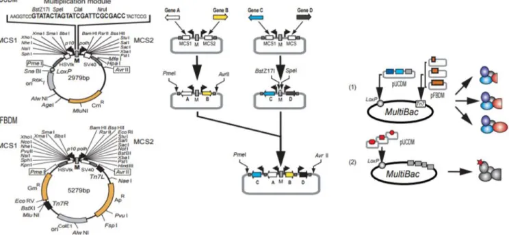
Figure 1.3 Multibac Expression system strategy showing the cloning approach of the system which enables high yield production of protein complexes. [71]
In multibac system given in figure above, two transfer vectors namely-pFBDM and pUCDM having a multiplication module (M) between two expression cassettes were designed and those cassettes were planned to driven under the control of p10 and polh viral promoters. PmeI and AvrII are the restriction sites that are used in the integration of expression cassettes in to another cassette containing more genes. In the assembly of the cassettes, PmeI& AvrII are compatible to BstZ17I&SpeI or NruI&SpeI restriction enzymes pairs so that one can assemble as many as possible genes in the transfer vector, theoretically. [71] Apart from this, two transfer vectors were constructed as having different mechanisms for gene integration to the bacmid. The clones are selected with simple blue/white screening according to resistant markers found in two vectors.
18
The production of multi-subunit protein complexes is inevitable in structural studies such as X-ray crystallography, functional domain-interaction studies and in exploring interaction surface determination. Multibac baculovirus system enables scientists to purify large-scale protein-complexes with optimum efficiency in terms of stoichiometry, functionality and purity.[ı] In addition to allowing proper protein folding and optimum gene expression from one baculovirus containing multiple genes, this system also allows the use of viruses in the mammalian cells to examine multi gene delivery. It is plausible to modify those transfer vectors (pFBDM, pUCDM) with mammalian promoters to drive expression in those cells as an alternative way to gene thèrapie studies. [71]
19
CHAPTER 2
MATERIALS:
2.1 BUFFERS and SOLUTIONS:
2.1.1 Western Blot, Staining and Nuclear Extract buffers:
1X SDS-PAGE Running Buffer 25 mM Tris, 192 mM glycine, 0.1% SDS
1X TAE 40 mM Tris (pH 7.6), 20 mM acetic acid,
1 mM EDTA
1X PBST 8mM Na2HPO4, 150mM NaCl, 2mM KH2PO4,
3mM KCl, 0.05% Tween20, pH 7.4 1X Tris-Glycine Transfer Buffer 25 mM Tris, 192 mM glycine
Western Blot stripping Buffer 62.5mM Tris-HCl (pH 6.7), 100mM beta-mercaptoethanol, 2% SDS
BC1000 (‘‘BC’’ followed by a numerical designation indicates the(variable) KCl concentration of buffers that, otherwise, have the same composition. eg:BC1000=1M KCl containing buffer) 20mM Tris– HCl (pH 7.9 at 4°C), 20% glycerol, 0.1mM EDTA, 0.5mM PMSF, 0.5 mM DTT, 1M KCl BC0 20mM Tris–HCl (pH 7.9 at 4°C), 20% glycerol, 0.1mM EDTA, 0.5mM PMSF, 2 mM DTT
Comassie Brilliant Blue (CBB) Staining Solution
0.1% CBB R-250 (w/v), 50% methanol(v/v), 10% glacial acetic acid(v/v), 40% H2O
20
2.1.2 Buffers for Immobilized Template Recruitment Assay using Streptavidin Dynabeads
2X B&W buffer 10mMTris-HCl (pH:7.5), 1mM
EDTA(pH:0.5), 2M NaCl
10X Assay Mix 0.2M HEPES-KOH (pH 8.2), 50mM MgCl2
Blocking Buffer 10X Assay Mix, 5mg/ml BSA, 5mg/ml PVP,
12.5mM DTT, 3% np-40
Wash Buffer 40mM HEPES, 4mM MgCl2, 4mM DTT,
100mM KCl, 0.1% np-40
Destaining solution 50% H2O, 40% methanol, 10% glacial acetic
acid
Acrylamide/Bisacrylamide solution (30%) 292g/L acrylamide 7.8g/L bisacrylamide 10% Ammonium Persulfate (APS) 100g/L APS (w/v)
4X SDS-PAGE sample loading buffer 240mM Tris-HCl (pH 6.8), 8% SDS(w/v) 40% glycerol(v/v), 0.04% bromophenol blue, 5% beta-mercaptoethanol
21 2.1.3 His-Tag Dynabeads Pulldown Buffers:
2X B&W 80mM HEPES KOH(pH:8.2), 600mM NaCl,
0.02% Tween-20
His-Elution Buffer 300mM Imidazole, 40mM HEPES KOH
pH:8.2, 10mM NaCl
2.2 MATERIALS
2.2.1 Cell Culture Media, Supplements and Culture Equipment:
Grace’s Insect Media (TNM-FH) Lonza Biowhittaker Cat. No: 04-649F
Gentamicin
Poloxamer SIGMA 16758
DMEM (1X) Gibco Life technologies Cat. No:
31885-023
Fetal Bovine Serum (FBS) Biowest Cat. No: S181T-500
Penicillin/Streptomycin Gibco Cat. No: 15140-122
100mm and 145 mm dishes
Corning Coster
6-well and 96 well plates Corning Coster
Plastic Pipettes (5/10/25 ml ) Corning Coster Spinner Flasks for Insect Cell Culture(10ml,
250 ml , 500ml )
22
2.2.2Antibodies Used in Western Blot and Immunoprecipitation
Med4 Home Made Med26 Home Made
Med6 Home Made Med27
Santacruz sc-390296
Med7 Home Made Med28 Home Made
Med12 Home Made Med29 (B-1) Santa Cruz
Biotechnology SC393800
Med13 Home Made Med30 Home Made
Med14 Abcam # ab170605 Rpb1(8WG16) Home Made
Med15 Proteintech
115661AP
Rpb5 Home Made
Med16 Santa Cruz Rpb6 Home Made
Med17 Home Made P53 Home Made
Med22 Home Made P62 Home Made
Med23 Home Made TAF100 Home Made
Med24 Home Made CDK7 Cell signaling
Med25 (A-7) Santa Cruz
Biotechnology SC393759 Anti-Flag Sigma F7425 ERα Cell Signaling mAb #8644 Cdk8 Home Made ccnc Home Made
23 2.2.3 Kits and other Tools :
BCA Protein Assay Kit Pierce BCA protein assay kit ThermoFisher Scientific Cat. No: 23227
Silver Stain Kit Pierce Silver Stain for Mass Spectrometry ThermoFisher Scientific Cat. No: 24600 Anti-flag M2 Affinity Agarose Beads Sigma #A4596
Flag Peptide Sigma #F3290
Dynabeads Histag Isolation Pulldown Novex Life Technologies Cat. no: 10103D Dynabeads M-280 Streptavidin Invitrogen ThermoFisher Scientific Cat. No:
11205D
Anti-HA M2 affinity Agarose Beads Thermofisher #88836
24
CHAPTER 3
METHODS:
3.1 Reconstitution and Purification of Partial Human Mediator complexes using Baculovirus Expression System
Mediator subunit cDNAs were cloned into pFBDM and pUCDM transfer vectors. Different tags (HA, histidine or Flag) were inserted into different subunits of the Mediator in order to enhance downstream purification. The individual cDNAs for subunits of the Mediator head module (MED6, MED8, MED11, MED18, MED19, MED20, MED22, and MED30) were inserted into the pFBDM and pUCDM transfer vectors and individual cDNAs for subunits of the middle module were also inserted into pFBDM and pUCDM transfer vectors (HA-MED7, MED4, MED21, His: MED10, MED31, MED9 and MED26). MED17 and Flag MED14 were integrated into different bacmids separately. Single viruses were generated by integrating the vectors in to bacmids followed by transfection of Sf9 cells with those bacmids. First generation (Po) viruses were amplified in Sf9 cells to second and third generation high titer P1 and P2 respectively. For protein production, Hi5 cells were infected with the particular viruses. (e.g :100 ml Hi5 cells were infected with (e6/ml) 1ml Head P2 +1ml f:Med17,18 to purify Head Module) In order to obtain near -stoichiometric complexes (one to one ratio proteins in the partial complexes) virus titrations were adjusted in each infection. 60-72 hours after infection, depending on the number of infected and dead cells, cells were collected and spin at 1500 rpm for 5 minutes. The pellet was resuspended in BC500 buffer (500mM KC, 20mM Tris– HCl (pH 7.9 at 4°C), 20% glycerol, 0.1mM EDTA, 0.5mM PMSF, 0.5 mM DTT) and homogenized by douncing 3 times using glass douncers. The lysate was centrifuged at 14000 rpm for 15 minutes. After centrifugation, the lysate was diluted to 300mM KCl by drop-wise addition of BC0.
25
The cell extracts were incubated with either anti-HA or anti-flag M2 agarose beads to pull down the partial complexes. Incubation was done overnight at 4°C by rotating. After overnight incubation, the beads were washed with 1ml BC300 containing 0.1% np-40 5 times. The complexes were eluted using 0.5mg/ml corresponding Flag or HA peptides at 4°C for 45 minutes by rotating. Elutions were repeated 3 times and each elute was analysed by CBB staining and WB.
3.2 The purified protein complexes using Baculovirus Expression System
Proteins/ Complexes
Number of Subunits in the complex Amount of Infected Hi5 cells Amount of infected Viruses Protein yield
Head (H)Module MED6, MED8, MED11, MED18, MED19, MED20, MED22, MED30, f:MED17,18 150 ml Hi5 (e6/ml) 750ul f:MED17,18 P2 + 1.5ml HEAD P2 1.2 ug/ul
Middle(M) Module HA:MED7, MED4,
MED21,His:MED10,MED31, MED9,MED26, f:MED14 100 ml Hi5 (e6/ml) 500ul f:MED14 P2 + 1ml MIDDLE P2 0.9ug/ul
H+M Module MED6, MED8, MED11, MED18, MED19, MED20, MED22, MED30, HA:MED7,
MED4,MED21, His:MED10, MED31, MED9, MED26, f:MED17,18 150 ml Hi5 (e6/ml) 1 ml f:MED17/18 P2, 1.5 ml HEAD P2, 1.5 ml MIDDLE P2 1ug/ul H+M+f:FLMED14 (core complex) H+M subunits, f:MED14, MED17,18 200 ml Hi5 (e6/ml) 1.5ml f:MED14 P2, 2.5 ml HEAD P2, 2.5 ml MIDDLE P2, 1.3ug/ul
26 2.5 ml MED17/18 P2 H+M+f:C’MED14 (core variant) H+M subunits, f:C’MED14, MED17,18 200 ml Hi5 (e6/ml) 1.5ml f:C’MED14 P2, 2.5 ml HEAD P2, 2.5 ml MIDDLE P2, 2.5 ml MED17/18 P2 1.2ug/ul H+M+f:N’med14 (core variant) H+M subunits, f:N’MED14, MED17,18 200 ml Hi5 (e6/ml) 1.5ml f:N’MED14 P2, 2.5 ml HEAD P2, 2.5 ml MIDDLE P2, 2.5 ml MED17/18 P2 1.3ug/ul
Kinase Module HA:CDK8,MED13,MED12, CCNC 200 ml Hi5 (e6/ml) 1ml HA:CDK8 P2, 2 ml MED13 P2, 2 ml MED12 P2, 2 ml CCNC P2 0.6ug/ul
Tail Module MED15,MED16,MED23,MED24, MED25,MED27/29, f:MED14 200 ml Hi5 (e6/ml) 1 ml from each subunits’ P2 viruses 0.6ug/ul f:p53 f:p53 150 ml Hi5 (e6/ml) 1.5 ml f:P53 P3 1.4ug/ul f:full length(FL)MED14 f: FL MED14 150 ml Hi5 (e6/ml) 2 ml f: FL MED14 P2 0.7ug/ul
F:N’MED14 f:N’MED14 150 ml Hi5 (e6/ml)
2 ml f: N’MED14 P2 0.7ug/ul
f:C’MED14 f:C’MED14 150 ml Hi5 (e6/ml)
27
f:ERα f:ERα 150 ml Hi5
(e6/ml)
2 ml f: ERα P2 1.1ug/ul
3.3 Purification of Endogenous Polymerase II and Human Mediator Complexes from HeLa Cells Stably Expressing f:rpb9 and f:nut2 Respectively
3.3.1 Cell Culturing:
HeLa cells stably expressing one of the subunits of Mediator complex(f:Med10/Nut2) and Pol II (f:rpb9) were kindly gifted from Roeder Lab (Rockefeller University, New York) The cells were grown in DMEM containing 10% FBS and 1% P/S also 300ug/ml geneticin for maintaining the stable cells in 145mm petri dishes in a 37°C incubator with 5 % CO2. Passaging was done by
washing the cells with 10ml 1X PBS after aspirating the media then trypsinizing with 2ml trypsin in 37°C incubator for 3 minutes. Cells were grown until they reached the sufficient amount for nuclear extract preparation.
3.3.2 Nuclear Extract Preparation:
Approximately 1.5 liter of HeLa cells stably expressing each f:rpb9 and f:nut2 were collected separately and centrifuged at 1500 rpm for 10 minutes at 4°C. The pellet was washed with ice-cold PBS two times, at each washing, centrifugating for 5 minutes at 1500 rpm at 4°C. The cells were lysed by douncing in the buffer containing 10mM Tris (pH:7.9) , 1.5mM MgCl2 , 10mM
KCl, 0.5mM DTT and 0.5 mM PMSF for three times. After that, lysate was centrifuged for 10minutes at 14000 rpm at 4°C and the pellet was saved. Nuclear pellets were resuspended in the buffer containing 20mM Tris (pH:7.9), 1.5 mM MgCl2 , 25% glycerol, 0.2mM EDTA, 0.5mM
DTT, 0.5mM PMSF and 0.3mM NaCl. Preparations were rocked for 30 minutes at 4°C by rotating and centrifuged for 15 minutes at 14000rpm at 4°C. The supernatant was quick frozen and store at -80°C for further purification.
28
3.3.3 Pull Down of Endogenous Pol II and Human Mediator Complex
Approximately 20 ml of each f:rpb9 and f:Nut2 nuclear extract were incubated with 150 ul anti-flag M2 agarose beads for overnight at 4°C by rotating. Before incubation, the beads were washed with 1ml BC300 + 0.1% np-40 for 5 times. After overnight incubation, the extracts were centrifuged at 1500 rpm for 5 minutes. The flow through was kept for Western Blot analysis. The beads were washed with BC300 +0.1% np-40 4 times. The last wash was done with BC200. The complexes were eluted by using 0.5 mg/ml flag peptide in BC200 for 45 minutes at 4°C by rotating. Elutions were repeated three times and each step, 0.5mg/ml flag peptide was used. Three elutions were pooled for each Mediator and Pol II complexes and analyzed by Silver Staining.
3.4 Silver Staining:
All steps in silver staining was performed in a clean tray with constant gentle shaking. Staining was done by using Pierce Silver Stain for Mass Spectrometry Kit according to manufacturer’s instructions. Before running the gel on SDS-PAGE, all apparatus were cleaned with ultrapure water in order to prevent any dirt during the staining procedure. After running the gel, it was washed with ultrapure water for 5 minutes two times. The gel was fixed with the solution containing 30% ethanol, 10% acetic acid and 60% water for 15 minutes. Fixing step was repeated two times and the gel was washed with 10% ethanol for 5 minutes two times. The gel was washed with ultrapure water for 5 minutes. Sensitizer working solution was prepared by mixing 1part Silver Stain Sensitizer with 500 parts ultrapure water (25ml water+50ul sensitizer). The gel was incubated in sensitizer solution for exactly 1 minute then washed with ultrapure water for 5 minutes two times. Enhancer solution was prepared by mixing 100 parts Silver Stain with 1part silver Stain Enhancer (0.25 ml of enhancer with 25 ml of stain) and incubated with the gel for 5 minutes. After enhancer, the gel was washed with ultrapure water for 20 seconds and then incubated with developer solution containing 0.25 ml of enhancer with 25 ml developer for 2-3 minutes until the bands appeared. When the desired band intensity was reached the developer was stopped by adding 5% acetic acid.
29 3.5 Protein Quantification
Protein quantification was done by using the BCA Assay Kit according to manufacturer’s instructions. 0.1 mg/ml BSA was prepared by diluting 2 mg/ml BSA stock. BSA standard solutions were prepared at eight different concentrations ranging from 0 to 0.1 ug/ul. 400 ul standard solutions were prepared as triplicates with a dilution factor of 100. The samples also diluted by 1:100 (4ul protein sample+ 396 ul ultrapure H2O) and prepared as triplicates. 100ul from standards
and samples were loaded to 96 well plate as triplicates. Working BCA solution was prepared by mixing reagents A and B in a 50:1 ratio and 100ul of it was mixed with the standards and with the samples. The plate was incubated at 37°C for 1-1.5 hours and absorbances were measured by using the SynergyHT microplate reader (Biotek,VT,USA) at 562nm. Using the absorbance values obtained from BSA standards and samples, a calibration curve was drawn and sample concentrations were quantified by using the standard curve equation.
3.6 SDS-PAGE
Before loading on the gel, concentrations of each protein sample was equalized by adding required amount(s) of ddH2O and 4X sample loading dye. The samples were denatured by boiling for 5
minutes at 95°C . Polyacrylamide stacking and separating gels were prepared by using the ingredients given in the table 3.6
Table 3.6 Reagents used in SDS-PAGE gel preparation
Reagent (for two gels )
7% separating gel 10% separating gel 12% separating gel 4% Stacking Gel H2O 7.65 ml 5.925 ml 4.875 ml 3 ml 1.5M Tris-HCl, pH8.8 3.75 ml 3.75 ml 3.75 ml 1.25 ml 0.5 M Tris-HCl pH6.8 20%(w/v) SDS 0.075 ml 0.075 ml 0.075 ml 0.025 ml
30 Acrylamide:bisacrylamide
(30% /0.8% w/v)
3.45 ml 4.95 ml 6ml 0.67 ml
10%(w/v) APS 125ul 125ul 125 ul 50 ul
TEMED 10ul 10ul 10ul 5 ul
Separating gel was poured between thick and thin glasses. In order to prevent any bubbles, the top of the gel was covered with isopropanol until the gel was fully polymerized. After polymerization, isopropanol was decanted and stacking gel was poured on top of the resolving gel and 15-well 1mm comb was placed to the stacking gel. Protein samples and appropriate protein marker was loaded to the gel and it was run first at 70V and after the samples entered to the resolving gel, it was run at 110 V for about 90 minutes.
3.7 Western Blotting:
After running the samples on SDS-PAGE, the proteins were transferred to PVDF membrane. The cassette was prepared by putting 4 layers of whatman paper on top of the sponge and then the gel and the membrane were placed on the cassette. Before transfer, PVDF membrane was activated by sinking in to 100% methanol for 1 minute. After setting the cassette, the transfer was performed at 330mA for 3 hours at 4°C. The membrane was blocked in 5% milk for 2 hours at room temperature by gently shaking. Primary antibody incubation (antibodies n 1% milk) was done either at 4°C overnight or 1 hour at room temperature by gently shaking. Membrane was washed with 1X PBST for 4 times with 5 minute intervals than secondary antibody (in 1% milk) incubation was done at room temperature for two hours with gentle agitation. The membrane was incubated with 1:1 ratio ECL solution for 2 minutes and development was performed in the dark room. The membrane was exposed to X-ray films with different periods of time ranging from 3 seconds to 30 minutes.
31
3.8 Immunoprecipitation for Pol II - Mediator Interaction by Using Purified Full length and truncated f:Med14 proteins
Purified full length f:Med14, amino terminus f:Med14 and carboxyl terminus f:Med14 were used in IP . Four reactions were designed by using 20ul A/G sepharose beads per reaction. Before starting, the beads were washed with 1ml BC300+0.1% np-40 for 5 times then 20ul beads were incubated with 3ul pol II 8WG16 antibody in 100 ul BC200 +0.025 % np-40.
After 4 hours at 4°C antibody incubation done in a rotator, the beads were washed with BC300+0.1% np-40 for 5 times. 12 ul purified Pol II was added to three reaction and 200ng full length and truncated f:Med14 proteins were added to remaining reaction in 100ul BC150+0.025% np-40 as a control. After 4 hours at 4°C incubation done in a rotator, the beads were washed with BC200+0.1%np-40 for four times at 4°C. The control beads were taken and 20ul 2X SDS sample loading buffer was added and kept at -20°C for WB analysis. 200 ng full length and truncated f:Med14 proteins added to three reactions sepearately in 100ul BC150+0.025 % np-40 and incubated for 4 hours at 4°C by rotating. After incubation, the beads were washed with BC200+0.05% np-40 for 5 times at 4°C and 20ul 2X SDS sample loading buffer was added to each reaction and kept at -20°C for WB analysis.
3.9 Immunoprecipitation (IP) for Pol II – Mediator Interaction by using Partial Mediator Complexes.
In order to see the direct interaction between Pol II and Human Mediator in the context of the complexes, the purified partial Mediator complexes were used in IP experiments. 5 reactions were designed by using Head, Head+Middle, Mediator core complex containing amino terminus of f:Med14, Mediator core complex containing carboxyl terminus of f:Med14 and finally Mediator core complex containing full length f:Med14. Since each complex compromising different number of subunits in them, before doing IP, each complex was checked by western blot and the concentration of each complex was adjusted accordingly. For each reaction, 20ul protein A/G sepharose beads were used. Before starting the experiment, the beads were washed with BC300 +0.1% np-40 for 5 times.