Laryngology
Malignant salivary gland tumours of the larynx:
a single institution review
Tumori maligni delle ghiandole salivari della laringe: un’unica review istituzionale
S. Karatayli-OzgurSOy1 2, J.a. BiShOp1 3, a.t. hillel1, l.M. aKSt1, S.r. BeSt11 Department of Otolaryngology, head and Neck Surgery, Johns hopkins School of Medicine, Baltimore, uSa; 2 Department of Otolaryngology, ufuk university, ankara, turkey; 3 Departments of pathology, Johns hopkins School
of Medicine, Baltimore, uSa SummAry
malignant salivary gland tumours of the larynx are very rare, with limited reports of clinical outcomes. We present the decade-long experi-ence of a single institution. A 10-year retrospective chart review of a tertiary head and neck cancer centre was performed. index patients were identified from a review of a pathology database, and reviewed by a head and neck pathologist. Patient demographics, presenting signs and symptoms, treatment modalities and clinical outcomes were extracted from electronic medical records. Six patients were included, with an age range of 44 to 69. All six had malignant laryngeal salivary gland tumours. Pathologies included: three adenoid cystic carcinoma (2 supraglottic, 1 subglottic), one mucoepidermoid carcinoma (supraglottic), one epithelial-myoepithelial carcinoma (supraglottic) and one adenocarcinoma (transglottic). All were treated with surgery (2 endolaryngeal, 4 open) and five of six with the addition of adjuvant therapy (4 radiotherapy, 1 concurrent chemoradiation). one patient had smoking history; no patients had significant alcohol history. With 4.5 years of median follow-up, none of the patients has had recurrence or local/distant metastasis. Salivary gland tumours of the larynx present in mid to late-age, and can be successfully managed with a multi-modality approach, resulting in excellent local and regional control rates.
Key WordS: Larynx • Cancer • Salivary • Chemotherapy • Surgery • Partial laryngectomy
riASSunTo
I tumori a istotipo salivare della laringe sono molto rari, con pochi report in letteratura in merito al loro andamento clinico. Nel presente manoscritto discutiamo un’esperienza di 10 anni presso una singola struttura. Abbiamo condotto una review retrospettiva della casistica di un centro di oncologia della testa e del collo di terzo livello. I pazienti sono stati individuati mediante analisi di un database e sono stati revisionati da un Anatomo Patologo testa collo. I dati inerenti la clinica, le modalità di trattamento e gli esiti sono stati prelevati da archivi elettronici. Sono stati inclusi sei pazienti nello studio, con un range di età dai 44 ai 69 anni. Tutti e sei erano affetti da neoplasie maligne a istotipo salivare della laringe. Gli istotipi includevano: tre carcinomi adenoido-cistici (2 sopraglottico, 1 sottoglottico), un carcino-ma mucoepidermoidale (sopraglottico), un carcinocarcino-ma epiteliale-mioepiteliale (sopraglottico), e un adenocarcinocarcino-ma (transglottico). Tutti sono stati sottoposti a trattamento chirurgico (2 chirurgie laser, 4 open) e 5 dei 6 pazienti sono stati successivamente sottoposti a terapia adjuvante (4 a radioterapia, 1 a radio-chemioterapia concomitante). Un paziente era fumatore; nessun paziente aveva storia di abuso di alcolici. A un follow-up con mediana di 4,5 anni nessuno dei pazienti ha presentato recidiva o metastasi locali o a distanza. I tumori a istotipo salivare della laringe si presentano solitamente in pazienti della seconda/terza età, e possono essere trattati con successo mediante approcci multimodali, con un ottimo controllo locoregionale di malattia.
PArole ChiAve: Laringe • Cancro • Salivare • Chemioterapia • Chirurgia • Laringectomia parziale
Acta Otorhinolaryngol Ital 2016;36:289-294
Introduction
malignant salivary gland tumours of the larynx are very rare neoplasms, which account for < 1% of all laryngeal malignancies 1. The most common malignant minor
sali-vary gland tumours are adenoid cystic (32-69%) and mu-coepidermoid carcinomas (15-35%) 2; adenocarcinomas
are less frequent, and epithelial-myoepithelial carcinomas are even more rare 3. unlike squamous cell carcinoma,
which is strongly associated with inhaled tobacco use, malignant salivary tumours in the larynx have no strong
association with smoking and appear to occur equally in both sexes. These tumours arise from subepithelial mu-cous glands in the larynx, which are found most common-ly in the subglottis and supraglottis 4. in the true glottis,
the possible areas of origin are floor of the ventricle and below the anterior commissure in the subglottis. Because these tumours do not arise on the free edge of the true vo-cal folds which are covered with thin squamous epitheli-um, there is often no voice change to detect these tumours at an early stage. They typically present in a subepithelial
fashion, and can grow to a large size before they present with dysphonia or airway symptoms.
The aim of this study is to present our experience over the last decade with these rare tumours to provide insight into the multimodality treatment that is typically required for long-term oncologic success.
Materials and methods
in an irB-approval protocol, the pathology archives of the Johns hopkins hospital and clinical records of pa-tients with laryngeal cancer were reviewed to identify patients with malignant salivary gland laryngeal lesions between January 2004 and december 2013. Age, gender, presenting symptoms, location of the tumour, pathology, Tnm classifications, treatment modality (surgery and/ or chemo radiotherapy) and disease status was extracted from patient charts.
Results
Six patients (3m, 3F) were included, with an age at diag-nosis range of 44 to 69 (mean: 56 years). Pathologic clas-sifications are summarised in Table i and included: three adenoid cystic carcinomas, one mucoepidermoid carci-noma, one epithelial-myoepithelial carcinoma and one adenocarcinoma, not otherwise specified. The most com-mon presenting symptom was dysphonia. half of patients presented with advanced stage disease, but all except the adenocarcinoma presented without regional or distant metastases. only one patient was a smoker (T4 subglottic adenoid cystic carcinoma).
Treatment modalities and oncologic results are summa-rised in Table i. All patients were treated primarily with
surgery, and negative surgical margins were obtained in all cases. The type of surgical approach varied with each case, but two were successfully managed with endola-ryngeal surgery, while four required an open surgical ap-proach. All but one patient underwent additional adjuvant therapy regardless of pathologic type or surgical margins (4 radiotherapy, 1 concurrent chemoradiation). After 4.5 years of median follow-up, none of the patients has had recurrence or local/distant metastasis. To highlight the unique presentation and management of these cases, three case studies with pathologic images are presented below.
Representative cases
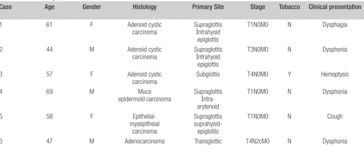
Case two: A 44-year-old male professor presented with dysphonia lasting two years. he denied any difficulty swallowing; however, he had a significant amount of coughing while eating. on fiberoptic laryngoscopy a la-ryngeal mass on the epiglottis was identified with normal vocal fold mobility (Fig. 1), and a staging CT scan dem-onstrated a large bulky supraglottic lesion on the laryn-geal surface of the epiglottis from the tip to the petiole, extending into the preepiglottic space, with fat planes preserved along the hyoid bone. direct laryngoscopy and biopsy was performed. The lesion was consistent with ad-enoid cystic carcinoma with both tubular and cribriform histology (Fig. 2).
With these findings, he underwent an open supraglottic laryngectomy. The final pathology demonstrated adenoid cystic carcinoma with perineural invasion and no defini-tive venous/lymphatic invasion and extensive involve-ment of the paraglottic space, staging the tumour as T3. Surgery was followed by post-operative adjuvant radia-tion therapy. At latest follow-up, 1.5 years after surgery, he was free of disease.
Table I. Patient demographics and tumour characteristics, treatments and uutcomes.
Case Age Gender Histology Primary Site Stage Tobacco Clinical presentation Surgical management Approach Surgical
margins Adjuvant therapy preservationLaryngeal Follow-up period Outcome
1 61 F Adenoid cystic
carcinoma Supraglottisİnfrahyoid epiglottis
T1N0M0 N Dysphagia Supraglottic
Laryngectomy Open - RT Y 3 y ANED
2 44 M Adenoid cystic
carcinoma Supraglottisİnfrahyoid epiglottis
T3N0M0 N Dysphonia Supraglottic
Laryngectomy Open - RT Y 13 m ANED
3 57 F Adenoid cystic
carcinoma Subglottis T4N0M0 Y Hemoptysis İpsilateral NDTL + Open - RT N 14 y ANED
4 69 M Muco
epidermoid carcinoma Supraglottis İntra-arytenoid
T1N0M0 N Dysphonia Partial
Laryngectomy Laser Endoscopic - RT Y 4.5 y ANED
5 58 F Epithelial-myoepitheial carcinoma Supraglottis suprahyoid-epiglottis T1N0M0 N Cough Supraglottic
Laryngectomy Laser Endoscopic - None Y 6 y ANED
6 47 M Adenocarcinoma Transglottic T4N2cM0 N Dysphonia TL +
Bilateral Selective ND, Levels 1-5
Open - RT / CT N 11.5 y ANED
Case four: A 69-year-old man presented with hoarseness and a history of slow and progressive voice change in the last 2 months. he denied pain, bleeding, cough, dysphagia, or otalgia. he was a non-smoker but had persistent gastro-oe-sophageal reflux. A suspicious mass was noted in the poste-rior commissure of the larynx on flexible laryngoscopy, and biopsy revealed a mucoepidermoid carcinoma in the larynx. he was then referred to our centre for management.
The lesion was centred in the posterior commissure, di-rectly between the arytenoids, approximately 1 cm in maximum diameter. A preoperative mri of the neck re-vealed a fullness of the posterior commissure with no
well-defined mass, and no pathological cervical lymphad-enopathy. The lesion was therefore completely excised endoscopically, using sharp instrumentation and a tissue shaver. Final pathology demonstrated ‘intermediate grade mucoepidermoid carcinoma’ (Fig. 3). All surgical mar-gins were negative and post-operative radiation was used as adjuvant treatment. on last follow-up, 8 years after treatment, there was no evidence of disease.
Case five: A 55-year-old woman complained of persistent cough of one year duration. She had presented one year prior for the same complaint and was noted at that time to have a small, benign appearing lesion of the epiglot-tis, which was diagnosed as presumptive epiglottic cyst. A chest x- ray performed as part of her work-up revealed
Table I. Patient demographics and tumour characteristics, treatments and uutcomes.
Case Age Gender Histology Primary Site Stage Tobacco Clinical presentation Surgical management Approach Surgical
margins Adjuvant therapy preservationLaryngeal Follow-up period Outcome
1 61 F Adenoid cystic
carcinoma Supraglottisİnfrahyoid epiglottis
T1N0M0 N Dysphagia Supraglottic
Laryngectomy Open - RT Y 3 y ANED
2 44 M Adenoid cystic
carcinoma Supraglottisİnfrahyoid epiglottis
T3N0M0 N Dysphonia Supraglottic
Laryngectomy Open - RT Y 13 m ANED
3 57 F Adenoid cystic
carcinoma Subglottis T4N0M0 Y Hemoptysis İpsilateral NDTL + Open - RT N 14 y ANED
4 69 M Muco
epidermoid carcinoma Supraglottis İntra-arytenoid
T1N0M0 N Dysphonia Partial
Laryngectomy Laser Endoscopic - RT Y 4.5 y ANED
5 58 F Epithelial-myoepitheial carcinoma Supraglottis suprahyoid-epiglottis T1N0M0 N Cough Supraglottic
Laryngectomy Laser Endoscopic - None Y 6 y ANED
6 47 M Adenocarcinoma Transglottic T4N2cM0 N Dysphonia TL +
Bilateral Selective ND, Levels 1-5
Open - RT / CT N 11.5 y ANED
TL-Total laryngectomy; ND- Neck dissection; RT- Radiation therapy; CT- Chemotherapy; ANED- Alive with no evidence of disease
Fig. 1. Flexible laryngoscopy showing a large mass of the epiglottis that did not impair vocal fold mobility, but did cause dysphonia through a mass effect on the supraglottic.
Fig. 2. This case of adenoid cystic carcinoma consists of tubules and cri-briform collections of cells with minimal cytoplasm and hyperchromatic, an-gulated nuclei. Prominent false ducts (asterisk) and subtle true ducts (arrow) are present, as is classic for this tumour type.
non-small cell lung carcinoma, for which she received chemotherapy with etoposide and cisplatin, radiation, and left lobectomy with mediastinal and hilar lymph node dis-section. despite treatment for lung cancer, her cough and throat irritation persisted and she therefore represented to clinic. She was a life-long non-smoker.
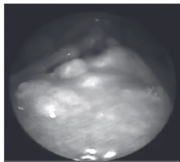
on flexible examination, the mass on the epiglottis was demonstrated again and was larger in size (Fig. 4). There-fore, endoscopic biopsy and excision of the mass was performed with Co2 laser. The lesion was centred on the
laryngeal surface of the epiglottis and completely excised to normal-appearing margins. The pathology of the le-sion returned as epithelial-myoepithelial carcinoma of the epiglottis (Fig. 5), with tumour extending to multiple cauterised specimen edges. Therefore, two months later, she underwent a repeat suspension microlaryngoscopy and Co2 laser excision of epiglottis with all final margins
negative for tumour. She has been regularly followed up for 6 years, and has no evidence of disease.
Discussion
in the larynx, salivary glands have a distinct anatomic dis-tribution 4, and occur mainly in the subglottis and
supra-glottis. relative rates of salivary malignancies for these subsites differ by series 2 5, but true vocal cord involvement
is rarely reported. This is in contradistinction to squa-mous cell carcinoma, which most commonly originates in the squamous epithelium covering the true vocal folds, and explains the difference in presenting symptoms and stage 6. most of our cases (67%) had supraglottic tumours
and therefore the most common presenting symptom was hoarseness, dysphagia and cough. it is interesting to note that the supraglottic tumours in our series mimic the
pat-terns of spread seen in traditional squamous cell carcino-ma – invasion in the preepiglottic but not to the true vocal cord, allowing for laryngeal preservation surgery. This sug-gests that the barriers to tumour spread within the larynx, such as the quadrangular membrane and conus elasticus, are resistant to tumours of all histologies. As in most other series, we report a male to female ratio of 1:1 1 2 7 8, with a
minority of patients having a history of smoking 2.
The predominant pathologic diagnosis in the literature is adenoid cystic carcinoma, consistent with the 50% rate reported in our series 2 5. in the early stages, salivary gland
tumours may not present with any symptoms due to slow submucosal spread. Two of three adenoid cystic carcino-ma cases were diagnosed as T3 and T4 tumours. one of
Fig. 3. Mucoepidermoid carcinoma consists of variable numbers of squa-moid cells, mucous cells and intermediate cells. This laryngeal intermediate-grade mucoepidermoid carcinoma consists primarily of uniform intermediate cells with abundant eosinophilic cytoplasm, with focal mucous cells (asterisk)
and squamoid cells (arrow). Fig. 4. Flexible laryngoscopy demonstrating an ulcerative mass on the la-ryngeal surface of the epiglottis with normal vocal fold mobility.
Fig. 5. This case of laryngeal epithelial-myoepithelial carcinoma was bipha-sic, with an inner layer of ductal cells (asterisk) tightly coupled with an outer layer of myoepithelial cells with clear cytoplasm (arrow).
these patients had perineural invasion. There are no stand-ardised therapeutic decisions with prospective studies due to the rarity and different histopathological types. Tumour type, grade, location and symptoms should be taken into consideration while choosing treatment options. Postop-erative radiotherapy is widely accepted as adjuvant mo-dality, especially in cases with a high-grade tumour, un-clear or positive margins, and perineural spread 8-12. All
three patients in our series with adenoid cystic carcinomas had adjuvant radiotherapy and none had recurrence. nod-al disease was not present in any patient in our study, and a low rate of nodal disease is consistent with previously published reports 1.
one of our patients had supraglottic mucoepidermoid carcinoma. About 60% of patients with mucoepidermoid carcinomas of the larynx are localised in the supraglot-tic area 13. in patients with high-grade mucoepidermoid
carcinomas, neck dissections have been suggested regard-less of clinical nodal status 13; others conclude that neck
dissections are not indicated in salivary gland tumours of the larynx unless nodal metastasis is clinically appar-ent 1 2 7 8. radiotherapy is also recommended in high-grade
cases because of the high incidence of local recurrence 14.
our patient underwent endoscopic excision, and given the patient’s intermediate grade pathology he also underwent adjuvant radiation therapy.
The most common site of epithelial-myoepithelial carci-noma (emC) is the parotid gland or submandibular gland. emC of the larynx is very rare. only four cases have been previously reported in the literature to date 3 15-17; one of
them being the present case 15. information about
demo-graphics, treatment and prognosis of emC in the larynx is scarce because of its rarity and there is no consensus for management. in our case, long-term disease control was achieved with surgical excision, but as highlighted in the case presentation, submucosal spread and an unusual pa-thology has the potential to create diagnostic uncertainty and inadequate initial resection if the surgeon is anticipat-ing a benign pathology. Therefore, our case required re-resection to achieve negative margins after final pathology from the surgical specimen revealed emC and positive margins.
Finally, one patient in the current series presented with advanced adenocarcinoma, not otherwise specified (noS) with significant local and nodal disease. only 0.35% to 0.5% of all laryngeal malignancies are adenocarcinomas noS, and are mostly seen in males 18, as in our case.
Ad-enocarcinomas noS of the larynx tend to metastasise to both regional and distant site 19-21 and therefore require
ag-gressive surgical management and adjuvant therapy. our patient did not have any recurrence or metastasis after to-tal laryngectomy and adjuvant chemoradiotherapy after a follow-up of 11.5 years.
Careful long-term follow-up for laryngeal salivary tu-mours is critical. distant metastases to the lung is a
hall-mark of adenoid cystic carcinoma and long-term follow up is mandatory, since recurrences may occur after more than 10 years after primary treatment 2 8 12 22. nevertheless,
adjuvant therapy may have a role in achieving long-term disease control, as the majority of our patients received adjuvant treatment and none had recurrences and/or local or distal metastasis after a median follow-up of 4.5 years.
Conclusions
Salivary gland malignancies are rare tumours of the lar-ynx that present in mid- to late-age, and can be success-fully managed with a multi-modality approach, resulting in excellent local and regional control rates.
This work was presented at the 10th Congress of the Eu-ropean Laryngology Society, April 9-12, 2014, Antalya, Turkey.
References
1 Batsakis Jg, luna mA, el-naggar AK. Nonsquamous
carcinomas of the larynx. Ann otol rhinol laryngol
1992;101:1024-6.
2 ganly i, Patel Sg, Coleman m, et al. Malignant minor
sali-vary gland tumors of the larynx. Arch otolaryngol head
neck Surg 2006;132:767-70.
3 moukarbel rv, Kwan K, Fung K. Laryngeal
epithelial-my-oepithelial carcinoma treated with partial laryngectomy. J
otolaryngol head neck Surg 2010;39:e39-41.
4 Bak-Pedersen K, nielsen Ko. Subepithelial mucous glands
in the adult human larynx. Studies on number, distribution
and density. Acta otolaryngol 1986;102:341-52.
5 nielsen TK, Bjørndal K, Krogdahl A, et al. Salivary gland
carcinomas of the larynx: a national study in Denmark.
Au-ris nasus larynx 2012;39:611-4.
6 levine hl, Tubbs r. Nonsquamous neoplasms of the larynx.
otolaryngol Clin north Am 1986;19:475-88.
7 veivers d, de vito A, luna-ortiz K, et al. Supracricoid par-tial laryngectomy for non-squamous cell carcinoma of the
larynx. J laryngol otol 2001;115:388-92.
8 del negro A, ichihara e, Tincani AJ, et al. Laryngeal
ad-enoid cystic carcinoma: case report. Sao Paulo med J
2007;125:295-6.
9 Wang mC, liu Cy, li Wy, et al. Salivary gland carcinoma
of the larynx. J Chin med Assoc 2006;69:322-5.
10 Zvrko e, golubović m. Laryngeal adenoid cystic carcinoma.
Actaotorhinolaryngol ital 2009;29:279-82.
11 Calis AB, Coskun Bu, Seven h, et al. Laryngeal
mucoepi-dermoid carcinoma: report of two cases. Auris nasus larynx
2006;33:211-4.
12 Tincani AJ, del negro A, Araújo PP, et al. Management of salivary gland adenoid cystic carcinoma: institutional
expe-rience of a case series. Sao Paulo med J 2006;124:26-30.
13 Prgomet d, Bilić m, Bumber Z, et al. Mucoepidermoid
car-cinoma of the larynx: report of three cases. J laryngol otol
14 gomes v, Costarelli l, Cimino g, et al.
Mucoepider-moid carcinoma of the larynx. eur Arch otorhinolaryngol
1990;248:31-4.
15 visaya Jm, Chu eA, Schmieg J, et al. Pathology quiz case 1.
Epithelial-myoepithelial carcinoma (EMC). Arch
otolaryn-gol head neck Surg 2009;135:832.
16 mikaelian do, Contrucci rB, Batsakis Jg. Epithelial-my-oepithelial carcinoma of the subglottic region: a case
pres-entation and review of the literature. otolaryngol head neck
Surg 1986;95:104-6.
17 yan y, Wang l, Ke J, Sun S, et al. Diagnosis and treatment of
nonsquamous cell neoplasms located in subglottis. lin Chung
er Bi yan hou Tou Jing Wai Ke Za Zhi 2014;28:182-5.
18 ebru T, omer y, Fulya oP, et al. Primary mucinous
adeno-carcinoma of the larynx in female patient: a rare entity. Ann
diagn Pathol 2012;16:402-6.
19 haberman PJ, haberman rS. Laryngeal adenocarcinoma, not otherwise specified, treated with carbon dioxide laser
excision and postoperative radiotherapy. Ann otol rhinol
laryngol 1992;101:920-4.
20 Fechner re. Adenocarcinoma of the larynx. Can J
otolaryn-gol 1975;4:284-9.
21 hilger AW, Prichard AJ, Jones T. Adenocarcinoma of the
lar-ynx--a distant metastasis from a rectal primary. J laryngol
otol 1998;112:199-201.
22 monin dl, Sparano A, litzky lA, et al. Low-grade mucoep-idermoid carcinoma of the subglottis treated with
organ-preservation surgery. ear nose Throat J 2006;85:332-6.
Address for correspondence: Simon r.A. Best, department of oto-laryngology, head and neck Surgery, Johns hopkins School of me-dicine, 601 n. Caroline Street, Baltimore, md 21287, uSA. Tel. 410 955 1654. Fax 410 955 0035