Reşat Dikme1, Mahmut Padak1, Mustafa Göz2, Mahmet Salih Aydın2, Ömer Göç1 1Vocational School of Health Services, Perfusion Techniques Program, 2Department of Cardiovascular Surgery, Faculty of Medicine, Harran University, Şanlıurfa, Turkey
Prognostic biomarker: ST2
New prognostic biomarker in cardiovascular field: ST2 (IL1RL1)
DOI: 10.4328/ACAM. 20008 Received: 21.03.2019 Accepted: 10.04.2019 Published Online: 13.04.2019
Corresponding Author: Reşat Dikme, Harran University Yenişehir Campus Vocational School of Health Services, Haliliye, 63300, Şanlıurfa, Turkey. GSM: +905354587844 F.: +90 4143183209 E-Mail: [email protected]
ORCID ID: 0000-0001-9157-7830
Abstract
As a result of natural immunity in the heart ST2 (IL1RL1) that protects the heart against excessive pressure load and tension, has two important isoforms (ST2L–membranebound, sST2-soluble). As ligand of ST2L and sST2 is interleukin 33 (IL33), the connection of ST2L to IL33 stimulates cardioprotective signal cascade whereas connection of sST2 to IL33 causes annihilation of these positive effects. As a high level of sST2 shows that the heart is under dense stress, this situation causes cell death, tissue fibrosis, decreased cardiac functions and an increase in progression rate of disease. That is why in cardiovascular dis-ease sST2 is accepted as a biomarker of poor prognosis. It was obtained that the incrdis-ease of sST2 over normal concentration multiplies negative situations related to cardiovascular diseases 3 times more. In 2013 in ACCF/AHA (American College of Cardiology Foundation/American Heart Association) guidelines, sST2 that was defined as “novel biomarker” for HF (Heart failure), is an independent biomarker from natriuretic peptides and cardiac troponins and is an important sign in cardiovascular diseases. ST2 is not affected by factors such as age, body mass index and kidney function disorder.
Keywords
ST2; Interleukin 1 Receptor-Like 1; sST2; Soluble sST2; Cardiovascular Diseases; IL1RL1
The Annals of Clinical and Analytical Medicine
Introduction
Interleukins (IL) are effective in the formation of cardiac remod-eling, they also show a cardioprotective effect by functioning as signal molecules between cardiomyocytes and fibroblasts during the proinflammatory period. Especially IL33 can be-have as a cytokine and transcriptional featured nuclear factor helps to protect heart and form signal among cells [1, 2]. ST2 (Interleukın 1 Receptor-Lıke 1, IL1RL1, T1, IL1RL1, Fit1, IL33R, DER4) plays a part in important steps in this period and inflam-matory response [3].
As a result of valvular heart disease, hypertension, coronary ar-tery disease (CAD) and myocardial infarction, pathophysiologi-cal processes such as cardiomyocyte hypertrophy and cardiac fibrosis occur. All these processes are given as a response to mechanical overload and wall stress is tried to be pulled to nor-mal limits in this way. As these responses are given deadly situ-ations show up mostly. When there is mechanical overload in heart, IL33 shows paracrine effect between cardiac myocytes and fibroblasts [1]. Although this cytokine is mainly produced in the heart by fibroblasts, they are produced by myocytes dur-ing cell necrosis [1, 4]. IL33, which’s gene expression is sup-ported by TNF- α (tumor necrosis factor α) and IL-1 β, prevent hypertrophy formation against pro hypertrophic factors [5]. In the studies done with rat, there are obtained findings of IL33 decreases heart hypertrophy and fibrosis when there is cardio-vascular overload. The same results are achived with IL33, that is applied to fibroblasts in a cell culture environment, similar to these data protecting heart [6].
During cellular stress with the liberalization of IL33, IL33 shows the effect by connecting to ST2. ST2, which was discovered in 1989, is the member of interleukin-1 (IL1) receptor protein family and it plays a central role in immune response and orga-nization of inflammatory response [7]. Many studies were done regarding IL1 family that is named as cytokines due to their intercellular communication capacities. These studies helped to clarify the connection between the rising body temperature and infection or inflammation. Among wide physiological role of IL1 family, there is stimulation and inhibition of cells.
The discovery in 1989 was made by two independent labs studying on growth and stimulated fibroblasts [8]. ST2 has been known as an orphan receptor (receptor associated with immune and inflammatory) for 15 years (1989-2005) however in 2002 it was obtained that it was secreted by cardiac cells as a response to myocardial stress and defined as IL33 receptor in 2005 [9]. ST2 that is in Q arm of the 2nd chromosome (2Q12) consists of 13 exons, 40536 base and 556 aa [7].
ST2 İsoforms
As a result of natural immunity ST2 that protects the heart against excessive pressure load and tension, has two important isoforms. These isoforms are membranebound form-ST2L and soluble sST2 (Figure 1).
As a membrane receptor ST2L is also known as IL1RL1-Beta whereas soluble receptor sST2 is known as IL1RL1-Alfa [2]. The third known isoform of ST2 is a variant form which is STV is de-fined as dominant in the gastrointestinal system [10]. Different forms of ST2 are formed with alternative splicing of the same mRNA. ST2 gene that is formed with the processing of alterna-tive splicing of 3 of the same mRNA and gene promoters, has proximal and distal promoters and is modified with transcrip-tional regulation [2].
sST2 was first used in 2002, it is a potential biological
bio-marker for the cardiac disease and this situation was empha-sized with a finding that sST2 mRNA is highly inducted after mechanical stress or treatment with IL-1β in cultured rat car-diomyocytes. In this study, sST2 concentrations were obtained from the blood of rats following myocardial infarction [5]. In a study done by Baba et al. two GATA patterns (1 and 2) were obtained in the upstream part of the region where transcrip-tion of the ST2 gene starts. In the same study especially in ST2 mRNA expression, it was found out that GATA2 provides the main control and as a result, GATA patterns were obtained to play an important role in the last expression of ST2 [11]. The data regarding inner control of differential transcription of sST2 and ST2L is not well-known and this situation is limited with hematopoietic cells (GATA2, neurotrophin p75 receptor) [11]. At sST2 levels, genetic factors affect 40% of interindi-vidual variability [12].
Membranedependent ST2L has 3 domains. One of the domains is “extracellular immunoglobulin G domain’’, the other one is “single transmembrane domain” and the last one is “intracel-lular domain”. Soluble sST2 does not have transmembrane and intracellular domain however it can move freely in the periphery cycle having with 9 aa C terminal sequence [2,13]. As ST2 act is complex and not quite unrealized, it is strictly connected with the effect style of IL33.
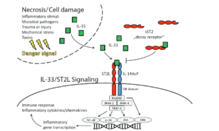
IL33 causes inflammatory gene transcription by bounding ST2L and as a result immune response is formed with stimulation of production of cytokines/chemokines. IL33 bounds to ST2L re-ceptor via IL1RAcP accessory protein. IL1RAcP accessory pro-tein increases bounding affinity of IL33 to ST2L [14]. As bound-ing of ST2L to IL33 shows the effect by stimulatbound-ing cardiopro-tective signal cascade (decrease of cardiac injury, prevention of apoptosis, the decrease of inflammatory effect, the decrease of hypertrophy and fibrosis, protection against mechanical warn-ing and damage), boundwarn-ing of sST2 to IL33 causes disappear-ance of these positive effects [1]. sST2 decreases the positive effects of IL33 by acting as a trap receptor (decoy receptor) [1]. By bounding IL33, sST2 decreases the usability of IL33 by inter-acting with sarcolemmal (cell membrane of muscle fiber) recep-tor. That is why one rise in sST2 can decrease the cardiopro-tective effect of IL33 over cardiac cells and create a negative prognostic effect over general cardiovascular risk profile [1]. According to multiple clinical studies, sST2 myocardium infarc-tion showed up as a clinically beneficial prognostic biologic biomarker in patients having cardiovascular diseases such as acute dyspnea and coronary failure with a low-risk community-based population [15]. According to the study done by Weinberg et al. increased sST2 levels just after myocardium infarction case, showed up as a negative prognostic factor [5].
Warning sequence occurs within the cell due to working of IL33/ST2L signal with NF-kB (nuclear factor kappa B), p38, JNK (jun N-terminal kinase) and ERK (extracellular signal-regulated kinases) pathway (Figure 2) [16]. IL33/ST2L also contributes to the immune response by excreting cytokines associated with TH2 in inflammatory diseases [17].
Kakkar et al. showed an increase of IL33 secretion from cyto-plasmic vesicles in the mechanical stress of living cells [16]. In another study parallel increased secretion of both ST2L and sST2 from cardiomyocytes and cardiac fibroblasts under bio-mechanic stress, was determined [18]. In a study done with rats, ST2 became the most stimulated molecule among thou-sands of gene transcription as a result of biochemical stress. In another study in models of ventricles overloaded with pressure, treatment with IL33 prevented hypertrophy whereas in hypoxia IL33 saved cardiomyocytes from apoptosis. In ischemia-reper-fusion MI IL33 treatment reduced infarction size, ventricular dilatation was refined, caspase 3 was repressed and apoptosis inhibitors increased [17]. IL33 can make these positive effects via ST2L. As a result of the blocking of ST2L by anti ST2L, the antihypertrophic and antiapoptosis effect of IL33 disappeared. In annihilation of the positive effect of IL33 by adding sST2, the same situation was observed. These studies present the importance of ST2.
Now the source of the sST2 cycle in healthy individuals and patients having different diseases cannot be obtained com-pletely. This is especially valid for cardiac disease. In a study done in neonatal rat heart myocytes, the source of the sST2 cycle in cardiac disease was found as myocardial derived [5]. In addition to this, newer studies showed that in human cardiac disease dominant source of sST2 may be vascular endothelium cells rather than human myocardium [19]. As all the studies are evaluated together it was found out that sST2 is produced by cardiac fibroblast, cardiomyocytes, macrovascular (aorta and coronary arteries) and microvascular endothelium cells in dam-age and stress condition.
sST2 Concentration Measurement
The level of sST2 can be obtained by making its quantitative measurement. In human serum/plasma for sST2 measurement, the first immunosorbent analysis related to enzyme was done in 2000 [20]. As high-level sST2 indicates that the heart is under stress, this leads to cellular death, tissue fibrosis in the heart as well as a decrease in cardiac functions and progress of the disease. That is why sST2 is accepted as a biomarker
of ingravescent prognosis in individuals in cardiovascular dis-ease [2]. As soluble sST2 is an urgent biomarker which is an important indicator of mortality, hospitalization requirement and negative results that may show up in patients with heart failure, it is a rather strong parameter for cardiovascular dis-eases. In recent years the reference values for sST2 in the cycle are derived from a subset of Framingham study (Framingham heart study) (Figure 3). According to this study there showed up important differences between males and females. In the study as reference range for sST2 in male sampling (n = 462) was found as 11-45 ng / mL, it was found as 9-35 ng / mL in female sampling (n=674) [21]. The results of the study regarding the reason for genderspecific differences and its possible results were not clarified [21].
In many previous studies as the normal average value of sST2 is 18 ng/ml, concentrations over 35 ng/ml strongly states in-creased risk in terms of cardiovascular diseases. It was ob-tained a value over 35, increases negative situations related to cardiovascular diseases 3 times more [10]. The basic reason for serum/plasma sST2 concentration difference generates from using different enzyme-linked immunosorbent assay (ELISA) kits. Today sST2 studies with ELISA method are generally done via 3 kits (Presage sT2 assay, R-D sT2 assay, MBL sT2 assay). The Penn Heart Failure Study (PHFS) revealed that high sST2 levels give a predictive idea about death, heart transplantation or hospitalization in patients having heart failure [23]. Accord-ing to the PHFS study, it was obtained that patients havAccord-ing sST2 value over 35 ng/ml carry 2.8 times higher risk of expos-ing negative results in 30 days compared to individuals havexpos-ing low sST2 concentration. According to the same study relative risks of exposing negative results in terms of cardiac diseas-es in the following 4 years in individuals having an sST2 level higher than 35 ng/ml, persist at 1.8 times. Since high sST2 level can reflect worsening of disease before changing symptoms of cardiac diseases visibly, treatment profile can be improved by evaluating sST2 level in plasma in patients having chronic heart failure.
According to new researches about ST2, HT, cardiac failure and cardiovascular mortality risk increases in asymptomatic indi-viduals having high sST2 values and in this way these people become a candidate for therapeutic interventions. According to an analysis done by the Framingham Heart Study Cohort, at least in 25% of asymptomatic individuals in terms of CAD, ST2 level gives important data. Among these patients, the ones having the highest sST2 values after an average of 11.3 years, sST2 levels were associated with 32% of cardiac failure and Figure 3. Serum/plasma concentration studies of s ST2 [22].
45% of death with increased risk. According to the results of another study, sST2 also shows itself as a strong ischemic bio-marker, especially it can predict cardiomyopathy etiologies. In these individuals making suitable risk classification by using sST2 level, will provide benefit.
In 2013; ST2 was defined as “novel biomarker” for HF (Heart failure) in ACCF/AHA (American College of Cardiology Founda-tion/American Heart Association) directory [24]. In clinical stud-ies done with patients having an acute myocardial infarction (AMI) or acute coronary syndrome, it was clearly revealed that increased sST2 is associated with disappointing results [25]. After AMI, early measuring sST2 in patients can be helpful in the prediction of recovery of negative left ventricle functionally. Although sST2 measuring in patients applied to ER with acute chest pain is not valuable for acute myocardium infarction or acute coronary syndrome diagnosis, lately sST2 is shown as an independent predictor about cardiovascular mortality and all long term reasons of disease in patients having stable CAD [26]. In the last term as sST2 becomes a biomarker independent from natriuretic peptides and cardiac troponins in the evalua-tion of HF, it is also an important sign in STEMİ, NSTEMİ, coro-nary artery, valvular diseases, cardiomyopathy, corocoro-nary bypass and cardiac surgery, acute cardiac allograft rejection and acute Kawasaki disease [27, 10].
Result
An optimum biologic biomarket that is used in diagnosis and prognosis should primarily be independent of other factors. For example, a biomarker that is used in cardiovascular diseases should not be in relation with other circumstances and should not be affected by mixing factors that can affect blood concen-trations. In order to become a reliable biomarker that is used in a disease, there should be an “optimal provision”. ST2 is not af-fected by factors such as age, body mass index and kidney func-tion disorder [27]. According to the results of studies done on in vitro stability of sST2, it stays stable in analytic room tem-perature for 48 hours, at 4°C for at least 7 days, at -20 °C and -80 °C for at least 1.5 years [28]. Increasing data about actions of sST2 and ST2L on cardiovascular system directed doctors thinking to evaluate sST2 plasma levels as a new marker re-garding cardiac failure and ischemic cardiac diseases. Defining of new biological biomarkers that can obtain ingravescent find-ings of clinical conditions of a disease earlier, stop or decrease negative results of disease is a chief step in clinical diagnosis development. Moreover, new biomarkers such as ST2 can be used to diagnose the benefits of early treatment and obtain a prognosis of disease [13].
Scientific Responsibility Statement
The authors declare that they are responsible for the article’s scientific content including study design, data collection, analy-sis and interpretation, writing, some of the main line, or all of the preparation and scientific review of the contents and ap-proval of the final version of the article.
Animal and human rights statement
All procedures performed in this study were in accordance with the ethical standards of the institutional and/or national re-search committee and with the 1964 Helsinki declaration and its later amendments or comparable ethical standards. No ani-mal or human studies were carried out by the authors for this article.
Funding: None Conflict of interest
None of the authors received any type of financial support that could be considered potential conflict of interest regarding the manuscript or its submission.
References
1. Sanada S, Hakuno D, Higgins LJ, Schreiter ER, McKenzie AN, Lee RT. IL33 and
ST2 comprise a critical biomechanically induced and cardioprotective signaling system. J Clin Invest. 2007;117 (6):1538–49.
2. Chiara C. Inflammation in cardiac disease: focus on Interleukin-33/ST2 path-way. Inflammation and Cell Signaling. 2014;1:e149.
3. Viereck J, Bang C, Foinquinos A, Thum T. Regulatory RNAs and paracrine net-works in the heart. Cardiovascular Research. 2014;102 (2): 290–301.
4. Demyanets S, Kaun C, Pentz R, Krychtiuk KA, Rauscher S, Pfaffenberger S. Components of the interleukin-33/ST2 system are differentially expressed and regulated in human cardiac cells and in cells of the cardiac vasculature. J Mol Cell Cardiol. 2012;60:16–26.
5. Weinberg EO, Shimpo M, De Keulenaer GW, Mac Gillivray C, Tominaga S, Solo-mon SD. Expression and regulation of ST2, an interleukin-1 receptor family mem-ber, in cardiomyocytes and myocardial infarction. Circulation. 2002;106:2961. 6. Zhu J, Carver W. Effects of interleukin-33 on cardiac fibroblast gene expres-sion and activity. Cytokine. 2012;58 (3):368–79.
7. Kakkar R, Lee RT. The IL33/ST2 pathway: Therapeutic target and novel bio-marker. Nat. Rev. Drug Discov. 2008; 7 (10): 827–40.
8. Tominaga S. A putative protein of a growth specific cDNA from BALB/c-3T3 cells is highly similar to the extracellular portion of mouse interleukin 1 receptor. FEBS Lett. 1989;258:301–4.
9. Schmitz J, Owyang A, Oldham E, Song Y, Murphy E, McClanahan TK, et al. IL33, an interleukin-1-like cytokine that signals via the IL-1 receptor-related pro-tein ST2 and induces T helper type 2-associated cytokines. Immunity. 2005;23 (5): 479–90.
10. Dieplinger B, Mueller T. Soluble ST2 in heart failure. Clinica Chimica Acta. 2015; 443, 57–70.
11. Baba Y, Maeda K, Yashiro T, Inage E, Kasakura K, Suzuki R, et al. GATA2 is a critical transactivator for the human IL1RL1/ST2 promoter in mast cells/baso-phils: opposing roles for GATA2 and GATA1 in human IL1RL1/ST2 gene expres-sion. J Biol Chem. 2012;287 (39):32689e32696.
12. Ho JE, Chen WY, Chen MH, Larson MG, McCabe EL, Cheng S, et al. Common genetic variation at the IL1RL1 locus regulates IL33/ST2 signaling. J Clin Invest. 2013;123 (10):4208e4218.
13. Weinberg EO. ST2 protein in heart disease: From discovery to mechanisms and prognostic value. Biomark Med. 2009; 3:495–511.
14. Liu X, Hammel M, He Y, Tainer JA, Jeng US, Zhang L. Structural insightsinto the interaction of IL33 with its receptors. Proc Natl Acad Sci U S A. 2013;110:14918– 23.
15. Wang TJ, Wollert KC, LarsonMG. Prognostic utility of novel biomarkers of cardiovascular stress: the Framingham Heart Study. Circulation. 2012;126:1596– 604.
16. Kakkar R, Hei H, Dobner S, Lee RT. Interleukin 33 as a mechanically respon-sive cytokine secreted by living cells. J. Biol. Chem. 2012; 287 (9): 6941–8. 17. Domingo A, Figal P, MD James L, Januzzi MD. The Biology of ST2: The Inter-national ST2 Consensus Panel. AmJ Cardiol; 2015;115[suppl]:3Be7B.
18. Matteo CM, Cortese F, Gesualdo M, Riccardi R, Di Nunzio D, Moncelli M, et al. A Novel Cardiac Bio-Marker: ST2: Molecules. 2013;18 (2): 15314-28.
19. Truong QA, Januzzi JL, Szymonifka J. Coronary sinus biomarker sampling com-pared to peripheral venous blood for predicting outcomes in patients with severe heart failure undergoing cardiac resynchronization therapy: the BIOCRT Study. Heart Rhythm. 2014;07:007.
20. Kuroiwa K, Li H, Tago K. Construction of ELISA system to quantify human ST2 protein in sera of patients. Hybridoma. 2000;19:151–9.
21. Coglianese EE, Larson MG, Vasan RS. Distribution and clinical correlates of the interleukin receptor family member soluble ST2 in the Framingham Heart Study. Clin Chem 2012;58:1673–81.
22. B. Dieplinger, T. Mueller / Clinica Chimica Acta 443. 2015;57–70. P.60. 23. Januzzi JLJr. ST2 as a cardiovascular risk biomarker: from the bench to the bedside. J. of Cardiovasc Transl Res. 2013; 6 (4):493–500.
24. McMurray JJ, Adamopoulos S, Anker SD, Auricchio A, Böhm M, Dickstein K, et al. ESC Guidelines for the diagnosis and treatment of acute and chronic heart fail-ure 2012: The Task Force for the Diagnosis and Treatment of Acute and Chronic Heart Failure 2012 of the European Society of Cardiology. Developed in collabo-ration with the Heart Failure Association (HFA) of the ESC. Eur Heart J. 2012;33 (14):1787–847.
25. Demyanets S, Speidl WS, Tentzeris I, Jarai R, Katsaros KM, Farhan S, et al. Soluble ST2 and interleukin-33 levels in coronary artery disease: relation to dis-ease activity and adverse outcome. PLoS One. 2014; 9(4):e95055.
26. Dieplinger B, Egger M, Haltmayer M. Increased soluble ST2 predicts long-term mortality in patients with stable coronary artery disease results from the Ludwigshafen Risk and Cardiovascular Health (LURIC) study. Clin Chem. 2014;60: 530–40.
27. Moore SA, Januzzi JL Jr. Found in translation soluble ST2 and heart disease. J Am Coll Cardiol. 2010;55:251e3.
28. Dieplinger B, Egger M, Poelz W, HaltmayerM, Mueller T. Long-term stability of soluble ST2 in frozen plasma samples. Clin Biochem. 2010;43:1169–70.
How to cite this article:
Dikme R, Padak M, Göz M, Aydın MS, Göç Ö. New prognostic biomarker in car-diovascular field: ST2 (IL1RL1). Ann Clin Anal Med 2019; DOI: 10.4328/ACAM. 20008.
![Figure 2. Intracellular pathways activated with interleukin-33/ST2L signal [10].](https://thumb-eu.123doks.com/thumbv2/9libnet/3723074.25627/3.914.93.442.224.441/figure-intracellular-pathways-activated-interleukin-st-l-signal.webp)
