Van Tıp Derg 26(2): 202-205, 2019 DOI: 10.5505/vtd.2019.79847
*Sorumlu Yazar: Müjgan Tek, TOBB Economy and Technology University Cardiology Department, Ankara, Turkey E-mail: [email protected], Tel: +90 (312) 292 99 00
Geliş Tarihi: 08.06.2018, Kabul Tarihi: 26.10.2018 KLİNİK ÇALIŞMA / CLINICAL RESEARCH
Effect of Vitamin D Status on Invasive
Electrophysiologic Parameters and Atrial Fibrillation
Inducibility
Vitamin D Düzeyinin İnvaziv Elektrofizyolojik Parametrelere ve Atriyal
Fibrilasyon İndüklenebilirliği’ne Etkisi
Müjgan Tek*, Basri Amasyalı, Mehmet Serkan Çetin, Aksüyek Savaş Çelebi, Erdem Diker,
Berkten Berkalp
TOBB Economy and Technology University Cardiology Department, Ankara, Turkey
Introductıon
Frequently attributed as a hormone of bone metabolism, Vitamin D (VitD) has multiple regulatory effects on other systems such as cardiovascular system. Deficiencies of VitD have been associated with coronary heart disease,
hypertension, and left ventricular hypertrophy (1). However the relationship of vitD status and invasive electrophysiologic properties has not been studied yet and also some controversial data arised (2-4). In this study we aimed to evaluate the
effects of vitD levels on invasive
electrophysiologic parameters and AF inducibility.
ABSTRACT
Objectives: Deficiencies of Vitamin D (VitD) has been
associated with coronary heart disease, hypertension and left ventricular hypertropy. However its effects on cardiac conduction system and atrial fibrillation (AF) predisposition have not been studied yet. In this study we aim to evaluate the effects of VitD on invasive electrophysiologic parameters and AF inducibility.
Materials and Methods: This retrospective
cross-sectional study included 135 patients. Study population was divided into three group as VitD sufficient, VitD insufficient and VitD deficient according to baseline vitD levels. Patients’ invasive electrophysiologic parameters and induced AF episodes were recorded.
Results: Corrected sinus node recovery time, baseline
cycle length, atrial-His interval, His-ventricular interval and Wenckebach cycle length were lengthened in vitD deficient group but they didn’t reach statistical significance. The rate of AF inducibility was twice as likely in VitD deficient group than sufficient group, however, it also didn’t reach statistical significance.
Conclusion: Baseline VitD levels were not associated
with cardiac electrophysiologic parameters and AF inducibility. To demonstrate the role of VitD in cardiac conduction system and AF inducibility thoroughly, further studies such as addressing VitD replacement are warranted.
Key Words: Vitamin D, electrophysiologic parameters,
atrial fibrillation inducibility
ÖZET
Amaç: Vitamin D (vit D) eksikliği, koroner kalp hastalığı,
hipertansiyon ve sol ventrikül hipertrofisi ile ilişkili bulunmuştur. Ancak kardiyak iletim sistemi ve atriyal fibrilasyon (AF) yatkınlığına etkisi henüz araştırılmamıştır. Bu çalışmada, VitD'nin invaziv elektrofizyolojik parametreler ve AF indüklenebilirliği üzerindeki etkilerini değerlendirmeyi amaçladık.
Gereç ve Yöntem: Bu retrospektif kesitsel çalışma 135
hastayı içermektedir. Çalışma popülasyonu, temel vitD düzeylerine göre VitD yeterli, VitD yetersiz ve VitD eksikliği olmak üzere üç gruba ayrıldı. Hastaların invaziv elektrofizyolojik parametreleri ve uyarılan AF epizodları kaydedildi.
Bulgular: Düzeltilmiş sinüs nodu iyileşme zamanı, bazal
döngü uzunluğu, atriyal-His aralığı, His-ventriküler aralık ve Wenckebach siklus uzunluğu, vitD eksikliği olan grupta uzamış, ancak istatistiksel olarak anlamlı bulunmamıştır. AF indüklenebilirlik oranı, VitD eksikliği olan grupta, yeterli gruba göre iki kat daha fazlaydı., ancak istatistiksel olarak anlamlı değildi.
Sonuç: Bazal VitD seviyeleri kardiyak elektrofizyolojik
parametreler ve AF indüklenebilirliği ile ilişkili değildi. VitD'nin kardiyak iletim sistemindeki rolünü ve AF'nin indüklenebilirliğini tam olarak göstermek için VitD replasmanını da içeren daha ileri çalışmalar gereklidir.
Anahtar Kelimeler: vitamin D, elektrofizyolojik parametreler, Atriyal fibrilasyon
Tek ve ark. / Vitamin D on Cardiac Electrophysiologic Parameters and Atrial Fibrillation Inducibility
Van Tıp Derg Cilt:26, Sayı:2, Nisan/2019 203
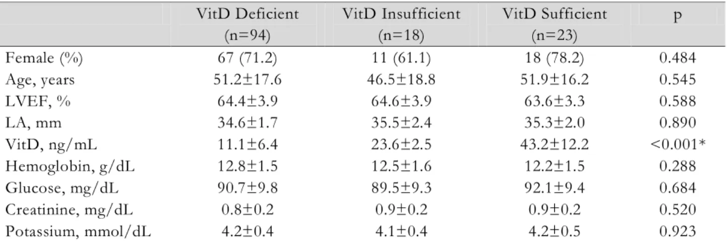
Table 1. Baseline Characteristics of Study Population VitD Deficient (n=94) VitD Insufficient (n=18) VitD Sufficient (n=23) p Female (%) 67 (71.2) 11 (61.1) 18 (78.2) 0.484 Age, years 51.2±17.6 46.5±18.8 51.9±16.2 0.545 LVEF, % 64.4±3.9 64.6±3.9 63.6±3.3 0.588 LA, mm 34.6±1.7 35.5±2.4 35.3±2.0 0.890 VitD, ng/mL 11.1±6.4 23.6±2.5 43.2±12.2 <0.001* Hemoglobin, g/dL 12.8±1.5 12.5±1.6 12.2±1.5 0.288 Glucose, mg/dL 90.7±9.8 89.5±9.3 92.1±9.4 0.684 Creatinine, mg/dL 0.8±0.2 0.9±0.2 0.9±0.2 0.520 Potassium, mmol/dL 4.2±0.4 4.1±0.4 4.2±0.5 0.923
*All pairwise correlations were significant at p<0.05
Abbreviations: LA, left atrium; LVEF, left ventricular ejection fraction; VitD, vitamin D
Materials and Methods
In this retrospective cross-sectional study, we included a total of 135 patients who were suffered with undiagnosed repetitive palpitations and referred to our clinic for diagnostic cardiac electrophysiologic study (EPS) between January 2015 and May 2017. Patients with structural heart disease, hypertension, diabetes, known sinus /atrioventricular nodal dysfunction, antiarrhytmic usage, thyroid or parathyroid diseases and calcium or vitD supplementation were excluded.
Patients’ electrophysiology reports were carefully reviewed and patients’ corrected sinus node recovery times (cSNRT), baseline sinus cycle lengths (BCL), Wenckebach cycle length, atrial-His (AH) and atrial-His-ventricular (HV) intervals and AF episodes were recorded. In all patients, AF was induced by burst atrial pacing and then categorized into sustained and nonsustained according to the duration (30 seconds).
Demographic information and vitD measures were recorded. All VitD measurements were within the one-month window before the procedure. According to the Endocrinology Society Guidelines, study population was divided into three group as VitD sufficient (VitD≥ 30ng/mL), VitD insufficient (VitD 21-29 ng/mL) and VitD deficient (VitD <20 ng/mL) (5).
Local ethics committee approved the study protocol and informed consent was taken from all patients.
Statistical Analysis: Continuous variables were presented as mean ± standard deviation,
categorical variables were presented as
percentages. Analysis of variance (ANOVA) test was used for statistical comparison of continuous
variables. χ2 test was used for comparison of
categorical variables. A two-tailed P < 0.05 was considered statistically significant. All statistical analyses were performed using the SPSS 15 (SPSS INC, Chicago, Illinois, USA). We also performed post hoc power analysis to preclude type II error. Effect size for the parameter which had the most difference between the VitD groups was specified and post hoc study power was determined with 0.05 type one error rate. Power analysis was carried out with G*Power 3.1.9.2.
Results
The study population was female predominant (71.1%) and had a mean age of 50. The demographic, laboratory and echocardiographic properties of groups were not statistically different (Table 1). cSNRT was increased 13% and 10% in VitD deficient and insufficient groups than sufficient group respectively. Similarly, BCL, Wenkebach cycle length, AH and HV intervals were increased in a similar but more attenuated fashion than cSNRT. However these differences did not meet statistical significance (Table 2). AF was two times likely inducible in VitD deficient patients than sufficient ones. However this difference also did not meet statistical significance (Table 2). With an effect size of 0.47 for cSNRT and with a type I error rate of 0.05, the study power was 99%.
Discussion
This study demonstrated that baseline VitD levels did not affect invasive electrophysiologic parameters and AF inducibility. To the best of our knowledge, this is the first study in the literature evaluating the effects of VitD on cardiac
Tek ve ark. / Vitamin D on Cardiac Electrophysiologic Parameters and Atrial Fibrillation Inducibility
Van Tıp Derg Cilt:26, Sayı:2, Nisan/2019 204
Table 2. Comparison of the Groups’ Electrophysiologic Parameters VitD Deficient (n=94) VitD Insufficient (n=18) VitD Sufficient (n=23) p cSNRT, msn 390.0±44.1 380.0±35.5 345.0±27.0 0.374 BCL, msn 769.8±168.2 746.5±108.6 725.6±83.9 0.420 Wenchebach, msn 331.5±44.0 326.6±34.9 319.1±12.7 0.389 AH, msn 86.8±21.1 84.3±14.6 84.7±7.1 0.813 HV, msn 44.3±7.7 44.0±5.6 42.6±2.4 0.564 Inducible AF, n (%) Nonsustained AF, n (%) Sustained AF, n (%) 16 (17.0 ) 10 (10.6 ) 6 (6.38) 2 (11.1) 0 (0.0) 2 (11.1) 2 (8.6) 2 (8.6) 0 (0.0) 0.584
Abbreviations: AF, Atrial fibrillation; AH, atrial-His interval; BCL, baseline sinus cycle length; cSNRT, corrected sinus node recovery time; HV, His-ventricular interval
conduction system and AF inducibility.
Deficiencies in VitD have been demonstrated as an emerging risk factor in cardiovascular diseases. Vitamin D exerts its cardiovascular functions by direct and indirect pathways. VitD receptors which are abundant in endothelium, vascular
smooth muscle and cardiomyocytes are
responsible for its direct cardiovascular effects while renin-angiotensin system (RAS) and inflammatory pathways are responsible for indirect effects. Studies have shown that vitamin D deficiency increases plasma renin activity, increases blood pressure, and has adverse effects on ventricular remodeling (4,6,7).
The mechanism of arrhytmias in VitD deficiency can be explained by primarily due to its effects on cardiac repolarization. Mineralocorticoid receptor activation causes an increase in the inward calcium channel current and a decrease in the transient outward potassium current. These changes prolong the repolarization interval and cardiac action potential. Also vitD have a direct effect on cardiac repolarization (8-10).
Previous studies investigated the possible mechanisms responsible for the relationship between VitD deficiency and AF. Deficiencies of VitD is associated with both structural and electrical atrial remodeling which are the consequences of increased TGFB1 expression,
enhanced atrial fibrosis, and conduction
heterogenity (11,12). Hanafy et al. demonstrated that replacement of VitD prolonged atrial action potential and therefore diminished AF inducibility and maximal AF rate in their animal study (13). Canpolat et al. showed that VitD deficiency prolonged atrial electromechanical delay which is an indicator for AF progression (14). Gode et al. reported that one ng/mL increase of VitD is associated with 15% decrease of postoperative AF
odds in patients undergoing coronary artery bypass surgery (15).
In our study, we evaluated the relationship between baseline VitD levels with invasive electrophysiologic parameters and AF inducibility and found that there were no significant differences existed. However, single measurement of vitD levels, seasonal variations (although our measurements were winter and spring time) may affect results. Also, apart from baseline levels, replacement of VitD may alter cardiac conduction times and AF inducibility.
In conclusion, baseline VitD levels were not
associated with cardiac electrophysiologic
parameters and AF inducibility. To demonstrate the role of VitD in cardiac conduction system and AF inducibility thoroughly, further studies such as addressing VitD replacement are warranted. Competing Interests: The authors declared no conflict of interest.
Funding: No funding was received for this research.
Acknowledgements: We are really grateful to cardiology staff of TOBB Economy and Technology University.
References
1. Norman PE, Powell JT. Vitamin D and cardiovascular disease. Circ Res 2014; 114(2): 379-393.
2. Deo R, Katz R, Shlipak MG, Sotoodehnia N, Psaty BM, Sarnak MJ, Fried LF, Chonchol M, de Boer IH, Enquobahrie D, Siscovick D, Kestenbaum B. Vitamin D, parathyroid hormone, and sudden cardiac death: results from the Cardiovascular Health Study. Hypertension 2011; 58(6): 1021-1028.
Tek ve ark. / Vitamin D on Cardiac Electrophysiologic Parameters and Atrial Fibrillation Inducibility
Van Tıp Derg Cilt:26, Sayı:2, Nisan/2019 205
3. Rienstra M, Cheng S, Larson MG, McCabe EL, Booth SL, Jacques PF, Lubitz SA, Yin X, Levy D, Magnani JW, Ellinor PT, Benjamin EJ, Wang TJ. Vitamin D status is not related to development of atrial fibrillation in the community. Am Heart J 2011; 162(3): 538-541.
4. Demir M, Uyan U, Melek M. The effects of vitamin D deficiency on atrial fibrillation. Clin Appl Thromb Hemost 2014; 20(1): 98-103. 5. Holick MF, Gordon CM. The Hormone
Foundation’s: Patient guide to vitamin D deficiency. J Clin Endocrinol Metab 2011; 96(7): 1-2.
6. Reddy Vanga S, Good M, Howard PA, et al. Role of vitamin D in cardiovascular health. Am J Cardiol 2010; 106: 798-805
7. Walters MR, Wicker DC, Riggle PC: 1, 25-Dihydroxyvitamin D3 receptors identified in the rat heart. J Mol Cell Cardiol 1986; 18: 67-72. 8. Kim HW, Park CW, Shin YS, Kim YS, Shin SJ,
Choi EJ, Chang YS, Bang BK. Calcitriol regresses cardiac hypertrophy and qt dispersion in secondary hyperparathyroidism on hemodialysis. Nephron Clin Pract 2006; 102: 21-29.
9. Perrier E, Kerfant BG, Lalevee N, Bideaux P, Rossier MF, Richard S, Gomez AM, Benitah JP. Mineralocorticoid receptor antagonism prevents the electrical remodeling that precedes cellular hypertrophy after myocardial infarction. Circulation 2004; 110: 776-783. Straus SM, Kors JA, De Bruin ML, van der Hooft CS, Hofman A,
Heeringa J, Deckers JW, Kingma JH, Sturkenboom MC, Stricker BH, Witteman JC. Prolonged QTc interval and risk of sudden cardiac death in a population of older adults. J Am Coll Cardiol 2006; 47: 362-367.
10. De Jong AM, Maass AH, Oberdorf-Maass SU, et al. Mechanisms of atrial structural changes caused by stretch occurring before and during early atrial fibrillation. Cardiovasc Res 2011; 89: 754-765. 11. Cardús A, Parisi E, Gallego C, Aldea
M, Fernández E, Valdivielso JM. 1, 25-Dihydroxyvitamin D3 stimulates vascular smooth muscle cell proliferation through a VEGF-mediated pathway. Kidney Int 2006; 69(8): 1377-1384.
12. Hanafy DA, Chang SL, Lu YY, Chen YC, Kao YH, Huang JH, Chen SA, Chen YJ. Electromechanical effects of 1, 25-dihydroxyvitamin d with antiatrial fibrillation activities. J Cardiovasc Electrophysiol 2014; 25(3): 317-323.
13. Canpolat U, Yayla Ç, Akboğa MK, Özcan EH, Turak O, Özcan F, et al. Effect of Vitamin D Replacement on Atrial Electromechanical Delay in Subjects with Vitamin D Deficiency. J Cardiovasc Electrophysiol 2015; 26(6): 649-655.
14. Gode S, Aksu T, Demirel A, Sunbul M, Gul M, Bakır I, Yeniterzi M. Effect of vitamin D deficiency on the development of postoperative atrial fibrillation in coronary artery bypass patients. J Cardiovasc Thorac Res 2016; 8(4): 140-146.