Effects of Vanadyl Sulfate on Liver
of Streptozotocin-Induced
Diabetic Rats
M
ERALK
OYUTURK,*
,1S
EVIMT
UNALI,
2S
EHNAZB
OLKENT,
3 ANDR
EFIYEY
ANARDAG21
Department of Histology and Embryology, Faculty of Medicine,
Kadir Has University, 80810-Gayrettepe, Turkey;
2Department
of Chemistry, Faculty of Engineering, Istanbul University,
34850-Avcilar, Turkey; and
3Department of Biology, Faculty
of Science, Istanbul University, 34459-Vezneciler, Istanbul, Turkey
Received April 1, 2004; Revised June 22, 2004; Accepted August 18, 2004.
ABSTRACT
The aim of this study was to investigate the microscopic and bio-chemical effects of vanadyl sulfate on liver tissue of normal and streptozo-tocin (65 mg/kg) diabetic rats. Vanadyl sulfate was administered by gavage at a dose of 100 mg/kg. Degenerative changes were observed in diabetic animals by light and transmission electron microscopes. Although there were individual differences in diabetic animals to which vanadium was given, some reduction of degenerative changes were detected. After 60 d of treatment, serum aspartate and alanine transaminase, alkaline phos-phatase, blood glucose levels, liver lipid peroxidation, and nonenzymatic glycosylation significantly increased, but liver glutathione levels signifi-cantly decreased in the diabetic group. On the other hand, treatment with vanadyl sulfate reversed these effects. As a result, it might be concluded that vanadyl sulfate has a protective effect on damage of liver of strepto-zotocin-induced diabetic rats.
Index Entries: Streptozotocin; liver tissue; vanadyl sulfate; light microscopy; electron microscopy; biochemistry.
INTRODUCTION
Diabetes mellitus is the world’s most severe endocrine disease involv-ing metabolic disorders characterized by hyperglycemia and inducinvolv-ing
Biological Trace Element Research 233 Vol. 104, 2005
0163-4984/05/10403–0233 $30.00
is an important tissue that regulates carbohydrate and lipid metabolisms (5,12). Vanadium compounds have been reported to inhibit lipogenesis (5,13) and gluconeogenesis (5,10,14) and to stimulate glycolysis (5,15) and glycogen synthesis (10,14) in the liver.
The present study was undertaken to examine the effects produced by vanadyl sulfate administration on the liver of control and streptozotocin (STZ)-diabetic rats. In addition, the purpose of this study was to investigate whether vanadyl sulfate has a protective effect on the liver of STZ-diabetic rats and whether this effect is related with the oxidative/antioxidative system.
MATERIALS AND METHODS
Preparation of Experimental Animals
Diabetes was induced by intraperitoneal injection of STZ in a single dose of 65 mg/kg body weight. STZ was dissolved in a freshly prepared 0.01 M citrate buffer (pH 4.5) (16).
Animals and Experimental Design
In this study, male, 6- to 6.5-mo-old, Swiss albino rats were used. The animals were clinically healthy and fed with laboratory pellet chow and given water ad libitum. The animals were randomly divided into four groups: group I included untreated, nondiabetic controls (n=13); group II included control animals given vanadyl sulfate (n=5); group III included STZ-diabetic animals (n=11); group IV included STZ-diabetic animals given vanadyl sulfate (n=11). Vanadyl sulfate was given by gavage tech-nique to rats in a dose of 100 mg/kg every day for 60 d. On d 60, the liver
tissues and blood samples were taken under ether anesthesia from animals that were fasted overnight.
Light Microscopic Study
The liver tissue samples were fixed in Bouin’s fixative and embedded in paraffin after having completed the routine follow-up. Sections of 5 µm thickness to which Masson tri-dye was applied were examined under a Carl Zeiss Ultraphot II Light Microscope.
Electron Microscopic Study
Small fragments of liver tissue were fixed with phosphate-buffered 2% glutaraldehyde. The tissues were postfixed with phosphate-buffered 1% osmium tetroxide. Embedding was performed in Epon 812 according to standard procedures.
Biochemical Study
Blood samples of rats were collected from the tail vein at 0, 1, 30, and 60 d. Fasting blood glucose levels (after a 18-h period of fasting) were determined by o-toluidine methods (17). Serum aspartate transaminase (AST) and alanine transaminase (ALT) activities were determined by Reit-man–Frankell methods (18). Serum alkaline phosphatase (ALP) activities were estimated by the two point method (19).
At the end of the experimental period, liver tissues were taken from overnight-fasting animals and sacrificed under ether anesthesia. The tis-sues were homogenized in cold with 0.9% saline by means of a glass homogenizer to make up a 10% (w/v) homogenate. The homogenates were centrifuged and the clear supernatants were used for lipid peroxida-tion, glutathione, nonenzymatic glycosylaperoxida-tion, and protein analysis.
Liver lipid peroxidation (LPO) and glutathione (GSH) levels were determined by the methods of Ledwozyw (20) and Beutler using Ellman’s reagent (21), respectively. Nonenzymatic glycosylation (NEG) levels were assessed by 2-thiobarbituric acid (22). Liver total protein levels measured by the method of Lowry using bovine serum albumin as standard (23).
Statistical Analysis
The results were evaluated using an unpaired t-test and analysis of variance (ANOVA) using the NCSS statistical computer package (24).
RESULTS
Light Microscopic Results
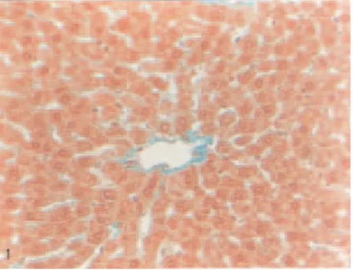
A normal histological appearance was observed in the liver tissue of untreated control and control groups given vanadium (Fig. 1). Vacuolization,
nuclei-containing perichromatin material, pycnotic nuclei, many large cyto-plasmic granules, rupturing in the epithelium of vein, moderately hyper-emia, and dilatations in sinusoids were observed in hepatocytes of diabetic group when compared to controls (Figs. 2 and 3). In hepatocytes of diabetic group given vanadium, although individual differences were present, it was noted that these degenerative changes were decreased except for less vacuolization and pycnotic nuclei in the hepatocytes of some animals (Fig. 4).
Electron Microscopic Results
Swelling in the cisternae of granular endoplasmic reticulum, pyc-notic nuclei, mitochondria surrounded by dilated granular endoplasmic reticulum cisternae, extensions in perinuclear area, an increase in smooth endoplasmic reticulum in some cells, a decrease in glycogen content, and cells with dark and light cytoplasms were observed in hepatocytes of the diabetic group by electron microscopy when compared with controls (Figs. 5–7). In the cytoplasmic appearance of vanadium-treated diabetic rats, swelling of granular endoplasmic reticulum cisternae, pycnotic nuclei, and an increase in smooth endoplasmic reticulum were detected (Fig. 8).
Biochemical Results
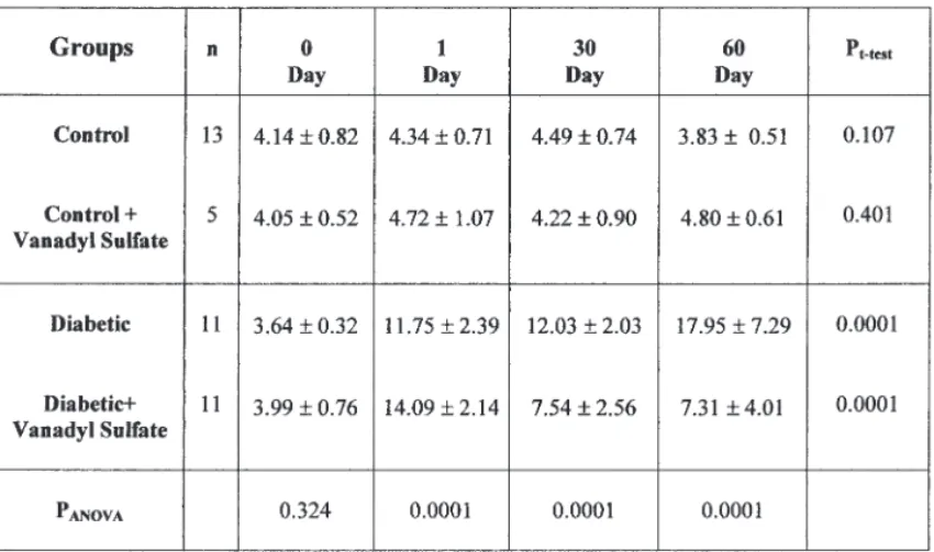
The mean blood glucose levels of the four groups are given in Table 1. According to Table 1, there was no significant difference in the blood
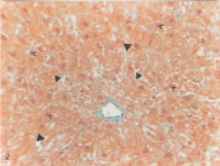
Fig. 2. Liver of a STZ-diabetic rat. Vacuolization (▲), pycnotic nuclei (>), cytoplasmic granulation (>), Masson’s tri-dye. Magnification: ×320.
Fig. 3. Liver of a STZ-diabetic rat. Sinusoidal dilations (d), moderately hyper-emia (h). Masson’s tri-dye. Magnification: ×320.
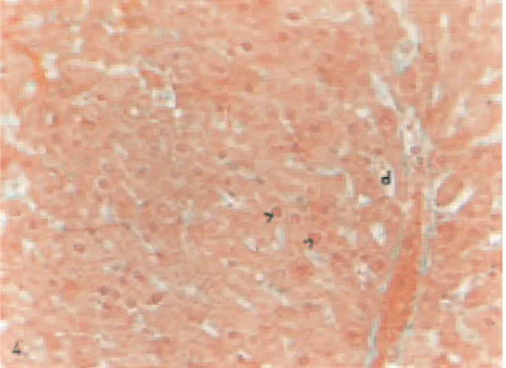
Fig. 4. Liver of a STZ-diabetic animal given vanadium. Less pycnotic nuclei (>) and sinusoidal dilations (d). Masson’s tri-dye. Magnification: ×320.
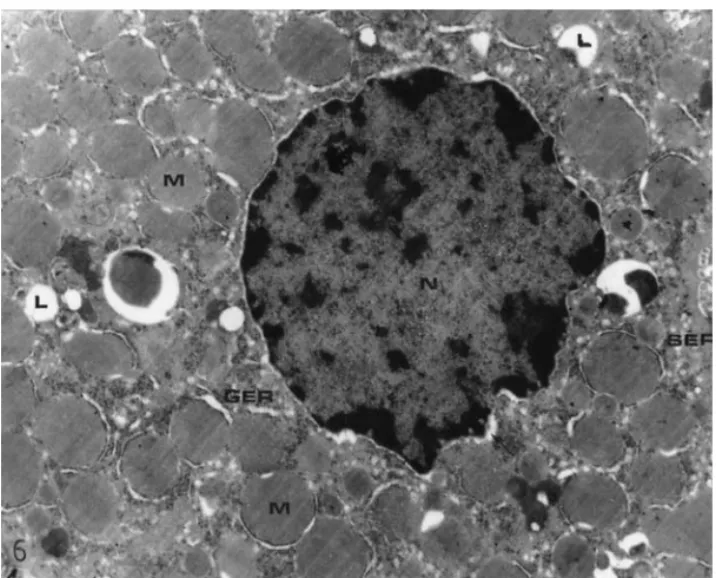
Fig. 6. An electron micrograph of the hepatocytes of a diabetic rat. Swelling in granular endoplasmic reticulum (GER), mitochondria surrounding with dilated granular endoplasmic reticulum (M), the dilation in perinuclear space (→), pyc-notic nucleus (N), an increase in smooth endoplasmic reticulum (SER) and lipid deposition (L). Bar=2 µm.
Fig. 7. An electron micrograph of the hepatocytes of a diabetic rat. An increase in smooth endoplasmic reticulum (SER) and lipid deposition (L). Bar=2 µm.
Table 1
Mean Levels of Blood Glucose for All Groups (mmol/L)*
Note: n = number of animals.
* Mean ± SD.
Fig. 8. An electron micrograph of the hepatocytes of a diabetic rat given vanadium. Swelling of granular endoplasmic reticulum (GER) and an increase in smooth endoplasmic reticulum (SER). Bar=2 µm.
glucose levels among the four groups on d 0 (pANOVA=0.324). After STZ
injection, a significant increase was observed in the blood glucose levels of diabetic rats at 1, 30, and 60 d compared to d 0 (pt-test= 0.0001). Vanadyl
sulfate treatment did not produce significant changes in the blood glucose levels in nondiabetic rats (pt-test= 0.401). The administration of vanadyl
sulfate to diabetic rats resulted in a significant decrease in the level of blood glucose (pt-test= 0.0001).
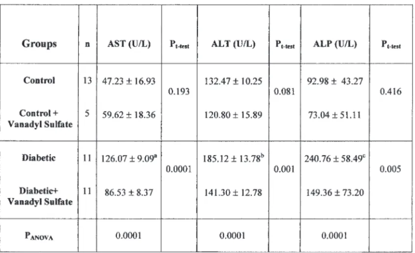
Table 2 gives the mean serum AST, ALT, and ALP activities of all groups. According to Table 2, a significant difference in the AST activities of all groups was observed at the end of the 60-d experiment (pANOVA=0.0001). Serum AST activities were significantly higher in
dia-betic groups when compared to controls (ap
t-test= 0.0001). Oral
adminis-tration of vanadyl sulfate for 60 d significantly decreased the serum AST activities in diabetic rats (pt-test= 0.0001).
The mean serum ALT and ALP activities are shown in Table 2. A sig-nificant difference in the serum ALT and ALP activities of all groups was observed at the end of the 60-d experiment (pANOVA= 0.0001). Serum ALT
and ALP activities were significantly higher in diabetic groups when com-pared with controls (b,cp
t-test= 0.0001). Vanadyl sulfate given to the
dia-Table 2
Mean Levels of Serum AST, ALT, and ALP Activities (U/L)*
Note: n = number of animals
* Mean ± SD; blood samples were taken from animals at d 60.
ap = 0.0001 vs control group. bp = 0.0001 vs control group. cp = 0.0001 vs control group.
betic rats lowered the serum ALT and ALP activities in a significant man-ner when compared with untreated diabetic rats (pt-test=0.001) and (pt
-test=0.005), respectively.
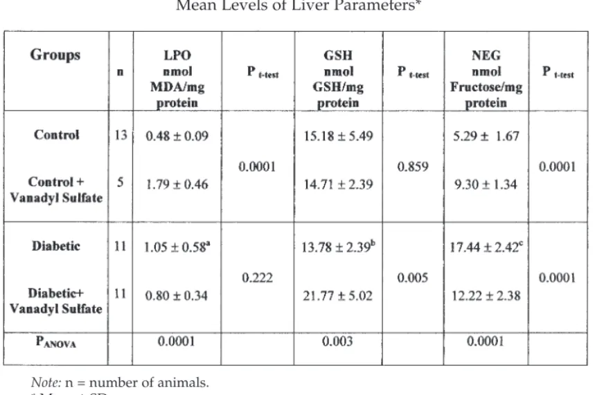
Table 3 shows the content of LPO, GSH, and NEG in the livers of nor-mal and experimental groups. In diabetic rats, a significant increase in liver LPO levels was observed (ap
t-test= 0.002). Administration of vanadyl
sulfate was found to reduce liver LPO levels in diabetic rats (pt-test=
0.222), but in the nondiabetic control groups, it significantly increased the liver LPO levels (pt-test= 0.0001).
The liver GSH levels were insignificantly reduced in the diabetic ani-mals as compared with the other groups (pANOVA= 0.003). In the diabetic
rats treated with vanadyl sulfate, the liver GSH levels significantly increased when compared with the diabetic group (pt-test= 0.005). Vanadyl
sulfate had no significant effect on liver GSH levels in the nondiabetic con-trol groups (pt-test= 0.859).
In the diabetic rats, NEG levels were significantly higher than those of the other groups (pANOVA= 0.0001) (Table 3). When given to the diabetic
rats, vanadyl sulfate lowered the liver NEG levels in a significant manner when compared with untreated diabetic rats (pt-test= 0.0001), but the liver
NEG levels significantly increased in nondiabetic animals (pt-test= 0.0001).
Note: n = number of animals.
* Mean ± SD.
ap = 0.002 vs control group. bp = 0.595 vs control group. cp = 0.0001 vs control group.
DISCUSSION
Diabetes mellitus encompasses a heterogeneous group of diseases with various etiologies. This disease is characterized by variable and chronic hyperglycemia and other disturbances in carbohydrate and lipid metabolisms as well as a variety of vascular and neurological complica-tions (25). Hyperglycemia is an important factor in the development and progression of the complications of diabetes mellitus and good glycemic control is necessary to prevent diabetic complications (26).
Vanadium is an important trace element that has an insulin-mimetic effect and plays an important role in lipid and protein metabolisms (9,27,28). The liver regulates normal levels of blood glucose with glycogenesis, glycogenolysis, glycolysis, and gluconeogenesis. Uptake of glucose is mainly related to insulin sensitivity of hepatocytes (12). In diabetes, the mechanism of some hypoglycemic agents such as vanadium remains unclear. Recent reports have indicated that vanadium compounds have insulin-mimetic properties (3,29,30). Vanadium’s effect of mechanism might be explained by its effects on the antioxidative system and oxidative dam-age in diabetes. Vanadium is found predominantly as vanadate (V), which is reduced to the vanadyl(IV) form. In plasma, vanadium exists in both oxi-dation states. These interactions can lead to numerous biological effects (31). Normoglycemic blood glucose levels were reported in the treatment of STZ-diabetic rats with different vanadate derivatives and vanadyl sul-fate (32,33). These reports indicate the improving effect of vanadyl sulsul-fate in diabetes by either replacing insulin or enhancing the effects of insulin. In this study, blood glucose levels were reduced in STZ-diabetic rats by the administration of vanadyl sulfate. This observation is in accordance with previous reports. These effects might be attributed to the insulin-mimick-ing effect of vanadium compounds (32,33).
Liver cell destruction shows itself as impairment in the permeability of AST and ALT, which are marker enzymes in the liver. Measurement of enzymic activities of aminotransferase (AST and ALT) and ALP is of clini-cal and toxicologiclini-cal importance, as changes in their activities are indica-tive of tissue damage by toxicants or in disease conditions (34). In STZ-diabetic rats, the activities of serum AST, ALT, and ALP were signifi-cantly increased by activities (163%, 40%, and 159%, respectively) relative to their normal levels (Table 2). Liver necrotization in STZ-diabetic rats supports our findings. Therefore, the increment of the activities of AST, ALT, and ALP in serum is mainly the result of the leakage of these enzymes from the liver cytosol into the bloodstream, which gives an indi-cation on the hepatotoxic effect of STZ (35). On the other hand, the admin-istration of vanadyl sulfate to STZ-diabetic rats caused a reduction in the activity of these enzymes in serum compared with the mean values of the diabetic group.
Lipid peroxidation, which is a primary damaging process, has been known to occur via oxidative destruction of membrane polyunsaturated
tive stress. Administration of vanadyl sulfate increased the content of GSH in the liver of diabetic rats.
Diabetes is characterized by a sequence of complications and the main alteration related to diabetic process is hyperglycemia. Oxygen free radi-cals have been implicated to be a causal issue in pathogenesis of diabetes (42). Also, the source of oxygen free radicals suggested the autoxidation of glucose and nonenzymatic protein glycation in diabetes (43,44). Oxidative damage as a result of free radicals is associated with vascular disease in people with type 1 and type 2 diabetes mellitus (45). There are several potential sources of increased free-radical production in diabetes, includ-ing autoxidation of plasma glucose, activation of leukocytes, and increased transition metal bioavailability (4). Several studies showed that the per-sistence of hyperglycemia causes increased production of reactive oxygen species, through glucose autoxidation and nonenzymatic glycation, sug-gesting an increased oxidative stress in diabetic animals. The increased oxidative stress in diabetic conditions might be caused not only by an accelerated production of reactive oxygen species but also by a decreased scavenging ability of those molecules (46).
Nonenzymatic glycation normally occurs at very slow rates in long-lived proteins (47). Serum protein glycation has been proposed to reflect the tissue accumulation of nonenzymatically attached glucose (48). Many proteins have been observed to undergo NEG. NEG of liver proteins causes alteration in their structures and functions. The levels of NEG were found to be increased in the STZ-diabetic groups with respect to untreated controls. Various means of preventing this increase have been investigated both in vitro and in vivo (49). Some transition metals such as nickel, chromium, and vanadium might act as catalysts of the oxidative deterio-ration of biological macromolecules (50). This might occur via formation of
reactive oxygen species and enhanced lipid peroxidation, depletion of sulfhydryls, and oxidative tissue injury (51). It has been reported that GSH, glycine, and various antioxidant vitamins and trace elements such as vitamins C and E, lipoic acid, vanadium, selenium, zinc, and chromium prevent the increase of tissue NEG levels (45). In our study, we found a sig-nificant increase in NEG of the liver protein in diabetic rats. The adminis-tration of vanadyl sulfate to STZ-diabetic rats reduced NEG.
The degenerative changes were observed in diabetic animals by light and electron microscopes. In our study, toxic effects by vanadyl sulfate treatments were not observed under microscopic evaluation and, in some cases, improving effects occurred. Although, there were individual differ-ences in diabetic animals given vanadium, some reduction of degenerative changes were detected. The partial decrease of the degeneration in the dia-betic group given vanadyl sulfate indicates that vanadium prevents the damage in the liver tissue of STZ-diabetic rats. As a result, it might be con-cluded that vanadyl sulfate has a protective effect on damage of liver of STZ-induced diabetic rats.
REFERENCES
1. W. B. Kannel and D. L. Mc Gree, Diabetes and cardiovascular disease. The Framingham Study, JAMA 241, 2035–2038 (1979).
2. H. King, R. E. Aubert, and W. H. Herman, Global burden of diabetes, 1995–2025: preva-lence, numerical estimates, and projections, Diabetes Care 21, 1414–1431 (1998). 3. J. Meyerovitch, Z. Farfel, J. Sack, and Y. Shechter, Oral administration of vanadate
nor-malizes blood glucose levels in streptozotocin-treated rats, characterization and mode of action. J. Biol. Chem. 262, 6658–6662 (1987).
4. E. Tsiani, N. Abdullah, and I. G. Fantus, Insulin-mimetic agents vanadate and per-vanadate stimulate glucose but inhibit amino acid uptake, Am. J. Physiol. Cell Physiol. 272,156–162 (1997).
5. K. Cusi, S. Cukier, R. A. DeFronzo, M. Torres, F. M. Puchulu, and J. C. Pereira Redondo, Vanadyl sulfate improves hepatic and muscle insulin sensitivity in type 2 diabetes, J. Clin. Endocrinol. Metab. 86, 1410–1417 (2001).
6. C. E. Heyliger, A. G. Tahiliani, and J. H. McNeill, Effect of vanadate on elevated blood glucose and depressed cardiac performance of diabetic rats, Science 227, 1474–1477 (1985).
7. M. Bendayan and D. Gingras, Effect of vanadate administration on blood glucose and insulin levels as well as on the exocrine pancreatic function in streptozotocin-diabetic rats, Diabetologia 32, 561–567 (1989).
8. M. H. Oster, J. M. Llobet, J. L. Domingo, J. B. German, and C. L. Keen, Vanadium treat-ment of diabetic Sprague–Dawley rats results in tissue vanadium accumulation and pro-oxidant effects, Toxicology 83, 115–130 (1993).
9. S. Verma, M. C. Cam, and J. H. McNeill, Nutritional factors that can favorably influence the glucose/insulin system: vanadium, J. Am. Coll. Nutr. 17, 11–18 (1998).
10. A. K. Srivastava, Anti-diabetic and toxic effects of vanadium compounds, Mol. Cell Biochem. 206, 177–182 (2000).
11. M. Barbagallo, L. J. Dominguez, and L. M. Resnick, Insulin-mimetic action of vanadate: role of intracellular magnesium, Hypertension 38, 701–704 (2001).
20. A. Ledwozwy, J. Michalak, A. Stepien, and A. Kadziolka, The relationship plasma triglycerides, cholesterol, total lipids and lipid peroxidation products during human atherosclerosis. Clin. Chim. Acta 155, 275–284 (1986)
21. E. Beutler, Glutathione in Red Cell Metabolism. A Manual of Biochemical Methods, 2nd ed., Grune and Stratton, New York, pp. 112–114 (1975).
22. K. M. Parker, J. D. England, J. D. Casto, R. Hessel, and P. E. Goldstein, Improved col-orimetric assay for glycosylated hemoglobin, Clin. Chem. 27, 669–672 (1981).
23. O. H. Lowry, N. J. Rosebrough, A. L. Farr, and R. J. Randall, Protein measurement with the folin phenol reagent, J. Biol. Chem. 193, 265–275 (1951).
24. J. L. Hintze, Number cruncher statistical system. Copyright North Kaysville, Utah, 84037 (801), 546–0445 (1986).
25. T. Storr, D. Mitchell, P. Buglyo, et al., Vanadyl–thiazolidinedione combination agents for diabetes therapy, Bioconjug. Chem. 14, 212–221 (2003).
26. N. Arun and N. Nalini, Efficacy of turmeric on blood sugar and polyol pathway in dia-betic albino rats, Plant Foods Hum. Nutr. 57, 41–52 (2002).
27. T. Jackson, A. I. Salhanick, J. D. Sparks, C. E. Sparks, M. Bolognino, and J. M. Amatrude, Insulin-mimetic effects of vanadate in primary cultures of rat hepatocytes, Diabetes 37, 1234–1240 (1954).
28. S. Tamura, T. A. Brown, R. E. Dubler, and J. Larner, Insulin-like effect of vanadate on adipocyte glycogen synthase and on phosphorylation of 95,000 dalton subunit of insulin receptor, Biochem. Biophys. Res. Commun. 113, 80–86 (1983).
29. S. Dai, K. H. Thompson, and J. H. McNeill, One-year treatment of streptozotocin-induced diabetic rats with vanadyl sulfate, Pharmacol. Toxicol. 74, 101–109 (1994). 30. N. Sekar, A. Kanthasamy, S. William, N. Balasubramaniyan, and S. Govindasamy,
Antioxidant effect of vanadate on experimental diabetic rats, Acta Diabetol. Lat. 27, 285–93 (1990).
31. E. Tsiani and I. G. Fantus, Vanadium compounds biological actions and potential as pharmocological agents, Trends Endocrinol. Metab. 8, 51–58 (1997).
32. G. Cros, J. J. Mongold, J. J. Serrano, S Ramanadham, and J. H. McNeill, Effects of vanadyl derivatives on animal models of diabetes, Mol. Cell Biochem. 109, 163–166 (1992).
33. R. M. Brand and F. G. Hamel, Transdermally delivered peroxovanadium can lower blood glucose levels in diabetic rats, Int. J. Pharm. 183, 117–123 (1999).
34. S. N. Singh, P. Vats, S. Suri, et al., Effect of an antidiabetic extract of Catharanthus roseus on enzymic activities in streptozotocin induced diabetic rats, J. Ethnopharmacol. 76, 269–277 (2001).
35. C. M. Navarro, P. M. Montilla, A. Martin, J. Jimenez, and P. M. Utrilla, Free radical scav-enger and antihepatotoxic activity of Rosmarinus, Planta Med. 59, 312–314 (1993). 36. A. W. Girotti, Lipid hydroperoxide generation, turnover, and effector action in
biologi-cal systems, J. Lipid Res. 39, 1529–1542 (1998).
37. R. Kakkar, S. V. Mantha, J. Radhi, K. Prasad, and J. Kalra, Antioxidant defense system in diabetic kidney: a time course study, Life Sci. 60, 667–679 (1997).
38. S. Horie, H. Ishii, and T. Suga, Changes in peroxisomal fatty acid oxidation in the dia-betic rat liver, J. Biochem. (Tokyo) 90, 1691–1696 (1981).
39. M. M. Kesavulu, B. K. Rao, R. Giri, J. Vijaya, G. Subramanyam, and C. Apparao, Lipid peroxidation and antioxidant enzyme status in Type 2 diabetics with coronary heart disease, Diabetes Res. Clin. Prac. 53, 33–39 (2001).
40. C. C. Shih, Y. W. Wu, and W. C. Lin, Antihyperglycaemic and antioxidant properties of Anoectochilus formosanus in diabetic rats, Clin. Exp. Pharmacol. Physiol. 29, 684–688 (2002).
41. S. Venkateswaran and L. Pari, Effect of Coccinia indica leaves on antioxidant status in streptozotocin-induced diabetic rats, J. Ethnopharmacol. 84, 163–168 (2003).
42. J. W. Baynes, Role of oxidative stress in development of complications in diabetes, Dia-betes 40, 405–412 (1991).
43. S. P. Wolff and R. T. Dean, Glucose autoxidation and protein modification. The poten-tial role of “autoxidative glycosylation” in diabetes, Biochem. J. 245, 243–250 (1987). 44. J. V. Hunt, C. C. Smith, and S. P. Wolff, Autoxidative glycosylation and possible
involvement of peroxides and free radicals in LDL modification by glucose, Diabetes 39, 1420–1424 (1990).
45. R. A. Anderson, A. M. Roussel, N. Zouari, S. Mahjoub, J. M. Matheau, and A. Kerkeni, Potential antioxidant effects of zinc and chromium supplementation in people with type 2 diabetes mellitus, J. Am. Coll. Nutr. 20, 212–218 (2001).
46. K. Tashima, A. Fujita, and K. Takeuchi, Aggravation of ischemia/reperfusion-induced gastric lesions in streptozotocin-diabetic rats, Life Sci. 67, 1707–1718 (2000).
47. M. Brownlee, H. Vlassara, and A. Cerami, Nonenzymatic glycosylation and the patho-genesis of diabetic complications, Ann. Intern. Med. 101, 527–537 (1984).
48. D. K. Yue, S. Mc Lennan, and J. R. Turtle, Non-enzymatic glycosylation of tissue pro-tein in diabetes in the rat, Diabetologia 24, 377–381 (1983).
49. R. Yanardag, S. Bolkent, O. Karabulut-Bulan, and S. Tunalí, Effects of vanadyl sulfate on kidney in experimental diabetes, Biol. Trace Elem. Res. 95, 73–85 (2003).
50. T. Wronska-Nofer, J. Wisniewska-Knypl, E. Dziubaltowska, and K. Wyszynka, Proox-idative and genotoxic effect of transition metals (cadmium, nickel, chromium, and vanadium) in mice. Trace Elem. Electrolytes 16, 87–92 (1999).
51. S. J. Stohs and D. Bagchi, Oxidative mechanisms in the toxicity of metal ions, Free Radic. Res. Commun. 9, 101–102 (1995).