ABSTRACT
Introduction: The purpose of this study was to compare the results of physical examinations (PEs) performed by a cardiologist with the results of point-of-care echocardiography for the diagnosis and management of patients during cardiac consultation rounds.
Patients and Methods: In all, 265 hospitalized consecutive patients from non-cardiac units (age, 62 ± 11; male, 56%) were included after complete cardiovascular (CV) examination by a consulting cardiologist. After the PE, the consulting cardiologist imaged each patient using a hand-carried cardiac ultrasound (HCU). All patients subsequently underwent a study with a standard echocardiographic device (SED) as the gold standard, performed on an upper-end platform. Defi nitive diagnosis, management change, and modifying diagnostic workup were also assessed.
Results: There were 196 CV fi ndings detected with an SED in this patient population. Of these, PE failed to detect 41% of the overall CV fi ndings and HCU missed 23% of the overall CV pathology. Overall, HCU had an effect on patient treatment decisions in 149 patients (56%); 42% had a change in medical therapy and 21% had a change in their diagnostic workup (most with changes in both).
Conclusion: HCU echocardiographic assessment during consultation rounds improved the detection of signifi cant CV pathology. Also, a direct assessment of cardiac function and anatomy at the bedside by an experienced cardiologist with HCU as a routine adjunct to PE results in an important change in clinical management and diagnostic workup during consultation.
Key Words: Hand-carried cardiac ultrasound; point-of-care screening
Kardiyak Konsültasyonlar Esnasında Tanı ve Tedavide Taşınabilir Ekokardiyografi Cihazları ile Birlikte Fizik Muayenenin Karşılaştırılması ÖZET
Giriş: Bu çalışmanın amacı, kardiyak konsültasyon esnasında tanı ve tedavi üzerinde ekokardiyografi sonuç-ları ile kardiyolog tarafından gerçekleştirilen fi zik muayene sonuçsonuç-larını karşılaştırmaktır.
Hastalar ve Yöntem: Kardiyak olmayan birimlerden yatırılan ardışık seçilmemiş 265 hasta danışman kar-diyolog tarafından tam kardiyovasküler muayene sonrası çalışmaya alındı. Fizik muayene sonrası danışman kardiyolog taşınabilir kardiyak ultrason kullanarak her hastayı görüntülenmiştir. Tüm hastalar daha sonra bir üst platformda gerçekleştirilen standart ekokardiyografi k cihazla altın standart olarak değerlendirildi. Kesin tanı, tedavi ve tanısal testlerdeki değişiklikler değerlendirildi.
Bulgular: Bu hasta popülasyonunda standart ekokardiyografi k cihazla 196 kardiyovasküler bulgu saptanmış-tır. Fizik muayene tüm bulguların %41’ini ve taşınabilir ultrason ise %23’ünü tespit etmede başarısız olmuş-tur. Taşınabilir kardiyak ultrason cihazı 149 hastanın tedavi kararında etkili olmuştur ve bunların %42’sinde tedavi değişirken %21’inde tanısal testler değişmiştir (hastaların çoğunda ikisi de değişmiştir).
Sonuç: Konsültasyonlar esnasında taşınabilir kardiyak ultrason ile değerlendirme önemli kardiyovasküler patolojilerin tespitini sağlamaktadır. Ayrıca, rutin fi zik muayeneye ek olarak deneyimli kardiyologlar tarafın-dan taşınabilir cihazla yatak başı kardiyak fonksiyon ve anatominin direkt değerlendirilmesi tedavi ve tanısal testlerde önemli değişiklikler ile sonuçlanmaktadır.
Anahtar Kelimeler: Taşınabilir kardiyak ultrason; bakım tarama noktası
Mehmet Ali Astarcıoğlu1, Taner Şen1, Celal Kilit1, Halil İbrahim Durmuş2, Adnan Doğan1, Türkan Paşalı Kilit1, Afşin Parspur1, Mehmet Yaymacı1, Muhammed Oylumlu1
1 Dumlupınar University Kütahya Evliya Çelebi Training and Research Hospital, Clinic of Cardiology,
Kütahya, Turkey
2 Kütahya Doç. Dr. Mustafa Kalemli Tavşanlı State Hospital, Clinic of Cardiology, Kütahya, Turkey
Comparison of Ultraportable Cardiac
Echocardiography with Physical Examination
for Diagnosis and Management during Cardiac
Consultation Rounds
Mehmet Ali Astarcıoğlu
E-mail: [email protected] Submitted: 18.10.2015
Accepted: 20.10.2015
@ Copyright 2016 by Koşuyolu Heart Journal. Available on-line at
www.kosuyoluheartjournal.com
23
Koşuyolu Heart Journal 2016;19(1):23-26 • DOI: 10.5578/khj.10403 ● ORIGINAL INVESTIGATION24
Koşuyolu Heart Journal 2016;19(1):23-26 ● Ultraportable Echocardiography During Cardiac ConsultationINTRODUCTION
Undiagnosed cardiac disease is known to cause serious perioperative complications and to increase morbidity and mortality during and after surgery(1). The decline in cardiovascular (CV) physical examination (PE) skills, especially for cardiac auscultation, has been well documented(2). Studies assessing PE skills have shown important mistake and oversight rates for physicians at all levels of training(3). Bedside echocardiography can bring important anatomical and hemodynamic information for the management of hospitalized patients. There are cardiac abnormalities that cannot be assessed by PE, such as left ventricular (LV) dysfunction and LV thrombus and vegetation. For all these reasons, hand-carried cardiac ultrasound (HCU) has potential as a diagnostic tool to provide information beyond PE at the point of care. The objective of this study was to investigate if the use of an HCU device during cardiac consultation rounds has an immediate infl uence on bedside treatment and diagnostic workup of patients at non-cardiac departments. Also, fi nding no abnormality on HCU would reduce the unnecessary test in patients being evaluated for a CV diagnosis.
PATIENTS and METHODS Study Population
We studied 265 hospitalized consecutive unselected patients (male, 56%), with a mean (SD) age of 62 ± 11 years, for whom a consultation by a cardiologist was requested. Critically ill patients were excluded.
Study Design
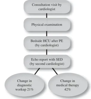
The main inclusion criterion for this study was the request from a physician in a non-cardiac department for cardiac evaluation of the patient. Routine treatment decisions regarding diagnostic workup and therapy were made from patient history, PE, 12-lead electrocardiogram (ECG), and a basic blood screen. All patients underwent two echocardiographic evaluations. First, HCU was performed by a cardiologist using the portable device Vscan (Vscan, GE Healthcare, Milwaukee, Wisconsin) and, consecutively, by a commercially available system (Vivid S5N; General Electric Medical Systems) by a second cardiologist, who was blinded to the initial assessment’s results (Figure 1).
The HCU examination included attempted two-dimensional (2D) imaging of parasternal long- and short-axis views, apical four- and two-chamber views, subcostal views, color Doppler fl ow mapping, and simple caliper measurements. Images were frozen and scrolled for review, and the measurements were performed online. The HCU imaging was limited to ≤ 15 min in duration. The HCU study focused on bedside assessment of LV global function [normal or reduced (LV ejection fraction < 40%)], LV regional wall motion abnormalities, identifi cation of ventricular enlargement, LV hypertrophy (septal thickness > 13 mm), presence or absence of pericardial effusion, cardiac source of embolism, endocarditis, and ventricular or atrial septal defect. Valve disease was classifi ed
as none, mild, moderate, or severe. After completion of the HCU study, defi nitive diagnosis, change in management, and workup tests were done, and associated immediate changes in clinical decisions were noted in a pre-designed form. Patients were also examined the same day by another cardiologist using standard echocardiographic device (SED), who was blinded to all HCU records.
HCU Device
Vscan is an HCU device with a unit size of 135 x 73 x 28 mm, weighing approximately 390 g, and a transducer size of 120 x 33 x 26 mm. The display is 3.5 inches, with a resolution of 240 x 320 pixels. It provides a black and white 2D mode for demonstrating structure in real time and color Doppler fl ow mapping for real time blood flow imaging. Vscan is equipped with phased-array transducer ranges from 1.7 MHz to 3.8 MHz. The device includes simple caliper measurements. All images were recorded on the system memory card for later review.
Statistical Analysis
Continuous variables are expressed as mean ± SD and compared using Student t test. Categorical variables are expressed as frequencies and compared using chi-square tests and, where appropriate, Fisher exact test. Statistical analysis was performed using SPSS version 13.0 (SPSS Inc., Chicago, Illinois, USA). A two-tailed p value of < 0.05 was considered signifi cant for all analyses.
RESULTS
Table 1 lists the reasons for cardiology consultation. HCU images were suitable for interpretation in 96% of the patients. In the remaining 4%, visualization was poor.
Figure 1. Flow chart for echocardiographic study. Echo report with SED (by second cardiologist)
Bedside HCU after PE (by cardiologist) Physical examination Consultation visit by cardiologist Change in diagnostic workup 21% Change in medical therapy 42%
25
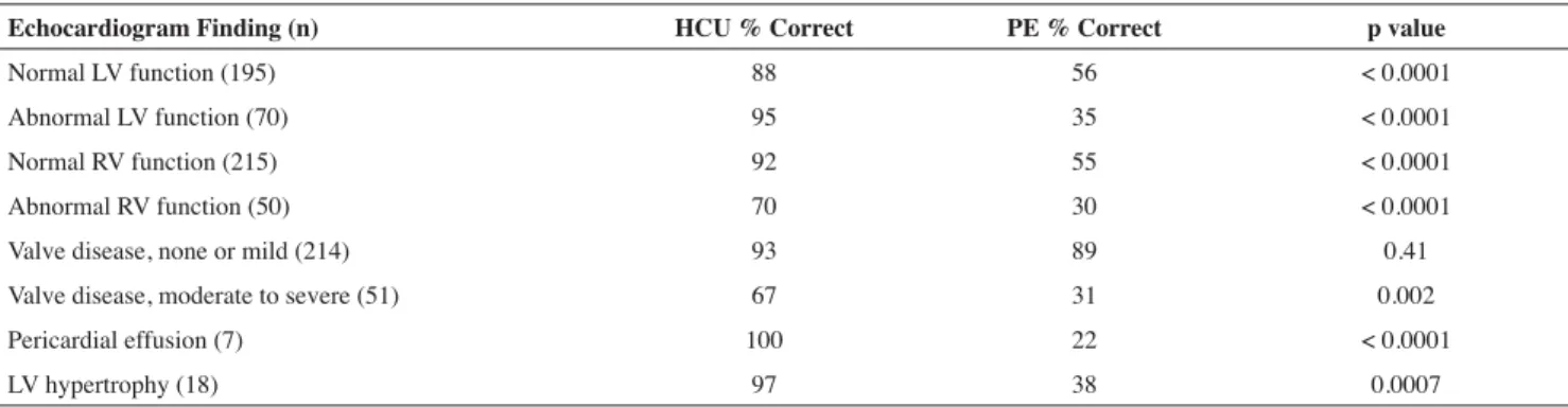
● Koşuyolu Heart Journal 2016;19(1):23-26 Astarcıoğlu MA, Şen T, Kilit C, Durmuş Hİ, Doğan A, Paşalı Kilit T, et al.There were 196 CV fi ndings detected with an SED in this patient population (Table 2). Of these, HCU correctly identifi ed 152 (77%) and physical examination correctly identifi ed 116 (59%, p< 0.0001). Reasons for requiring an SED evaluation were lack of pulsed wave and continuous wave Doppler to estimate the severity of stenotic lesions and for diagnosis of pulmonary hypertension. There was an increase in valvular fi ndings, ventricular systolic dysfunction, and hypertrophy detection with HCU over PE.
In all, 149 of the 265 patients (56%) had a change in management (both medical therapy and diagnostic workup), 42% had a change in medical therapy, and 21% had a change in their diagnostic workup. In two cases, fi ndings detected with the HCU device had an immediate impact on clinical management. Cardiology consultation was requested for preoperative risk assessment prior to cataract surgery in a 77-year-old man. He was asymptomatic and had no previous cardiac examination. Bedside HCU showed endocarditis of aortic valve, which prompted a TEE (The SED did not add any information). The diagnosis of endocarditis was established by TEE, and the patient had valve operation prior to cataract surgery. A 25-year-old pregnant woman presented with progressive shortness of breath at 28 weeks of pregnancy. HCU revealed enlargement of the right ventricle and a large mobile thrombus in the right atrium moving into the right ventricle. TTE showed moderate tricuspid regurgitation and pulmonary
artery hypertension. The diagnosis was made as right heart thrombosis with pulmonary embolism. After evaluation of the treatment options with the patient and her family, the decision was made in favor of thrombolytic therapy with low-dose and prolonged infusion of tissue-type plasminogen activator. TTE performed after thrombolysis showed a signifi cant decrease in the right ventricular size and complete lysis of the thrombus in the right heart.
DISCUSSION
This study demonstrates that HCU provides important information in a variety of CV diseases than PE at the bedside on cardiology consultation rounds. Furthermore, fi nding no signifi cant abnormality on HCU is also likely to reduce additional testing for patients being evaluated for suspected CV disease.
HCU provided information at the bedside that supplemented routine clinical data, such as the patient history, physical examination, and 12-lead ECG. Furthermore, this HCU information infl uenced patient treatment in more than half of the consultation visits by altering medical therapy, planned further diagnostic workup, or both. Subsequent echocardiographic study using a full-size echocardiographic system demonstrated a close overall agreement for estimation of ejection fraction, LV hypertrophy, regional wall-motion abnormalities, and pericardial effusion.
Several previous studies reported that HCU showed good concordance of diagnostic capability with SED in terms of assessing LVEF and LV dimension measurement and detecting pericardial effusion and valve disease(4-9). Also, they are being placed in emergency or critical care settings to assist in the evaluation of hemodynamically compromised patients(10-12).
The capability and simplicity of this new HCU make it ideally suited for bedside use during consultation rounds. Because HCU is superior to PE by increasing the number of diagnosis, and also decreasing the number of medical errors, and reducing the use of unnecessary tests. HCU was superior to PE in the majority of CV diseases. One of the most important
Table 1. Reasons for a cardiac consultation request from non-cardiac department (n= 265)
Preoperative risk assessment 83
Heart failure/dyspnea 67
Coronary artery disease/chest pain 40
Rhythm abnormalities 32
Valvular disease/murmur evaluation 18
Hypertension 16
Other 9
Table 2. Correct diagnosis by HCU and PE with SED as the reference
Echocardiogram Finding (n) HCU % Correct PE % Correct p value
Normal LV function (195) 88 56 < 0.0001
Abnormal LV function (70) 95 35 < 0.0001
Normal RV function (215) 92 55 < 0.0001
Abnormal RV function (50) 70 30 < 0.0001
Valve disease, none or mild (214) 93 89 0.41
Valve disease, moderate to severe (51) 67 31 0.002
Pericardial effusion (7) 100 22 < 0.0001
LV hypertrophy (18) 97 38 0.0007
26
Koşuyolu Heart Journal 2016;19(1):23-26 ● Ultraportable Echocardiography During Cardiac Consultationcontributions of HCU to the bedside examination is the high yield of LV function and valve disease.
This study describes that HCU was markedly superior in identifying the presence of moderate to severe mitral and tricuspid regurgitation, but not aortic stenosis. Also, HCU was superior to PE for assessment of LV function in the 107 patients referred to the echocardiography laboratory with chest pain or dyspnea as the primary or secondary indication. Both normal and abnormal LV functions were much better assessed by HCU than by PE.
Limitations
The HCU that was used for this study had no spectral Doppler modalities to obtain hemodynamic data.
CONCLUSION
During consultation rounds, an HCU can help to make an instant diagnosis at the bedside, leading to a shortened time to diagnosis with effi cacy equal to that of an SED.
CONFLICT of INTEREST
The authors reported no confl ict of interest related to this article.
AUTHORSHIP CONTRIBUTIONS Concept/Design: MA, TŞ, CK
Analysis/Interpretation: MA, HD, MY, AP Data acquisition: MA, TK, AD, AP, MO Writing: MA, TŞ, MO, HD
Critical revision: CK, TK, AD, HD Final Approval: All of authors
REFERENCES
1. Reich DL, Bodian CA, Krol M, Kuroda M, Osinski T, Thys DM. Intraoperative hemodynamic predictors of mortality, stroke, and myocardial infarction after coronary artery bypass surgery. Anesth Analg 1999;89:814-22.
2. Mangione S, Nieman LZ. Cardiac auscultatory skills of internal medicine and family practice trainees. A comparison of diagnostic profi ciency. JAMA 1997;278:717-22.
3. St Clair EW, Oddone EZ, Waugh RA, Corey GR, Feussner JR. Assessing housestaff diagnostic skills using a cardiology patient simulator. Ann Intern Med 1992;117:751-6.
4. Fukuda S, Shimada K, Kawasaki T. Pocketsized transthoracic echocardiography device for the measurement of cardiac chamber size and function. Circ J 2009;73:1092-6.
5. Egan M, Ionescu A. The pocket echocardiograph: a useful new tool. Eur J Echocardiogr 2008;9:721-5.
6. Prinz C, Dohrmann J, van Buuren F. Diagnostic performance of handheld echocardiography for the assessment of basic cardiac morphology and function: a validation study in routine cardiac patients. Echocardiography 2012;29:887-94.
7. Vourvouri EC, Poldermans D, De Sutter J, Sozzi FB, Izzo P, Roelandt JR. Experience with an ultrasound stethoscope. J Am Soc Echocardiogr 2002;15:80-5.
8. Spencer KT, Anderson AS, Bhargava A. Physician-performed point-of-care echocardiography using a laptop platform compared with physical examination in the cardiovascular patient. J Am Coll Cardiol 2001;37:2013-8. 9. Prinz C, Voigt JU. Diagnostic accuracy of a hand-held ultrasound scanner
in routine patients referred for echocardiography. J Am Soc Echocardiogr 2011;24:111-6.
10. Culp BC, Mock JD, Ball TR, Chiles CD, Culp WD. The pocket echocardiograph: a pilot study of its validation and feasibility in intubated patients. Echocardiography 2011;28:371-7.
11. Nguyen VT, Ho JE, Ho CY, Givertz MM, Stevenson LW. Handheld echocardiography offers rapid assessment of clinical volume status. Am Heart J 2008;156:537-42.
12. Rugolotto M, Chang CP, Hu B, Schnittger I, Liang DH. Clinical use of cardiac ultrasound performed with a hand-carried device in patients admitted for acute cardiac care. Am J Cardiol 2002;90:1040-2.