INVESTIGATION OF NOVEL RNAi AND
NANOPARTICLE APPROACHES FOR THEIR
ANTI-PROLIFERATIVE AND DRUG-SENSITIZING
EFFECTS IN BREAST CANCER
A DISSERTATION SUBMITTED TO
THE GRADUATE SCHOOL OF ENGINEERING AND SCIENCE OF BILKENT UNIVERSITY
IN PARTIAL FULFILLMENT OF THE REQUIREMENTS FOR THE DEGREE OF
DOCTOR OF PHILOSOPHY IN
MOLECULAR BIOLOGY AND GENETICS
By
Ermira Jahja
August 2017
INVESTIGATION OF NOVEL RNAi AND NANOPARTICLE APPROACHES FOR THEIR ANTI-PROLIFERATIVE AND DRUG-SENSITIZING EFFECTS
IN BREAST CANCER By Ermira Jahja
August 2017
We certify that we have read this dissertation and that in our opinion it is fully adequate, in scope and in quality, as a thesis for the degree of Doctor of Philosophy.
________________________ Özlen Konu Karakayalı (Advisor)
________________________ Sreeparna Banerjee ________________________ Özgür Şahin ________________________ Dönüş Tuncel ________________________ Hilal Özdağ
Approved for the Graduate School of Engineering and Science
__________________ Ezhan Karaşan
Abstract
INVESTIGATION OF NOVEL RNAi AND NANOPARTICLE
APPROACHES FOR THEIR ANTI-PROLIFERATIVE AND
DRUG-SENSITIZING EFFECTS IN BREAST CANCER
Ermira Jahja
Ph.D. in Molecular Biology and Genetics Advisor: Özlen Konu Karakayalı
August 2017
Drug resistivity remains a major challenge in treating different cancer types. Among several strategies adapted to increase drug sensitivity in breast cancer cells, in the present thesis I studied an RNAi molecule targeting cholinergic receptor nicotinic alpha 5 subunit (CHRNA5) and a red-emitting oligomer nanoparticle, the two agents which I experimentally identified as negative regulators of cell proliferation.
Cholinergic signaling is implicated in several different pathologies including cancer. Nicotinic acetylcholine receptors (nAChRs) are shown to be involved in regulation of cell proliferation, however they are mainly studied as mediators of nicotinic activity. CHRNA5 subunit has been shown to have roles in acetylcholine (ACh) production/stability, drug addiction and susceptibility to lung cancer. Few studies of lung and gastric cancers as well as high throughput RNAi screens show CHRNA5 as a modulator of cell proliferation. In the present study multiple CHRNA5 isoforms were cloned from MCF7 breast cancer cells (ER positive, TP53 positive) as in the case of lung cancer; moreover, a significant antimitotic effect of CHRNA5 RNAi application was demonstrated in MCF7 breast cancer cells. Similar effect of CHRNA5 silencing was only partially observed in BT-20 and MDA-MB-231 cells (ER negative, P53 mutant), yet in a seeding density-dependent manner. For the first time in literature the transcriptomic changes associated with CHRNA5 RNAi in the MCF7 cells were studied by microarrays from which differentially expressed gene lists were used to obtain the affected pathways. Additional assays confirmed the
reduction in cell viability, DNA synthesis, G1 growth arrest, and changes in cytoskeleton complementing the microarray studies. Use of camptothecin (CPT) and doxorubicin (DOXO) in the absence or presence of CHRNA5 siRNA in MCF7, led to identification of CHRNA5’s role in drug sensitivity. Comparisons between CHRNA5 siRNA and public microarray datasets revealed common genes/networks between topoisomerase (TOPO)/cyclin-dependent kinase (CDK) inhibitors and CHRNA5 depletion profile in MCF7 cells. mRNA-miRNA network analysis of differentially expressed common gene sets between TOPO inhibitors and CHRNA5 RNAi treatment identified potential common regulatory miRNAs.
In an independent study the anti-cancer as well as drug sensitivity associated effects of a novel CB7-capped, red-emitting conjugated oligomer nanoparticle (Red-CON) were characterized in MCF7 and MDA-MB-231 cells. Red-CON in its encapsulated form exhibited low toxicity and good efficacy as a drug delivery system. This nanoparticle formulation might serve well for future clinical and less toxic chemotherapeutic regimens.
Keywords: Breast cancer, CHRNA5, proliferation, motility, drug resistance,
Özet
MEME KANSERİNDE ÖZGÜN RNAi VE NANOPARTİKÜL
YAKLAŞIMLARININ ANTİ-PROLİFERATİF VE
İLAÇ-HASSASİYETİNE YÖNELİK ETKİLERİNİN İNCELENMESİ
Ermira Jahja
Moleküler Biyoloji ve Genetik, Doktora Tez Danışmanı: Özlen Konu Karakayalı
Ağustos 2017
Kanser tedavisinde ilaç özdirenci önemli bir sorundur. Bu tez kapsamında meme kanserinde ilaç hassasiyetini arttırmak için uyarlanmış bazı stratejilerin arasından, deneysel olarak hücre proliferasyonunun negatif regülatörü olduğunu bulguladığım iki molekülü, kolinerjik reseptör nikotinik alfa 5 altbirimini (CHRNA5) hedef alan RNAi ve kırmızı ışık yayan oligomer nanopartiküllerini, meme kanser hücrelerinde çalıştım.
Kolinerjik sinyalizasyon kanser dahil birçok değişik patolojide önemli yer tutar. Nikotinik asetilkolin receptörleri (nAChRs), hücre proliferasyonun regülasyonundaki rolleri gösterilmiş olmakla birlikte genelde, nikotinik aktivitenin aracıları olarak görülmüştür. CHRNA5 altbiriminin asetilkolin (ACH) üretimi/stabilitesi, ilaç bağımlılığı ve akciğer kanserinin duyarlılığında rol oynadığını gösterilmiştir. Daha önce akciğer ve gastrik kanserler üzerinde yapılan bazı çalışmalarda, ve yüksek çıktılı RNAi verileri ile, CHRNA5’in hücre proliferasyonunun da modülatörü olduğu gösterilmiştir. Bu tez çalışmasında ise, akciğer kanserinde olduğu gibi, MCF7 (ER pozitif, P53 positif) meme kanser hücrelerinden de birden fazla CHRNA5 izoformu klonlanmış olup MCF7 meme kanser hücrelerinde CHRNA5 RNAi uygulamasının antimitotik etki gösterdiği bulgulanmıştır. CHRNA RNAi’ın etkisi, BT-20 ve MDA-MB-231 (ER negatif, P53 mutant) hücrelerinde ise hücre ekim yoğunluğuna bağlı olarak kısmen görülmüştür. Literaturde ilk defa bu tez çalışması ile, MCF7 hücrelerinde CHRNA5 RNAi ile ilgili transkriptomik değişikliklerin profili cizilmiş, farklı ifade edilen genlerin listesi ve etkilenen yollaklar belirlenmiştir. Ek tahliller,
hücre yaşayabilirliği ve DNA sentezinde azaltma ve G1’de hücre siklusunda tıkanmanın yanısıra hücre iskeletinde de değişiklikler olduğunu göstermiştir. Kamptotesin (CPT) ve doxorubisin (DOXO), CHRNA5 siRNA’nın varlığı ya da yokluğunda MCF7 hücrelerinde çalışılmış olup bulgular, CHRNA5’in ilaç hassasiyetindeki rölünü göstermektedir. CHRNA5 siRNA profilinin halka açık microarray verileri ile karşılaştırılması sonucunda topoisomeraz (TOPO)/CDK inhibitor ve CHRNA5 RNAi profilleri arasındaki anlamlı ortak genler ve gen ağları ortaya çıkarılmıştır. TOPO inhibitörü ilaçlar ve CHRNA RNAi uygulamasında ortak etkilenen genlerin mRNA-miRNA ağ analizi ise potansiyel ortak regülatör miRNA’ların tespit edilmesine yol açmıştır.
Bağımsız bir diğer çalışmada yeni CB7-ekli, kırmızı ışık yayan konjuge oligomer nanopartikül (Red-CON), MCF7 ve MDA-MB-231 hücrelerinde, anti-kanser ve ilaç duyarlılığı açısından karakterize edilmiştir. Red-CON kapsüllenmiş formunda kullanıldığında ilaç dağıtma sistemi olarak düşük toksisite ve iyi etkinlik göstermiştir. Bu nanopartikül formulasyonun, gelecekteki klinik ve toksiksistesi düşük kemoterapi rejimlerinde faydalı olabileceği düşünülmektedir.
Anahtar sözcükler: Meme kanseri, CHRNA5, proliferasyon, motilite, ilaç direnci,
Acknowledgement
Firstly, I want to express my sincere gratitude to my advisor Dr. Özlen Konu for accepting me in her group, giving freedom to learn from my errors during initial lab experiences, for her scientific advices, guidance throughout my Ph.D. studies and her patience in reading and revising my thesis. Besides my advisor, I would like to thank my thesis committee members: Dr. Sreeparna Banerjee for continuous support, insightful comments during my TIKs and the detailed revision of my thesis; Dr. Özgür Şahin for his invaluable advise; Dr. Dönüş Tuncel for making it possible to have a very fruitful and at the same time enjoyable collaboration; Dr. Hilal Özdağ for revising my thesis and giving very helpful suggestions. I also want to thank Drs. Ihsan Gürsel and Tamer Kahraman for their help in analyzing the PI data as well as Dr. Işık Yuluğ and IY group members, in particular, Dr. Gurbet Karahan, Dr. Nilüfer Sayar and Buse Özel, for sharing products and protocols.
I’m especially thankful to those persons who have contributed to my personal and professional time in Bilkent. I’m grateful to Huma Shehwana who has been a wonderful friend and apart from sharing very joyful times, she has continously been supportive and caring. Thank you for encouraging me when I lost motivation to encourage myself. I’m greatful to Bilge Kılıc and Abdullah Ünnü who provided a safe, caring and family environment in MBG; Zeynep Ayyıldız, Seçil Demirkol and Şahika Cıngır for their friendship and nice time we had together, as well as Özlem Ünal for being so kind and hardworking. I want to show my gratitude to my cousins and all my Albanian friends in Turkey who have made good times more enjoyable and hard times easier to handle.
I would like to acknowledge Huma Shehwana also for her help in microarray analysis and contributions in the bioinformatics studies; Dr. Ender Avcı for helpful discussions and supervision; Şahika Cıngır for collaboration in performing several cell culture and expression analysis experiments; Sıla Özdemir and Ilgın Cağnan for their help in optimization studies; Ayşe Gökçe Keşkuş for her support in my assistantship duties as well as all other present and former Konu lab members for the supportive environment of our group: Azer Acıkgöz, Seniye Targen, Başak
Özgürsoy, Murat Yaman, Fatma Dinçaslan, Said Tiryaki and Bircan Çoban. I want to thank Drs. Ayça Ergül and Mehtap Yılmaz for sharing protocols and inspiring ideas as well as Dr. Jousheed Pannakalathil for his hard efforts in our collaborative work. I especially thank my family who are the source of my achievements. I thank my beloved father who trusted, supported and encouraged me to follow my dreams. Thanks for being a genuinely excellent father. I would like to show my deepest gratitude to my mother for her emotional support throughout my studies and being nearby in my most difficult times, as a new mother and Ph.D student. I’m so lucky to have a mother who always gives, without expecting anything in return. I also want to show my sincere gratitude to my parents-in-law for being there when I needed help. The best gift I had during my Ph.D studies were my two cute kids, Amina and Imran, who have made my life much more beautiful. Thank you for tolerating a part-time mother. I would like to thank another of my life blessings, my husband Bledar, for showing persistent support, encouragement, patience and unconditional love. Without you being a great partner and father, I wouldn’t have managed to end this up.
This study was funded by grants from The Scientific and Technological Research Council of Turkey (TÜBİTAK). Grant no. 111T316 (to Dr. Ozlen Konu) supported the CHRNA5 RNAi studies (e.g., cloning, mRNA microarray experiments and expression studies and functional assays) while grant no. 114S367/COST BM1406 (to Dr. Ozlen Konu) supported the mRNA-miRNA network studies. In addition, the study performed with the RED-CON nanoparticles was a collaboration project with Dr. Donus Tuncel, who received funding from TUBITAK-TBAG 112T704 and COST Action TD1004. I have been supported during my PhD studies by Bilkent University, Molecular Biology and Genetics Department, and the TUBITAK grants 111T316 and 114S367.
Contents
Abstract ... ii Özet ... iv Acknowledgement ... vii Abbreviations ... xvii Chapter 1 Introduction ... 11.1 Breast cancer treatment and resistance ... 1
1.1.1 General background on breast cancer treatment... 1
1.1.1.1 Treatment of estrogen receptor (ER)-positive patients ... 2
1.1.1.2 Treatment of human epidermal growth factor 2 receptor (HER2)-positive patients ... 3
1.1.1.3 Treatment of triple negative breast cancer (TNBC) patients ... 3
1.1.2 Drug resistance in breast cancer patients ... 4
1.1.2.1 Molecular mechanisms of drug resistance ... 4
1.1.2.2 Breast cancer subtype-specific drug resistance ... 5
1.1.3 Topoisomerase enzymes as anti-cancer drug targeting ... 5
1.1.3.1 Topoisomerase mode of action ... 5
1.1.3.2 Topoisomerase inhibitors ... 6
1.1.3.3 Topoisomerase I inhibitors ... 7
1.1.3.4 Topoisomerase II inhibitors ... 8
1.1.4 Camptothecin and doxorubicin in breast cancer research and treatment ... 8
1.1.4.1 Camptothecin mode of action in breast cancer cell lines ... 8
1.1.4.2 Doxorubicin mode of action in breast cancer cell lines ... 9
1.1.5 Nanomedicine in breast cancer treatment ... 10
1.1.5.1 Properties of nanoparticles used in cancer treatment ... 10
1.1.5.2 Usage of Conjugated Polymer Nanoparticles for medical applications ... 11
1.1.5.3 Combinatorial treatments using nanoparticles as drug carriers ... 12
1.2 siRNAs as breast cancer candidate drugs ... 14
1.2.1 General background on RNAi machinery ... 14
1.2.1.1 RNAi molecules ... 14
1.2.2 RNAi delivery methods ... 16
1.2.2.2 RNAi transfection modalities ... 17
1.2.3 RNAi machinery and cancer ... 19
1.2.3.1 Role of miRNAs in tumor progression and cancer treatment ... 19
1.2.3.2 Advances in research about endogenous siRNAs ... 20
1.2.3.3 In vitro RNAi screening ... 21
1.2.3.4 In vitro RNAi studies in breast cancer ... 21
1.2.4 New advances in siRNA research and human trials ... 22
1.2.4.1 Advances in siRNA developing methods ... 22
1.2.4.2 In vivo RNAi screening ... 23
1.2.4.3 siRNA-based human clinical trials ... 24
1.3 Nicotinic Acetylcholine Receptors and CHRNA5... 25
1.3.1 Acetylcholine ... 25
1.3.1.1 Neuronal acetylcholine synthesis and release ... 25
1.3.1.2 Acetylcholine role in CNS ... 26
1.3.1.3 Non-neuronal acetylcholine ... 27
1.3.2 Cholinergic receptors ... 28
1.3.2.1 Cell membrane receptors ... 28
1.3.2.2 Role of ion channels in cancer ... 28
1.3.2.3 Expression of cholinergic receptors ... 29
1.3.2.4 Cholinergic receptors in CNS ... 30
1.3.2.5 Cholinergic receptors at the neuromuscular junction ... 31
1.3.2.6 Cholinergic receptors in non-excitable tissues ... 32
1.3.2.7 Cholinergic receptors and cancer ... 33
1.3.2.8 Cholinergic receptors as drug targets ... 35
1.3.3 CHRNA5 ... 36
1.3.3.1 CHRNA5 role in CNS ... 36
1.3.3.2 CHRNA5 role in cancer ... 37
1.4 Genes investigated in the context of CHRNA5 depletion in this study ... 38
1.4.1 MAP1B ... 39 1.4.2 CLDN1 ... 39 1.4.3 GJA1 ... 39 1.4.4 GADD45A ... 40 1.4.5 GPNMB ... 40 1.4.6 CDKN1A ... 41 1.4.7 ANLN ... 42 1.4.8 BIRC5 ... 42 1.4.9 WDHD1 ... 43
1.5 Microarray Technology in Cancer ... 43
1.5.1 Microarray technology ... 43
1.5.1.1 Microarray platforms ... 43
1.5.1.2 Affymetrix microarrays and data processing ... 44
1.5.2 Affymetrix GeneChip data analysis ... 45
1.5.2.1 Affymetrix GeneChip data processing ... 45
1.5.2.2 Microarray data analysis standardization ... 45
1.5.2.3 Statistical analysis of microarray data ... 46
1.5.2.4 Biological interpretation of microarray data ... 46
1.5.3 Microarray studies in cancer... 48
1.5.3.1 Microarray technology impact in cancer field ... 48
1.5.3.2 Impact of microarray technology in breast cancer treatment and diagnosis ... 49
1.5.3.3 Microarray studies in breast cancer research ... 49
1.6 Aims and Rationale ... 51
Chapter 2 Materials and Methods ... 53
2.1 Materials ... 53
2.1.1 Laboratory reagents and kits used during experimental procedures ... 53
2.1.2 Products and reagents used in cell culture procedures ... 55
2.1.3 RNAi molecules used in experimental procedures ... 56
2.1.4 Antibodies used in Western blot and immunofluorescence studies ... 56
2.1.5 Primers used in the study ... 57
2.1.6 Laboratory equipments used in the study ... 58
2.2 Solutions and Media Preparations ... 59
2.3 Methods ... 60
2.3.1 Maintenance and handling of cell lines ... 60
2.3.2 Competent E.coli cell preparation from DH5α strain ... 61
2.3.3 RT-PCR ... 61
2.3.4 Cloning of CHRNA5 isoforms ... 62
2.3.5 Isoform sequencing and isoform specific primer design ... 63
2.3.6 RNA extraction and cDNA synthesis... 63
2.3.7 siRNA transfection ... 64
2.3.8 Microarray and data analysis... 64
2.3.9 Primer design and primer efficiency ... 65
2.3.10 RT-qPCR and expression analysis ... 65
2.3.12 DNA replication using BrdU staining ... 67
2.3.13 Cell cycle progression using PI staining and FACS analysis... 67
2.3.14 Apoptosis detection using CDD assay ... 68
2.3.15 Trypan blue excision assay of flowthrough ... 69
2.3.16 SDS-PAGE ... 69
2.3.17 Western blot ... 70
2.3.18 Cell morphology based on phalloidin staining ... 70
2.3.19 In Vitro wound healing assay ... 71
2.3.20 Comparative transcriptome analysis of CHRNA5 siRNA-1 and chemotherapeutic drugs... 72
2.3.21 Prediction of regulatory miRNA networks from CHRNA5 siRNA-1 and TOPO drugs microarray datasets ... 72
2.3.22 Nanoparticle-based MTT assay in breast cancer cell lines ... 73
2.3.23 Nanoparticle-based cell imaging in breast cancer cell lines ... 73
2.3.24 Statistical Analysis ... 73
CHAPTER 3 RESULTS ... 75
3.1 CHRNA5 cloning and isoform identification ... 75
3.2 RNAi studies and microarray analysis ... 77
3.2.1 CHRNA5 siRNA treatment in breast cancer cell lines... 77
3.2.2 siRNA treatment optimization ... 78
3.2.3 RNAi validation studies ... 80
3.2.4 Transcriptional modulations by CHRNA5 RNAi based on microarray analysis ... 81
3.2.5 Microarray data verification by RT-qPCR ... 82
3.2.6 RT-qPCR analysis of BT-20 and MDA-MB-231 cell lines ... 83
3.3 Functional analysis of CHRNA5 ... 84
3.3.1 Cell viability using MTT assay ... 84
3.3.2 Testing of DNA replication using BrdU staining ... 86
3.3.3 Cell cycle progression using PI staining and FACS analysis ... 87
3.3.4 Apoptosis using CDD kit ... 87
3.3.5 Trypan blue staining of cells in suspension ... 88
3.3.6 Cell morphology using phalloidin staining ... 89
3.3.7 Western blot analysis ... 93
3.3.8 In vitro would healing assay ... 95
3.3.9 Microarray data expression analysis of actin-binding proteins ... 96
3.5 mRNA-miRNA networks of CHRNA5 depletion upon filtering with drug
expression profiles ... 100
3.5.1 Extraction of common signatures of CHRNA5 RNAi and drug expression profiles and STRING analysis ... 100
3.5.2 Constructing mRNA-miRNA networks of common expression profiles obtained from CHRNA5 RNAi and TOPO inhibitor studies .... 104
3.6 Red conjugate oligomer nanoparticle (Red-CON) characterization ... 106
3.6.1 Cytotoxicity study of Red-CON ... 106
3.6.2 Red-CON usage as a drug delivery agent ... 106
3.6.3 Red-CON usage as an imaging agent ... 107
Chapter 4 Discussion ... 109
4.1 CHRNA5 in breast cancer cells ... 109
4.1.1 CHRNA5 targeting via siRNA ... 109
4.1.2 CHRNA5 RNAi role in cell proliferation ... 109
4.1.3 CHRNA5 RNAi role in MCF7 morphology and motility ... 110
4.1.4 CHRNA5 RNAi as an inducer of cell death ... 113
4.1.5 CHRNA5’s role in cholinergic signaling ... 114
4.1.6 CHRNA5 role in drug sensitivity ... 114
4.1.7 Common miRNA networks of CHRNA5 and TOPO drugs ... 116
4.2 Red-CON as a drug delivery and imaging agent ... 117
Chapter 5 Future Perspectives ... 118
REFERENCES ... 120
List of figures
Figure 1.1: Activity of topoisomerase enzymes and their inhibitors ... 7
Figure 1.2: Structure of CB7-capped Red-CON ... 12
Figure 1.3: RNAi machinery ... 16
Figure 1.5: α3/α5/β4 gene cluster ... 36
Figure 1.6: CHRNA5 isoforms ... 38
Figure 3.1: Agarose gel electrophoresis of RT-PCR amplified CHRNA5 variants ... 76
Figure 3.2: Cloning of CHRNA5 isoforms ... 76
Figure 3.3: Graphical representation of CHRNA5 primer pairs and siRNA molecules used in the present study ... 77
Figure 3.4: Optimization of CHRNA5 siRNA-1 doses in MCF7 cells ... 79
Figure 3.5: CHRNA5 isoform expression and downregulation using RNAi ... 79
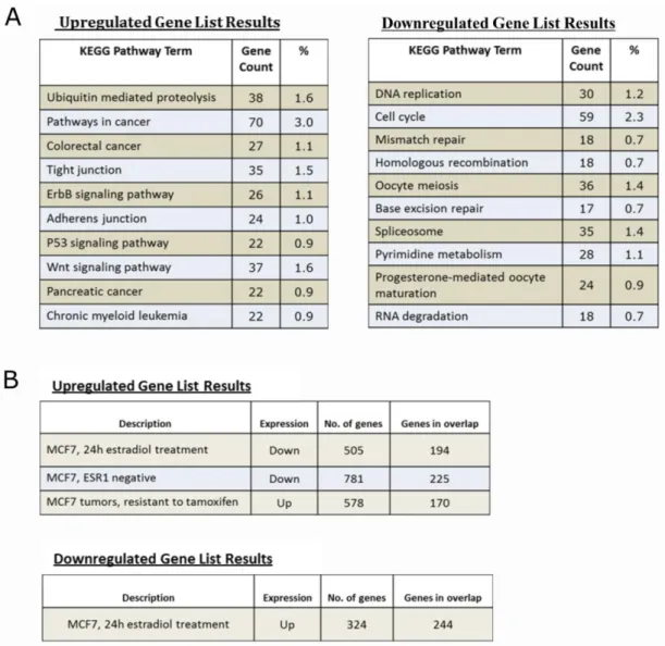
Figure 3.6: Microarray and related validation studies ... 80
Figure 3.7: Microarray data annotation ... 82
Figure 3.8: RT-qPCR confirmation of genes affected by CHRNA5 RNAi ... 83
Figure 3.9: RT-qPCR expression analysis of selected genes upon siRNA-1 treatment in BT-20 and MDA-MB-231 ... 84
Figure 3.10: Effects of CHRNA5 depletion on MCF7 cell viability ... 85
Figure 3.11: Cell viability of siRNA-1 treated BT-20 and MDA-MB-231 cell lines ... 86
Figure 3.12: Effects of CHRNA5 RNAi on DNA synthesis ... 87
Figure 3.13: Effects of CHRNA5 RNAi on cell cycle, apoptosis and detachment of MCF7 cells ... 89
Figure 3.14: Effects of 72h CHRNA5 depletion on MCF7 cell morphology ... 91
Figure 3.15: Effect of 72h CHRNA5 depletion on MCF7 cell morphology ... 92
Figure 3.16: Effects of 120h CHRNA5 depletion on MCF7 cell morphology ... 93
Figure 3.17: Validation of microarray data using RT-qPCR and Western blot analysis ... 95
Figure 3.18: Effects of CHRNA5 RNAi on MCF7 cell motility ... 96
Figure 3.19: Effect of CHRNA5 RNAi on the expression of actin regulatory genes ... 97
Figure 3.20: Effects of CHRNA5 depletion on the sensitivity of MCF7 cells to TOPO inhibitors ... 99
Figure 3.22: Microarray comparisons between CHRNA5 siRNA-1 and chemotherapeutic drug profiles ... 102 Figure 3.23: Common signatures between topoisomerase inhibitors and
siRNA-1 treatment ... 105 Figure 3.24: Effects of red-emitting nanoparticle on breast cancer cell viability .. 107 Figure 3.25: Characterization of red-emitting NP compartmentalization and
List of tables
Table 2.1: Products used for experimental procedures ... 53
Table 2.2: Products used for cell culture procedures ... 55
Table 2.3: Nucleic acids used for RNAi studies... 56
Table 2.4: Antibodies used in WB and IF studies ... 56
Table 2.5: Primer pairs used in RT-qPCR studies ... 57
Table 2.6: Laboratory instruments used for experimental procedures ... 58
Table 2.7: Solutions and buffers used in the study ... 59
Table 2.8: Reaction conditions of RT-qPCR ... 66
Table 2.9: SDS-polyacrylamide gel preparation ... 69
Table 3.1: STRING network statistics of expression profile comparison between siRNA-1 and individual drug treatments in MCF7 cells ... 103
Table 3.2: Significant KEGG pathways showing common regulation between siRNA-1 and individual drug expression profiles ... 103
Table 3.3: miRNET statistics of common differentially expressed genes in siRNA-1 and drug treatments (DOXO, SN38) in MCF7 cells ... 104
Table 3.4: Candidate miRNA molecules suggested to affect the functional properties of CHRNA5 and TOPO drugs ... 105
Abbreviations
CHRNA5 Cholinergic receptor nicotinic apha 5 SSB Single strand break
TOPO Topoisomerase ACh Acetylcholine
DOXO Doxorubicin ChAT Choline acetyltransferase
CPT Camptothecin AChE Acetylcholinesterase
NP Nanoparticle nAChR Nicotinic acetylcholine receptor
CON conjugated oligomer-based nanoparticles PR Progresterone receptor
CB7 Cucurbituril 7 CNS Central nervous system
ER Estrogen receptor RMA Robust multi-array average SCC Squamous cell carcinoma TNBC Tripple negative breast cancer SERD Selective estrogen receptor ECM Extracellular matrix
downregulators HER2 Human epidermal growth factor
receptor ERP Enhanced Retention and Permeability 2
Chapter 1
Introduction
1.1 Breast cancer treatment and resistance
1.1.1 General background on breast cancer treatment
Breast cancer therapies have undergone revolutionary progress, especially during the last twenty years, due to better understanding of breast cancer biology. The work of Halsted and colleagues [1] has shown that breast cancer is a disease which can result in metastasis through the lymphatic system. This implied the necessity of breast cancer surgery and radiation therapy as curative means. Another evolutionary finding was by Sir George Beatson who has shown that many breast cancers in premenopausal women could regress if ovaries were removed [2]. Discovery of estrogen receptor (ER) in 1960 was followed by the finding that ER could be measured and therapeutic response could be predicted [3]. Thus numerous ER or estrogen targets were subsequently developed. The first selective ER targeting agent was Tamoxifen; the first molecularly targeting drug in oncology. However, it was found that not all ER-positive breast cancers responded to endocrine manipulation while ER-negative tumors were totally out of the tamoxifen spectrum [4]. Human epidermal growth factor receptor 2 (HER2) over-expression (major cause of breast cancer patient mortality) on the other side was followed by the production trastuzumab (humanized monoclonal antibody), development of other monoclonal antibodies and small molecule receptor kinase inhibitors, all of which targeted HER2 protein [5]. The finding that some breast cancer patients lacked both PR, ER expression and HER2 overexpression (nowadays defined as “triple-negative”), made breast cancer research focus on the treatment of patients not benefiting from either of the therapies. These cancers were characterized by high proliferation, poor prognosis, low differentiation and high metastatic potential, leading to therapeutic uncertainity [6].
Apart from therapeutic approaches following the ER and HER2 status of breast tumors, there are other means of cancer treatment which focus on targeting the general hallmarks of cancer biology, such as angiogenesis; defined as new blood vessel formation within/around tumor mass. Bevacizumab (a humanized monoclonal antibody), targets VEGF/VEGFR entity as a major player in vascularization however, still only a subgroup of breast cancer population benefit from this treatment. Yet it is not possible to determine which group will benefit or not from this drug, making it a non-targeted therapy [7].
Current treatment of breast cancer depends on its subtype, based on the oncogenic signaling pathway that derives it. Surgery, radiotherapy, hormonal therapy and chemotherapy are the current options for breast cancer treatment, separately or in combination [8]. The current chemotherapeutic agents for breast cancer include taxanes (i.e., docetaxel, paclitaxel), anthracyclines (i.e., doxorubicin, epirubicin) [9], anti-metabolites (i.e., capecitabine, gemcitabine) [10], microtubule inhibitors and/or stabilizers (i.e., vinorelbine, ixabepilone) [11] and platinum-based antineoplastic drugs (i.e., cisplatin, carboplatin) [12].
Contemporary breast cancer research has improved our understanding of breast cancer as being not a single disease; rather a mixture of diseases with heterogeneous histology. Thus breast cancer outcomes have improved due to treatments which include improved old and new approaches. Nowadays most oncologists focus on personalized therapy by providing the right treatment, at the right time, to the right tumor type. Moreover, new therapeutic ways of attacking cancer cells more precisely, use drug combinations (sometimes included in one formulation), with less side effects to normal tissues [13].
1.1.1.1 Treatment of estrogen receptor (ER)-positive patients
ER-positivity includes the most prevalent invasive type of breast cancers, showing variable clinical outcomes and responses to anticancer therapy [14]. The current standard treatments of ER+ breast cancers are based on using aromatase inhibitors, selective estrogen receptor downregulators (SERDs) and tamoxifen [15]. Aromatase inhibitors (such as anastrozole and letrozole) are drugs which lower the estrogen
production by targeting the aromatase enzyme that produces it. SERDs are agents that inhibit ERapha expression, while tamoxifen is an ER modulator which antagonizes the receptor function in breast tissue [15]. Luteinizing hormone-releasing hormone (LHRH) agonists, as chemicals which prevent ovaries to produce estrogen and progesterone, are important for progesterone receptor (PR)-positive breast cancer therapy [16]. All of the above mentioned drugs enter in the class of ‘endocrine/hormonal therapy’. However, endocrine therapy is characterized more by inducing a reversible dormant state to breast cancer cells rather than causing cell death [17].
1.1.1.2 Treatment of human epidermal growth factor 2 receptor (HER2)-positive patients
HER2 positivity cover 15-20% of early stage breast cancer patients, and is associated with increased tumor aggressiveness, low disease-free and overall survival, as well as increased breast metastasis rates [18]. Current treatment of HER2-positive breast cancer patients include small molecule inhibitors, monoclonal antibodies and antibody drug conjugates. In addition, combinational therapy shows better disease outcomes when compared to monotherapies. For instance, the addition of trastuzumab to neoadjuvant (pre-operative) chemotherapy doubles the probability of obtaining a complete pathological response and reduces the relapse risk [19]. Administration of aromatase inhibitors in combination to HER2-based therapy significantly improves treatment outcome in hormone-receptor positive women. Anti-HER2 vaccination is also a developing approach of assisting HER2-overexpressing tumors, with likeliness to improve treatment and avoid the side effects of current therapies [20]. Systemic chemotherapy on the other side, is applied for women having higher proliferative tumors [21].
1.1.1.3 Treatment of triple negative breast cancer (TNBC) patients
TNBC includes 10-20% of invasive breast cancers. It encompasses different subtypes; basal-like, normal breast-like, claudin-low, BRCA1-deficient breast tumors, all of which are more frequently seen in younger African-American women
expression and HER2 amplification, there has been no targeted therapy available for this subgroup of patients, which caused an increasing demand for personalized therapy. Currently, chemotherapy is the only approved treatment for TNBCs, which serve solely for pain relief and prolonging survival. It includes anthracyclines, taxanes, anti-metabolites and microtubule inhibitors/stabilizers, as single-agent regiments or combined therapy [23]. Retrospective studies have shown that TNBCs are more sensitive and responsive to chemotherapeutic agents (probably due to their high proliferation rate) and increased neoadjuvant response as compared to other tumors [24]. Similar to other breast cancer subtypes, Doxorubicin- and Paclitaxel- based regiments are the standard treatments for TNBCs [25]. They are administered either alone or in a combinatorial way (especially when quick response is necessary). TNBCs, which are highly associated with BRCA-1 gene mutations, are quite sensitive to platinum agents as well [25]. Recent research on TNBC treatment includes a large phase III clinical study of targeted therapy using Bevacizumab (anti-angiogenic agent against VEGF), which might be given in the future as an adjuvant in addition to chemotherapy [26]. Vaccines (as a means of immunotherapy) are also under investigation for treating metastatic TNBCs [23].
1.1.2 Drug resistance in breast cancer patients
1.1.2.1 Molecular mechanisms of drug resistance
Development of cancer cell resistance toward anticancer agents is a major challenge in cancer treatment. Cancers may consist of cells with stem cell-like properties and/or with intrinsic ability to resist therapies [27] due to: loss of target protein expression after continuous therapy [28], induction of G0 phase [29], overexpression of some proteins related to drug efflux and metabolism [30], upregulation of some pro-apoptotic genes, activation of prosurvival signaling [31], and regulation of DNA repair mechanisms [32]. Moreover, these stem cells are suggested to posses self-renewal and tumor initiation abilities, which make cancer treatment even more difficult. Due to molecular heterogeneity of tumors, drug resistance can also arise from a small population of cells acquiring resistance as a selection mechanism [33]. In addition to tumor-derived cells, tumor microenvironment (consisting of ECM,
fibroblasts, immune cells and blood vessels in solid tumors) might also contribute to drug resistance by providing growth, resistance and refuge to metastatic cancer cells [34]. More interestingly, same microenvironment might render the tumor cells more responsive to other agents (named “synthetic lethality”), which seems to be cancer- and therapy type-specific [35].
1.1.2.2 Breast cancer subtype-specific drug resistance
Drug resistance in advanced breast cancer is the cause of incurability, however, early-stage breast cancer resistance is of great concern due to the possibility of being cured. For the ER+ breast cancers, endocrine therapy resistance is categorized either as intrinsic (not showing any response to endocrine therapy) or acquired (developing resistance after a response phase) [36]. Resistance to hormonal therapy seems to be agent-specific; after resistance developed from one drug, another endocrine therapy approach might show effectiveness [37]. Mechanisms of drug resistance in HER2+ patients on the other hand, involve absence of the extracellular binding domain [38], loss of HER2 amplification during treatment [39], activation of other HER-family receptors as a compensatory mechanism [40], and deregulation of several signaling pathways [41] . Combining of HER2 inhibitors (which show synergistic effects) is a strategy followed in order to overcome drug resistance [42].
1.1.3 Topoisomerase enzymes as anti-cancer drug targeting
1.1.3.1 Topoisomerase mode of action
During DNA replication and transcription process, double helix DNA needs to be uncoiled however, its structure restricts DNA of a free rotation inside cell nucleus. Thus, strand separation by helicase enzymes induces the formation of positive supercoiling at the front side and negative supercoiling at the back side of DNA replication and transcription [43]. The positive supercoiling (if not processed), stalls DNA replication and transcription progress, while supercoiling at the back side might induce development of abnormal DNA structure, which might be an obstacle for normal DNA functioning [44]. During normal DNA replication and transcription
process, DNA tension is avoided with the help of large proteins/enzymes named as ‘topoisomerases’ [45].
Human genome encodes six topoisomerases, which are classified either as Type I and Type II. During their catalytic functions, Type I enzymes make a single cleavage in one of the DNA strands, while type II topoisomerases cleave both strands [46]. Both enzyme groups similarly perform DNA cut by a nucleophilic attack and subsequent binding of their catalytic residue to the phosphate end of the phosphodiester backbone takes place [44]. After the relief of supercoiling by topoisomerization, DNA is religated and its sequence if left unchanged. One of the topoisomerase subtypes;TOP1mt, shows similar activity to topoisomerase 1 in uncoiling of the mitochondrial DNA [47].
1.1.3.2 Topoisomerase inhibitors
Topoisomerases are further categorized into subtypes according to their mode of activity. Apart from their role in DNA relaxation during transcription and translation, they also show some other functions (i.e., as splicing factors) [48, 49]. Except topoisomerase IA and IIB, other subtypes are already clinical therapeutic targets [44].
Topoisomerase (TOPO) inhibitors of the mammalian enzymes bind to DNA-topoisomerase complex and block the DNA religation step of the enzyme-catalytic action, resulting in single or double strand breaks [50] (Figure 1.1).
Figure 1.1: Activity of topoisomerase enzymes and their inhibitors.
Schematic representation of topoisomerase enzymes and TOPO inhibitors of class I and II. Arrows indicate the reversible processes of DNA cleavage and relegation during normal enzyme activity. DNA damage is induced when A-C) TOPO I inhibotors bind to topoisomerase I and D-F) TOPO II ihibitors bind to topoisomerase II enzymes. This figure was reprinted with permission from ScienceCentral. Young Ho Seo, Dual Inhibitors Against Topoisomerases and Histone Deacetylases, (2015) Journal of Cancer Prevention, https://www.e-sciencecentral.org/articles/SC000010991.
1.1.3.3 Topoisomerase I inhibitors
Topoisomerase 1 inhibitors include the camptothecin (CPT) and non-camptothecin types of drugs. CPT was first discovered by M. E. Wall and M. C. Wani (1966) from the wood bark of the Chinese tree Camptotheca acuminate, in an attempt to find anticancer drugs by screening of the natural products [51]. Few of the camptothecin derivatives are already approved for clinical use, i.e., topotecan and irinotecan [52]. Although having a wide range of application as anticancer drugs, CPT derivatives show limitations in usage due to their chemical instability at physiological pH, caused by the E-ring presence in their molecular structure [52]. Additional CPT derivatives have been designed to improve the clinical tolerability however could not overcome the chemical instability of camptothecins [47]. Non-camptothecin synthetic topoisomerase I drugs such as indenoisoquinolines, dibenzonaphthyridinones and
aromathecins have recently been developed to improve drug stability and to form more stable cleavage complexes [53, 54].
1.1.3.4 Topoisomerase II inhibitors
Topoisomerase II inhibitors poison Top2α and Top2β after which a TOP2-DNA cleavable complex is formed, named as ‘TOP2cc’, which is considered as a barrier to the normal progression of replication and transcription process [55]. TOPII inhibitors not only cause DNA double strand breaks (DSB), but also single strand breaks (SSB) [56]. They are classified into two major groups. The first group includes the most clinically used ‘poisons’ which elevate the level of TOP2-DNA covalent bond, such etoposide, doxorubicin and mitoxantrone [57]. The second class consists of ‘catalytic inhibitors’ which block the catalytic activity of TOPII but do not increase the TOP2-DNA complex levels, such as bisdioxopiperazines [57]. Doxorubicin, a TOP2 poison, was firstly isolated from Streptomyces peucetius species [58]. It is an anthracycline antibiotic and at the same time widely used for many kind of malignancies like childhood solid tumors, breast cancer and leukemias. However it came up as being toxic also to normal cells due to the production of reactive oxygen species (ROS), and causing of cardiotoxicity [59].
1.1.4 Camptothecin and doxorubicin in breast cancer research and treatment
1.1.4.1 Camptothecin mode of action in breast cancer cell lines
Chemotherapy is the dominant approach in treatment of cancer and usage of conventional drugs/their derivatives is still the most used medication means. In breast cancer therapy related research, there are several studies showing camptothecin and doxorubicin effects on breast cancer cell lines. A 2005 study conducted by Lamparska et al., showed that camptothecin exposure to MCF7 cells resulted in apoptosis within a short time period (60min) while in the long run of 24h drug administration, continuous development of autophagy occurred at a slower rate [60]. Moreover, BID knockdown in CPT treated MCF7 cells, induced a shift from apoptosis to autophagy, suggesting that BID might serve as a molecular switch in the way of cell decision between the two
cell death mechanisms [60]. In another study conducted by the same research group, electron microscopy imaging of 6h CPT administration in MCF7 cells revealed a heterogeneous population of cells having both apoptotic- and autophagy- related morphological features [61]. MCF7 cells treated 8-16h by 0.15 uM CPT accumulated p53 protein in the nuclei of cells, with a rapid increase of ~20 fold in S-phase cells [62]. In another study, characterization of apoptotic response to CPT of several breast cancer cell lines revealed that MCF7 (p53 wild type) was among the resistant ones, indicating that p53 might not be required for CPT-induced apoptosis [63]. P53-independent effect of CPT was also observed in multiple breast cancer cell lines in which CPT induced degradation of WRN, a helicase enzyme having role in DNA repair, genome stability and cellular senescence [64].
1.1.4.2 Doxorubicin mode of action in breast cancer cell lines
Similar to CPT, doxorubicin (DOXO) studies show not a single mechanism of breast cancer cell death induced by this drug. Doxorubicin-resistant MDA-MB-231 cell line, when exposed to an autophagy (shown to negatively associate with drug resistance) inhibitory molecule, became more sensitive to doxorubicin treatment and the cell death mode shifted from apoptosis to necrosis [65]. Doxorubicin resistance of MDA-MB-231 cell line has been linked to NF-kB expression which is also a metastasis inducer [66]. Restoration of p53 in this cell line showed impairment of NF-kB expression induced by doxorubicin, implying p53-dependent cytotoxicity of this drug [66]. On the other hand, MCF7 cells treated with doxorubicin showed upregulation of p21 level and increases in cellular senescence [67]. However, another study conducted in both MCF7 and MDA-MB-231 cell lines showed p53-status independent effect of doxorubicin in inducing apoptosis [68]. Doxorubicin uptake and drug response in MCF7 cells was correlated with cellular fluidity [69] as well as with diverse drug formulations to improve its biopharmaceutical and physicochemical properties [70, 71]. In order to increase efficacy and overcome resistivity of breast cancer cells to chemotherapeutic effect of doxorubicin, several studies have been conducted including combined treatments of this drug with other molecules. Combination of estrogen and doxorubicin showed enhanced apoptotic action of the drug on estrogen-independent MCF7 cells (MCF7/LS) via suppression
doxorubicin was similarly increased upon co-treatment with proteasome inhibitor [73]. In addition, many combinatorial treatments with natural products (reported from in vitro and in vivo studies), showed reduced cardiotoxic effects and/or higher drug efficacy of doxorubicin [74-76].
1.1.5 Nanomedicine in breast cancer treatment
1.1.5.1 Properties of nanoparticles used in cancer treatment
Nanotechnology has contributed vastly to the chemotherapeutic drug delivery system. Contemporary cancer treatment studies focus on the preparation of the drug formulations which are less toxic to normal tissues and which specifically target tumor cells [77]. Novel drug carriers are developed for delivering of already existing as well as newly designed drug molecules and at the same time for overcoming the pharmacological and physiological barriers of old therapies. Small size formulations of drug encapsulating complexes, increase in their drug loading efficiencies, as well as combination of different drugs/molecules in one complex are the characteristics of good nanoparticles (NPs) [78]. Such designs are also expected to show improved drug uptake and specificity to cancer cells, decrease resistivity to chemotherapeutic agents, lower toxicity to normal cells and increase efficacy in cancer therapy [79]. Since the gaps between the endothelial cells in the tumor vasculature surrounding cancer cells range from 100nm to 780nm, NPs for drug delivery are accordingly designed in nm size [80]. NPs are particularly coated with hydrophobic molecules to prevent binding to plasma proteins and gain favourable clearance properties from circulatory system [81]. Construction of NPs include usage of different materials (i.e., polymer, lipid, metal, ceramic), size and shapes (i.e., liposomes, spheres, emulsions, nanotubes), depending on their intended use [80]. NPs are further categorized into organic (e.g., liposomes, polymeric, micelles, etc.) and inorganic (e.g., silica, gold, iron oxide, etc.). In addition, such drug-encapsulating NPs should also be degradable after targeting cancer cells [80]. Drug targeting approach might be passive (passively entering the tumor interstitium and cell) [82], active (based on molecular recognition of cancer cells, i.e., being coupled to a ligand) [83] or a combination of the two [84]. In addition to the above mentioned advantages of
nanocapsules, recently designed NPs show sustained drug release, enhanced retention and permeability (EPR) in cancer cells as well as imaging functions [85]. EPR effect is the gold standard for newly designed NP-based formulations. Due to their molecular weight higher than 40-50kDa, macromolecular systems show selective accumulation of drug complexes in tumor site as well retention for longer periods [86]. This is due to higher vascular density around solid tumors with large gaps between endothelial cells [86]. Given these advantages, NPs have long been considered as having the potential to dramatically change clinical treatments by improving the present therapies or introducing new ones.
1.1.5.2 Usage of Conjugated Polymer Nanoparticles for medical applications
Conjugated polymer nanoparticles (CPNs) are another class of nanoparticles, conjugated polymer (CP) counterpart of which, is produced by one of the polymerization reactions (e.g., oxidative polymerization, Heck coupling, etc.) using different types of conjugated polymers (PF, PPV, PPE, PT). Their properties are significantly affected by the surface charges and functional groups [87]. CPNs are characterized by striking abilities of light-harvesting and light-emitting properties which make them very useful and multifunctional expecially for fluorescent targeted imaging, diagnosis, gene therapy and drug delivery [88]. Moreover, recently designed multifunctional nanoparticles show high photostability, low cytotoxicity and quantum yield, good biocompatibility, small size and ability to sensitize reactive oxygen species. Their imagining capabilities have shown importance in monitoring the delivery process by tracking the assembly and dissasambly of their cargo (e.g., oligonucleotides) [89].Thus, CPNs have been recently used to kill microorganisms or tumor cells as well as to label cells in vitro and in vivo [90]. Furthermore, conjugated oligomer-based nanoparticles (CONs) are a class of NPs given less focus in literature, but having comparable or even more advantages over CPNs. For instance, CONs show comparable cellular uptake and higher fluorescent quantum yields as well as faster release of their cargo compared to their polymeric counterparts [91]. CONs have also shown promising results as oligonucleotide nanocarriers for future drug delivery usage [92]. However, cellular toxicity is a major concern in CON design due to their high positive charge. To improve cellular compatibility,
complexes, such as Cucurbituril (CB); a macrocycle composed of glycoluril units, having a hydrophobic cavity and two hydrophilic portals [93]. For instance, Pennakalathil et al., (2014) introduced a CB7-capped, red-emitting CON (Red-CON) which showed to be pH-responsive and possess both cellular imaging and drug delivery potential [94] (Figure 1.2). The oligomeric nanoparticle composed of amine groups, showed precipitation in human blood serum, however when capped by CB7 (a water soluable CB), it became non-precipitative in blood serum, at least for a 24h incubation. In addition, CB7-capping only mildly increased the oligomer NP size, keeping its size-dependent properties unchanged [94]. As an example of CONs’ potential advantages, a very recent study has presented a multifunctional fluorescent oligomer with very promising results in both nonionizing real-time imaging of sentinel lymph node (SLN) (for early detection of breast cancer metastasis) and photothermal therapy (PTT) (for specific killing of metastatic cells) [95].
Figure 1.2: Structure of CB7-capped Red-CON.
Illustrative representation of Red-CON structural composition, capping as well as potential cellular internalization and drug delivery abilities. This figure was reprinted with permission from ACS publications. Copyright (2014) American Chemical Society. Pennakalathil et al., Red Emitting, Cucurbituril-Capped, pH-Responsive Conjugated Oligomer-Based Nanoparticles for Drug Delivery and Cellular Imaging, (2014) Biomacromolecules.
1.1.5.3 Combinatorial treatments using nanoparticles as drug carriers
Co-delivery of doxorubicin and other chemotherapeutic drugs in liposome encapsulated formulations is reported in several recent in vivo mice studies [78, 96], as well as patients of phase II and phase III trials [97, 98]. Newly developed liposomes can transport and release active drugs also by using the pathophysiology of tumor microenvironment as well as by targeting cancer cell-specific receptors, i.e., estrogen-anchored [99] and pH-sensitive formulations [99, 100].
Drug formulations designed for targeting of a specific aberrant pathway in tumor cells have increased interest in the last years. In addition, due to very effective survival mechanisms developed by cells to resist drug toxicity, RNAi (using small interfering RNA; siRNA) approach is an alternative in treating tumors. siRNA therapy (either alone or in combination to other molecules) is performed using non-viral carriers such as NP-based formulations. There are studies showing siRNA librariy screens including hundreds of siRNA candidates against cell cycle proteins in combination with conventional anticancer drugs for breast cancer therapy usage, in drug-resistant and -sensitive breast cancer cell lines and mouse xenografts [101-103].
After extensive research on NP-based drug formulations, some of them are now FDA approved either for therapeutic or diagnostic purpose. Cancer nanoparticle medicines are now used to treat different cancer types and stages [104]. In addition, intravenously delivered organic nanoparticle formulations show substantial success in gene therapy applications and delivery of small molecule drugs for cancer treatment [105]. Most of the approved nanoparticle drugs include liposomal encapsulations of anticancer drugs [105]. The first liposomal cancer nanomedicine to be approved by FDA (1995) was Doxil (PEGylated liposomal doxorubicin) [106], followed by other drugs such as DaunoXome (liposomal daunorubicin) [107], Myocet (non-PEGylated Doxorubicin) [108] and Abraxane (albumin-bound paclitaxel NP) [109]; all of which, compared to the free drug delivery process, posses the ability to preferentially accumulate in the tumor microenvironment due to their enhanced retention and permeability (ERP) [105]. In addition, several nanoparticles are now used as imaging agents, i.e., Feridex and Resovist (imaging of liver lesions) [110], Ferumoxtran (imaging of lymph node metastasis) [111], Optison and SonoVue (as ultrasound contrast agent) [112]. Current human trials are on the way to approve several other formulations, majority of which will be used for cancer treatment [105].
1.2 siRNAs as breast cancer candidate drugs
1.2.1 General background on RNAi machinery
A vast majority of human genome encodes for genes which are not translated to protein. Only 1.2% of genome is translated to protein [113]. Around 60 years from now, the first non-coding RNA (tRNA) was identified and its primary structure was characterized [114]. After 20 years, uridine-rich small nuclear RNAs (snRNA) were discovered [115]. These RNAs, in association with ribonucleoprotein particle (RNP), were later shown to be involved in spicing process of pre-mRNA. Most of the small nucleolar RNA (snoRNAs) interestingly were found to be processed from released introns [116]. Around 15 years later, Wightman et al. showed the regulatory mechanism of lin-14 gene (involved in the development of C. elegans) [117], after which two other studies directed by Ambros and Ruvkun, found lin-14 gene product to be a non-coding RNA of 22 nucleotides which regulated its own gene expression by binding to the 3’ UTR [118, 119]. In year 2000, Pasquinelli et al., identified a small temporal RNA involved in C.elegans development, which was also conserved in humans [120]. This brought extensive discoveries of other small regulatory RNAs in humans and model organisms, now termed as ‘microRNAs’. Together with these small-RNA class entities, long non-coding RNAs (lncRNAs) which size ranges up to several kilobases, are on investigation [121].
1.2.1.1 RNAi molecules
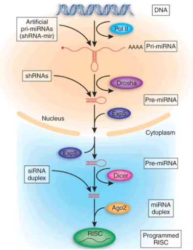
A microRNA (miRNA) molecule is a 21-25 nucleotides long non-coding RNA strand which is byproduct of a larger nuclear RNA precursor molecule. In brief, miRNAs are synthesized from the DNA strand by RNA polymerase II as pri-miRNAs, after which (while still in nucleus) they are processed by Drosha proteins into a hairpin structure called as ‘miRNA’ and are then transported to cytoplasm [122]. Afterwards, pre-miRNAs are cleaved by Dicer protein which render the now called ‘mature miRNA’ or ‘siRNA’ in an ~21-25 nucleotide, 3’ overhangs containing structure. Single-strand miRNAs then bind to nucleoprotein complex RISC (Figure 1.3) and either in perfect or partial complementarity with certain mRNA strands, induce the degradation of the transcript or their translation repression, respectively [122].
Small interfering RNAs (siRNAs) are the canonical affectors of RNAi mechanism which initially were recognized as exogenous elements involved in the defense mechanisms of plants [123]. They are double stranded, long and perfectly based-paired RNA strands which are either introduced from nucleus to cytoplasm as such or enter cell from the outside environment [124] (Figure 1.3). Endogenous small-interfering RNAs (endo-siRNAs) are mainly transcribed from the processed pseudogenes (DNA copies with no introns, reversed transcribed from their parental gene transcipts) [125], but also can originate from the diced genomic transcripts, convergent RNA transcripts or from natural sense-antisense transcript pairing [126]. Similar to miRNAs, endo-siRNAs are processed by Dicer after loaded into Ago-2 and form the RISC complex. During the RNAi pathway, the single strand siRNA guides RISC complex to the target transcript where it binds with perfect complementarity. The slicer activity of this complex induces a phosphodiester bond breakage of the target RNA; 10-11 nucleotides from its 5’ base-paired start sit [127]. After this, other nuclease enzymes bind and complete the target transcript degradation process. RISC is now released and ready to cleave other targets [127]. Apart from the perfect complementarity, many siRNAs bind imperfectly to targets, similarly to miRNAs. Most of these imperfectly matches include their ‘off-targets’ binding events [128]. The 2-8 bases in the 5’ region of RNAi molecules include the seed-region; the primary target-recognition site of siRNA. The degree of siRNA off-target gene silencing is determined by the strength of base-pairing between its seed region and the off-target transcript, affected also by other auxiliary factors [129]. Exogenously transfected RNAi molecules on the other hand, are nucleic acid molecules directly transfected to cells either as synthetic double strands, endonuclease-prepared or as short hairpin RNA strand (shRNA) precursor [130] (Figure 1.3). siRNA treatments are not 100% efficient and only transiently downregulate their target gene expression. However, for studying the short term effects of gene knockdown this method is sufficient and even advantageous, especially for targeting of essential genes [130]. RNAi screening has held an important role in identifying new genes as well as resolving many questions on biological networks and processes such as cell viability, cell morphology, drug resistance, etc [131].
Figure 1.3: RNAi machinery.
The mechanism of processing endogenous and exogenous RNAi molecules by the cell. This figure was reprinted with permission from Nature Publishing Group. Cullen, RNAi the natural
way, (2005) Nature Genetics.
1.2.2 RNAi delivery methods
1.2.2.1 RNAi transfer systems
Gene delivery of RNAi is used either in research procedures for downregulating the target gene or for gene therapy purpose. Two of the main nucleic acid classes mostly used in RNAi transfection experiments include 2-10 kilobase plasmid DNA or 19-25 base-composed short double-strand RNA (either in the form of siRNA or miRNA) [132]. When transfected to cells, two RNA molecule characteristics should be taken into account; 1) cell compartment where they are expected to function (cytoplasm or nuclei) and 2) their length (base or kilobase magnitude) [133]. Delivery methods of nucleic acids include a) non-pathological viruses which are mostly used for in vivo studies, b) viral-like particles and c) non-viral techniques [134]. Viruses are
specifically designed to include the nucleic acid sequence of interest into the viral genome and release their cargo into the cytoplasm of the infected cell. The viral genetic material then enters nucleus, integrates into the host genome and starts being expressed using the host transcriptional machinery [134]. Different viral types are currently used for this purpose including retroviruses, lentiviruses, adenoviruses, adeno-associated viruses, parvoviruses and herpes simplex viruses. Eventhough displaying high transfection efficiency, usage of viral formulations show health concerns based on the host immune response induction and insertional mutagenesis. Other obstacles of using viral particles include challenges like complexity of their design, their packaging capabilities as well as the expensive procedures for their production [135]. Virus-like particles on the other hand, include recombinant viruses which except their capsid do not contain any other viral genetic information. The advantage of this technique is the easy packing possibility of the siRNA or plasmid DNA by spontaneous capsid self-assembly, via chemical dissociation and reassociation process. Most of these viruses are used as a vaccination tool, while several others are available in the market for research purpose such as: human polyoma JC virus, papillomavirus and polyomavirus [136].
1.2.2.2 RNAi transfection modalities
Cell membrane (a polar lipid bilayer) doesn’t internalize negatively charged nucleic acid molecules spontaneously. This is the reason why many methods for delivering of nucleic acids to cells have been established. Non-viral nucleic acid delivery methods include 1) mechanical, 2) physical and 3) chemical methods. Mechanical method includes microinjection, particle bombardment and single-walled carbon nanotubes. By microinjection nucleic acid is directly delivered to a single cell using glass capillaries. Eventhough efficient, this is a difficult procedure for in vivo and cell culture application studies, since it includes large number of cells [137]. Particle bombardment on the other hand, is a delivery method by which gold or tungsten nanoparticles loaded with nucleic acids are given a high velocity in order to cross the cell membrane of many cells simultaneously. This method is mainly used for vaccination and plant studies [138]. Single walled carbon nanotubes are composed of one-dimensional layers of hexagonal carbon nanostructure. By addition of positively
nucleic acid to the host cell. This method has shown positive results of siRNA gene targeting even in in vivo studies [139]. Electroporation technique, as a means of physical transfection method, includes application of an electric field to cells. This treatment causes short-term depolarization and pore formation on the cell membrane, thus makes it possible for molecules like siRNA and plasmid DNA to be internalized into the cell. This technique is efficient and non-toxic, as far as multiple parameters are optimized and kept constant, such as: length of the pulse, number of pulses and temperature [140]. Permeabilization of cell membrane is also achieved by laser – beam-mediated-gene delivery (as a means of physical transfection method), which is performed by the application of a focused high-energy laser light. Eventhough showing very promising results on transfection efficiency and cell toxicity, laser-beam-mediated-gene delivery is quite challenging due to its manual application which makes it more appropriate for studies including limited cell number [141]. On the other hand, chemical method for nucleic acid delivery includes: 1) the widely used calcium phosphate method, 2) polymers, 3) PAMAM dendrimers and 4) lipid formulations. In the first technique, sodium phosphate and calcium chloride are mixed with nucleic acid molecule after which calcium phosphate crystals are formed. These crystals bind the nucleic acid and carry it to the cell. This method has a limited application because of being highly sensitive to pH changes and its cell type-dependent transfection efficiency [142]. Polymers used for transfection studies are mostly made up of synthetic polyethylenimines (PEIs). Composed of positive amine charges, PEIs formulations range from those carrying small oligonucleotides (such as siRNAs), to the ones carrying large plasmids; which are taken up by cells via endocytosis. Polymer efficiency and toxicity is based on the formulation size of the molecule [143]. Similar to PEIs, PAMAM dendrimers are polymers composed of amine groups, more appropriate for binding and transferring of plasmid DNA rather than short oligonucleotides. Several commercial PAMAM dendrimer formulations which have water solvable, biocompatible and non-immunogenic properties are now available for research purpose [144]. Lipid molecules on the other hand, are widely used for siRNA and plasmid DNA gene delivery purpose. They carry nucleic acids either by entrapping them inside the internal aqueous region, or by binding of nucleic acid on the lipid bilayer surface. Lipid designing and formation is a very flexible process specifically adapted to the purpose of usage by modifying their surface charge, size, composition and morphology. Another advantage of lipids as nucleic
acid carriers is their good stability due to being protected from the cell nucleases cleavage [145]. As a subclass of lipids, cationic lipids are widely used both in in vitro and in vivo studies of siRNA and plasmid transfection. Their overall positive charges make it possible for them to bind the cell membrane and induce membrane perturbation and fusion. Although showing no immunogenic responses and insertional mutagenesis in the host genome, these lipids don’t show transfection efficiency as high as the viral particles. Moreover these lipids are inactivated in the presence of serum (both from in vivo and in vitro cell studies) [146]. Currently there are several cationic lipid formulations available in the market both for siRNA and plasmid DNA delivery, such as HiPerfect and Attractene Transfection Reagents, respectively. Specialized lipid-based formulations are elegant designs (mostly for in
vivo gene delivery). By modifying their functional groups, such lipid formulations
can be specialized for specific targeting and/or including pH-sensitive platforms for siRNA and plasmid-DNA drug designs [147].
1.2.3 RNAi machinery and cancer
1.2.3.1 Role of miRNAs in tumor progression and cancer treatment
To better understand the RNAi impact in human health, we should consider that human cells express approximately 100 types of endogenous miRNAs which are thought to regulate around 30% of the transcriptome. They regulate many cellular processes such as reproduction, wound healing, cardiovascular remodeling, pain, as well as show significant role in different pathologies such as epilepsy, cerebrovascular diseases, hypertension and diabetes [148, 149]. miRNAs are also key regulators of cancer initiation and progression, with important implications in metastasis process [150]. Extensive and increasing research on miRNA deregulation in cancer has revealed several of them as tumor suppressors, oncogenes or having both roles; depending on the cellular context and/or cancer type. Current studies also focus on exploiting miRNA signatures for their possible usage as prognostic, diagnostic, theranostic (treatment monitoring) markers and personalized therapy [151]. The fact that a single miRNA has multiple targets could be advantageous and indicates that modulating one miRNA’s activity might open door to innovative therapies in breast
cancer. Targeting of miRNAs which show oncogenic activity could be an approach toward personalized therapy. This can be achieved either by natural/synthetic agents or oligonucleotides targeting overexpressed miRNAs; all of which have been experimentally demonstrated. Another benefit of targeting the endogenous RNAi machinery could be that of preventing future metastatic potential of cancer or treatment of an already developed metastasis [152]. In breast cancer, several miRNAs have been associated with cell cycle, cell morphology, cancer stem cell regulation and metastasis. Overexpression of miRNAs in breast cancer specimens is often an indication of genomic abnormalities. On the other hand, miRNA expression can be evaluated even from clinical specimens like fine-needle aspirates [153]. Accordingly, there has been intensive research conducted, focusing on the possibility of using circulating miRNA molecules for diagnosis and prognosis purpose. Few circulating protein molecules have already been used as biomarkers for metastatic breast cancer however they didn’t show high sensitivity for diagnosis of primary breast cancers [154]. Using the circulatory miRNAs from microscale serum volumes of breast cancer patients has recently identified circulating miRNA signatures which might well sever as biomarkers for early breast cancer diagnosis [154].
1.2.3.2 Advances in research about endogenous siRNAs
Endogenous siRNAs (endo-siRNAs), are not as extensively investigated as the miRNA molecules. Endo-siRNAs share many similarities to the other small-RNA family members (miRNAs and piRNAs), which makes it difficult to separate and trace them specifically [155]. Most of the endo-siRNAs studies include their characterization in the invertebrate model organisms, while the vertebrate studies show not a clear mechanistic aspect and physiological role of endo-siRNAs, with discrepancies among several research groups [156]. In different organisms, pseudogenes-derived endo-siRNAs have been implicated to their ancestor-gene regulation [157]. In C.elegans studies, endo-siRNAs have shown role in chromosome organization and gene regulation [158, 159]. Drosophila-related studies have revealed endo-siRNAs role in mitochondrial metabolism, transposon regulation and ovarian cell gene regulation [160, 161]. Similarly, two nature papers in 2008 showed that endo-siRNAs regulate gene expression in mouse oocytes [162, 163] while other studies implicated endo-siRNAs in murine reproductive and nervous system [164, 165].