DOI: 10.1501/commuc_0000000181 ISSN 1303-6025
© 2015 Ankara University DIFFERENT APPROACHES FOR BREAST CANCER: VOLTAGE GATED
POTASSIUM CHANNELS AND MICRORNAS Çağrı ÖNER1, Ertuğrul ÇOLAK2, Didem TURGUT COŞAN1 1Eskişehir Osmangazi University, Medical Faculty, Department of Medical Biology,
ESKİŞEHİR/TURKEY, [email protected]; [email protected]
2Eskişehir Osmangazi University, Medical Faculty, Department of Biostatistics and Medical
Informatics, ESKİŞEHİR/TURKEY; [email protected]
(Received: June 15, 2015; Accepted: October 05, 2015 )
ABSTRACT
Micro RNAs and voltage-gated potassium channels (VGPCs) both play critical roles in the development of cancer. We aimed to reveal the diversity of miR-126/126*, which effects angiogenesis and vascular development through the inhibition of VGPCs.
In this study, potassium channel inhibitors, including tetraethylammonium (5mM), 4-aminopyridine (5mM), margatoxin (1µM) and astemizole (2µM), were applied to MCF-7 and MDA-MB-231 breast cancer cell lines. After totally isolating RNA from the cells, Real-Time Polymerase Chain Reaction was used in order to identify gene expressions. OneWay ANOVA was used for variation analyses, while Tukey HSD and Tamhane were used to assess whether multiple comparisons were statistically significant (P < 0.001). Our results showed an increase in miR-126/126* expressions after the channel inhibition of MCF-7 and MDA-MB-231 cell lines (P < 0.001). miR-126/126* expressions were increased using TEA, 4-AP and astemizole in both cell lines. miR-126/126* expressions were only increased through the use of margatoxin in MCF-7.
miR-126/126* may interact with voltage-gated potassium channels. In our study, the inhibition of K channels using K channel blockers resulted in an increase of miR-126/126* expression. Therefore, our data suggested that there could be another perspective between K channels and non-coding RNAs in the development of breast cancer.
1.
INTRODUCTION
Ion channels, which are the main signaling complex, are expressed in most tissues and have different cellular activity. Therefore, many of the pathophysiological conditions in cancer include ion channels (1). Voltage-gated potassium channels [VGPCs] are fundamental to cellular physiology and play a number of key functions, such as differentiation, proliferation, apoptosis, adhesion, migration and control of volume. These functions are linked to the formation of primary and metastatic tumors (2). Ion channels are expressed in various tissues and show various activities. This is particularly clear in voltage-gated ion channels which allow K+, Cl- and Na+ ions to be expressed extensively (3). Ion channels play an
important role in electrical signaling and it is vital (2). Ion channels are main signaling complexes which have different cell activities and are expressed in various tissues. Thus, ion channels are the most observed pathophysiological conditions involved in cancer (3). Furthermore, it has been suggested that the functional changes or increases of voltage-gated potassium channels have shown oncogenic effects in normal cells. It has been shown that K channels play a key role in breast cancer cell proliferation, cell cycle progression, apoptosis, and metastasis (4, 5). Potassium channels may be of particular interest in the future with regards to new anti-tumor therapies. Thus, it was an important precondition in determining specific therapy targets and potential biomarkers in cancer. Recently, studies into breast cancer and potassium channels have progressed significantly. Potassium channel activity significantly increases in breast and various tumors in comparison to normal tumors. It has already been shown that these channels increased oncogenic functions and cell proliferation in carcinogenesis and later stages (6).
K channel blockers/toxins are K channel inhibitors which block the physiological functions of K channels, such as cell volume control and the transportation of solids. 4-aminopyridine (4-AP), tetraethylammonium (TEA), margatoxin and astemizole are the most widely known K channel blockers. 4-AP is a non-specific potassium channel inhibitor (7, 8). TEA is a selective blocker of intermediate-conductance
Ca-activated K channels (BKca) when applied at various doses (9). Margatoxin is a kind of scorpion venom which blocks Kv1.3 and Kv1.6 channels in particular (10). Astemizole is an antihistamine which is especially effective on the function of the Kv11.1 channel. As a result of research, it is expected that channel blockers may inhibit proliferation, migration, metastasis, and integrin-mediated cell adhesion (11, 12). Channelopathies, the variations of K channel function and density, can have profound pathophysiological consequences in various diseases, such as cancer (13-15). The blockage of voltage-gated Kv1.3 channels decreases the proliferation of both T lymphocytes (16, 17) and HEK293 cells (18) but it increases the proliferation of neural progenitor cells (19). The blockage of ATP-sensitive K channels also decreases the proliferation of primary rat hepatocytes and several human cancer cell lines (20) but it increases the proliferation of islet cells (21).
MicroRNAs are the small regulatory RNA molecules which play a part in physiological and pathological processes such as apoptosis and stress response, and the proliferation, differentiation, development and regulation of target genes expression (22, 23). They have a nucleotide length of 18–29 and consist of two distinct mechanisms which are interrupted by the endonucleases Drosha and Dicer. miRNAs play an important role in cell development and differentiation by using translational inhibition or degradation of mRNA mechanisms (24). miR-126 and its complementary miR-126*, which are located in the 3’-untranslated region (3’-UTR) of epidermal growth factor such as 7 (Egfl-7), affect signaling, angiogenesis, vascular development, and regulation by vascular endothelial growth factor (VEGF) in various cancers (31).
The changes in cancer make it possible to bind only certain molecules. miRNAs have been shown in studies to be effective in cancer and metastasis. Additionally, studies into K channels have revealed that these channels may be effective in cancer and metastasis. The aim of this study is to determine the interaction between K channels and miR-126 and its complementary miR-126*, which are known to be tumor suppressors in breast cancer and metastasis.
2. MATERIALS AND METHODS
2.1. Cell Culture and Toxin Releasing: MCF-7 (weak invasive) and
MDA-MB-231 (strong invasive) breast cancer cell lines (ATCC,
Washington D.C., USA) were cultured using Dulbecco’s Modified
Eagle’s Medium (DMEM; Gibco, United Kingdom) containing 10%
Fetal Bovine Serum (FBS; Gibco, United Kingdom) and incubated
at 37
°C containing 5% CO
2in 25 cm
2flasks (Greiner, Cellstar,
Germany). To affect the channels, DMEM without phenol red was
used. When the applications had been carried out, cells were
collected from the bottom of the flasks using trypsin (Gibco, United
Kingdom). These cells were counted and plated in 6-well plates
(Orange Scientific, Braine-l’Alleud, Belgium). After an incubation
period of 24 hours, the cells were treated with 5 mM
4-aminopyridine (4 - AP; Sigma, St. Louis, USA), 5 mM
tetraethylammonium (TEA; Sigma, St. Louis, USA), 1 µM
margatoxin (Sigma, St. Louis, USA) and 2 µM astemizole (Sigma,
St. Louis, USA), these being potassium channel toxins. After 48
hours later, the Total RNA was isolated.
2.2. Total RNA Isolation: The Total RNA was isolated from cells using
the Paris kit (Ambion, Carlsbad, USA) procedure.
2.3. Real-Time Polymerase Chain Reaction (RT-PCR): The RNAs
isolated from the cells were converted to cDNA through reverse
transcription (Bioneer Accupower, Republic of Korea). miR-126
and miR-126* (Alpha DNA, Montreal, Quebec) expressions were
determined using a Real-Time Polymerase Chain Reaction
(RT-PCR; Stratagene MXpro3000, UK). ΔΔCT values were calculated
from the obtained data. Glyseraldehide-3-phosphate (GAPDH;
Alpha DNA, Montreal, Quebec) was used as an internal control in
the calculation of ΔΔCT.
2.4. Statistical Evaluation: The expressions of miR-126/126* were
evaluated statistically using OneWay ANOVA for variation
analyses, and Tukey HSD and Tamhane tests were used for multiple
comparisons.
3.
RESULTS
An SPSS IBM 20.0 was used for statistical evaluation. The
Kolmogorov-Smirnov test was used to test for normality, and all of the samples
demonstrated a statistically normal distribution. OneWay ANOVA was used
for variation analyses, and Tukey HSD and Tamhane tests were used to test
if multiple comparisons were statistically significant. Our results showed an
increase in miR-126/126* expressions after K channels of the benign breast
cancer cell line MCF-7 and the MDA-MB-231 malign breast cancer cell line
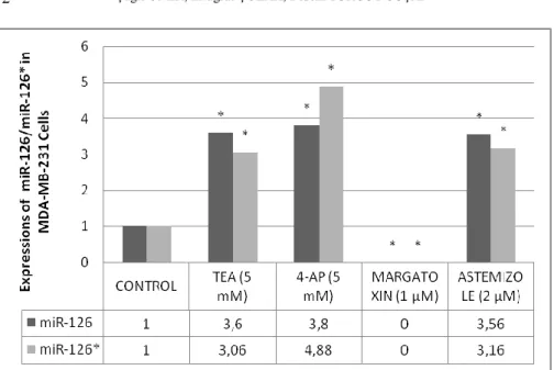
were inhibited by TEA, 4-AP and astemizole (P < 0.001). miR-126/126*
expressions increased in both MCF-7 (5.86; 3.68) and MDA-MB-231 (3.60;
3.06) cells when 5 mM TEA was applied to these cells. Furthermore, when 5
mM 4-AP was applied to MCF-7 and MDA-MB-231 cells, the expression of
miR-126/126* increased (6.23; 3.57/3.80; 4.88). Our data shows that the
application of 2 µM astemizole brought about an increase in miR-126/126*
expressions in MCF-7 (5.97; 3.92) and MDA-MB-231 (3.56; 3.16). It was
determined that the application of margatoxin (1 µM) brought about an
increase in both miRNA expressions (6.15; 3.77) in MCF-7 cells (P < 0.001;
As shown in Figure 1, 2).
4.
DISCUSSION
Channelopathies, the variations of K channel function and density, can have
profound pathophysiological consequences in various diseases, such as
cancer (13-15). The changes in cancer make it possible to bind only certain
molecules. In our study, the interactions between K channels and
miR-126/126*, which are known to help in tumor suppression in breast cancer,
were investigated. We aimed to determine the relationship between
miR-126/126* and K channel activities. miR-miR-126/126* decrease in various
cancers due to activity which suppresses tumors.
Recent studies show that K channels play an important role in the cell cycle,
apoptosis, proliferation, and metastasis in breast cancer cells (1, 4, 25, 26).
In another study, it was shown that the potassium flow density interacts with
a metastatic character in strong metastatic PC-3 prostate cancer cells as
opposed to weak metastatic LNCaP prostate cancer cells (13). It could,
therefore, be related to VGPC staining being increased in clinical metastatic
PCa when compared to normal patients’ tissue samples (3). Furthermore,
these channels’ molecular structures have not yet been determined (27). It is
either EAG1 gene expression or the inhibition of channel activation which
decreases tumor cell proliferation in vivo and in vitro as a result of antisense
oligonucleotides, siRNA, monoclonal antibodies or non-specific (Kv10.1)
EAG1 channel inhibitors. Therefore, EAG1 has been suggested as a target
for cancer diagnosis (28).
miR-126, which shows a low expression in breast, colorectal, lung,
pancreatic and prostate cancer tissues, was analyzed immunohistochemically
(IHC) and was detected in endothelial and immune cells (29). In another
study, miR-21, -106, -126, -155, -199 and -335 expression were examined,
and miR-126, -199a and -335 expressions were found to be low when
compared to tumor type (malignant, benign, and tumor grade), and the
amount of estrogen and progesterone. To determine miR-126 expressions in
a study, transfecting oncogene v-Src to Cx43KO mouse embryonic brain cell
lines brought about a decrease in miR-126/126* gene expressions (30). In a
study into Hsa- miR-126 and hsa-miR-183 in metastatic non-small cell lung
cancer (NSCLC) cells, it was observed that miR-126 expression led to a
decrease of the CRK protein in lung cancer (31). In a proliferation study,
MCF-7 breast cancer cells which were treated with anti-miR-126 showed an
86% proliferation and survival level (23). In a study using MCF-7 and
MDA-MB-231 cell lines, it was found that miR-126 affects insulin receptor
1 (IRS-1) and brought about a decrease in cancer cell growth (32). Through
the use of the immunohistochemistry (IHC) and florescent in situ
hybridization (ISH) methods, a decrease in miR-341 and -126 expressions
and an increase in miR-21 and -155 expressions were found to occur in
breast, colorectal, lung, pancreas and prostate cancerous tumors, while
miR-126 and miR-155 expressions were determined in endothelial and immune
cells, and miR-34a and miR-21 expressions were determined in cancer cells
(29). miR-21, -106a, -126, -155, -199a and -335 expressions were analyzed
according to tumor type (malign, benign and tumor grade) and
estrogen/progesterone levels. Although miR-21, -106a, and -155 expressions
were high, miR-126, -199a and -335 expressions were found to be low.
Considering this data, it suggests that miR-126, -199a, and -335 expressions
decrease in breast cancer (29). Wang et al. (2010) suggested that these six
miRNAs could be used as biomarkers in breast cancer prognosis and
diagnosis through the consideration of multiple parameters (33). It has been
reported that miRNAs which are found in blood may have the potential to be
used as diagnosis, treatment, and biological indicators of cancer (31).
Non-coding RNAs, one of the epigenetic mechanisms, can be a determinant for the development of cancer. In addition to research into epigenetic mechanisms and cancer studies, there are no adequate studies into K channels and non-coding RNAs, particularly into miRNAs. In a study, inhibition of Kir6.1/SUR2B channel, one of ATP sensitive potassium channels which plays an important role on vascular development, by methylglyoxyline caused an increase of miR-9a-3p expression about 240% on smooth muscle cells (34). The relationship between miRNAs and K channels in pathophysiological diseases, such as those of the heart, neurodegenerative diseases, and diabetes, has been investigated. In a study of cardiac arrhythmias, it was found that miR-1 regulates Kir2, a subunit of the K channel (35). In another study, Xiao et al. (2007) determined that miR-133 inhibits the HERG K channel in diabetes (36). In another study, it was determined that potassium channels and miR-190 play crucial role in development of pulmonary arterial hypertension vasculazation. In the same study, it was suggested that activity of KCNQ5 potassium channel could be affected by miR-190 (37). In a study the interaction between miR-1 and KCNE1 and KCNB2 potassium channels was observed. When potassium flow was increased by patch-clamp method, decrease of KCNE1 and KCNB2 potassium channels and increase of miR-1 expression was determined. In the same study, it was suggested that KCNE1 and KCNB2 could be a potential target of miR-1 (38). In a study, low concentrations of potassium intake caused a decrease of miR-194 expression in kidney (39). Lin and collueges determined that damage of cornea caused an increase of miR-205 expression in human corneal cells, and as a result of this increase inhibition of KCNJ10 potassium channel, target of miR-205, was observed (40). It was observed that miR-129 inhibited its potential targets HuD RNA binding protein and Kv1.1 potassium channel during neuronal activity while mTORC1 was active (41).
Ru et al. determined that (2015) a potassium channel blocker Quinidine effected miRNA expression in glioma (42). In another study, it was suggested that human ether-a-go-go related potassium channel (HERG1) was a potential target of miR-96 in pancreatic cancer (43). In a study, increase of miR-34a caused a decrease of Eag1
potassium channel activity and proliferation in osteosarcoma (44). In SHSY5Y cells, tumor suppressive miR-34 expression increased when hEAG potassium channel inhibited (45). Bai et al. (2013) reported that increase of Eag1 (KCNH1) potassium channel expression caused a decrease of miR-296-3p expression in glioblastoma. (46). Inhibition of Eag1 potassium channel by Taxol, a chemotherapy drug in glioblastoma, and inhibition of miR-155 by anti-miR-155 caused anti-proliferative and anti-development effect in glioblastoma cells (47).
It was determined that K channel blockers negatively affect the proliferation,
migration, metastasis and inhibition of integrin-mediated cell adhesion in
cancer (31). Cell volume and changes in K ion concentration are related to
an increased K flow through potassium channels on plasma membranes
(48-51). It was thought that a decrease in K ions regulate critical events in early
phases of apoptosis (28, 50). Previous studies indicated that K channel
blockers contribute to the development of metastasis through the inhibition
of integrin-related cell adhesion in the development of extracellular
matrices, proliferation, migration, and metastasis (28). In one study, it was
determined that 4-AP, a non-specific potassium channel inhibitor, inhibited
cell proliferation in conjunction with ERK1/2 activation and also increased
the activation of p38 in the MCF10A cells (52). In another study, Kv1.3, a
voltage-gated
K
channel,
protein
expression
was
investigated
immunohistochemically in 60 breast cancer samples and, although there
were no stains in normal breast epithelial cells, all of the breast cancer
tissues were stained (100%), while 60% of cells were high, 30% of cells
were higher, and 22% of these cells were low. In the same study, K channel
blockers stopped the proliferation of MCF-7 breast cancer cells at a level of
90% (53). Yang et al. (2011) used 4-tetraethylammonium (TEA, a
non-specific K channel inhibitor) to inhibit KV1.3 channels in breast cancer cell
lines, and it was determined that the repression of cell proliferation occurred
(35). Furthermore, TEA not only controls Kv10.1 proliferation, but it was
also shown that normal cells could change into cancer cells through the loss
of contact inhibition and increased cell division when in vitro expression
was increased. When it was injected into subcutaneous layer of nude mice, it
was seen that vivo tumor growth was possible (54).
Cancer has become more common in recent times. Although there are
studies looking into a cure for cancer, the specific treatments for many types
of cancer have not yet been found. Perhaps it is more realistic to find the
formula for living with cancer, rather than trying to wholly defeat human
cancer. For these we must first of all block the metastasis of cancer.
Revealing the condition of the cells in cancer and metastasis is an important
step towards determining the future treatment strategy. Our study has
determined that K channel blockers were found to be more effective in
providing an increase of miR-126/126* expressions in the poorly invasive
breast cancer cell line MCF-7 when K channel functions had been cut off by
K channel blockers. In recent studies, it has been surmised that K channel
blockers may inhibit proliferation, cell cycle, migration, metastasis, and
adhesion. K channels are also known to have an oncogenic potential in cases
of breast cancer.
miR-126 and its complementary miR-126* are molecules which play a part
in the formation of breast cancer and its metastasis, and they have also been
shown to have tumor suppressor properties in cellular movements like
migration, invasion, and adhesion in these cell lines. In previous studies, it
had been determined that both K channel blockers and miR-126/126* are
involved in different mechanisms in the formation of breast cancer. In this
study we investigated whether these two different mechanisms affect each
other. We have revealed that the possible relationship between potassium
channels and cancer can also be observed in other miRNAs, such as
miR-126/126*.
In conclusion, it was observed that while we blocked partial K flow by inhibiting different K channel blockers, mi126/126* expressions were increased in comparison with the control. The increase of miR-126/126* expressions might decrease the risks of breast cancer. It was also observed that Non-coding RNAs may be interacting with voltage-gated potassium channels. Further studies into cancers may be needed to verify our results.
TABLES AND FIGURES
Figure 1. The expression of miR-126/126* in MCF-7 breast cancer cell line.All obtained data was evaluated according to control group.*p<0.001 .
Figure 2. The expression of miR-126/126* in MDA-MB-231 breast cancer cell line. All obtained data was evaluated according to control
group.*p<0.001(TEA:Tetraethylammonium; 4-AP: 4-aminopyridine)
ACKNOWLEDGMENTS
This study was supported from Eskisehir Osmangazi University, Commission of Scientific Research Projects (Project Number: 201111032).
CONFLICTS OF INTEREST
Çağrı Öner; corresponding and first author, takes part in editing the study, experimental steps (cell culture and toxin releasing, total RNA isolation, reverse transcription and RT-PCR gene expression) and evaluation of results.
Ertuğrul Çolak; takes part in statically evaluation of results.
Didem Turgut Coşan; author, takes part in editing the study, experimental steps (cell culture and toxin releasing, total RNA isolation, reverse transcription and RT-PCR gene expression) and evaluation of results.
REFERENCES
[1] W. J. Brackenbury, A. M. Chuioni, J. K. J. Diss and M.B.A. Djamgoz. The neonatal splice variant of Navl.5 potentiates in vitro invasive of MDA-MB-231 human breast cancer cells, Breast cancer research and treatment, 101 (2007) 149-160.
[2] H. O. Ahidouch, A. Ahidouch. K+ Channel Expression in Human Breast Cancer Cells: Involvement in Cell Cycle Regulation and Carcinogenesis, J. Membrane Biol., 221 (2008) 1-6.
[3] M. Abdul, N. Hoosein. Reduced Kv1.3 potassium channel expression in human prostate cancer, J. Membr. Biol., 214 (2006) 99-102.
[4] A. S. Borowiec. IGF-1 Activates HERG K Channels Through an Akt-Dependent Signaling Pathway in Breast Cancer Cells: Role in Cell Proliferation, Journal of Cellular Physiology, (2007) 690-701.
[5] L. Zhang, W. Zou, S. S. Zhou and D. D. Chen. Potassium channels and proliferation and migration of breast cancer cells, Sheng Li Xue Bao, 61 (2009) 15-20.
[6] M. Abdul, A. Santo and N. Hoosein. Activity of potassium channel-blockers in breast cancer, Anticancer Res., 23 (2003) 3347-3351.
[7] L. Y. Jan, Y. N. Jan. Voltage-gated and inwardly rectifying potassium channels, J. Physiol., 505/2 (1997) 267-282.
[8] G. J. Kaczorowski, M. L. Garcia. Pharmacology of voltage-gated and
calcium-activated potassium channels, Curr Opin Chem Biol, 3/4
(1999) 448-458. doi: 10.1016/S1367-5931(99)80066-0
[9] J. M. Quayle, M. T. Nelson and N. B. Standen. ATP-sensitive and inwardly rectifying potassium channels in smooth muscle, Physiol Rev, 77/4 (1997) 1165-1232.
[10] S. H. Jang, S. Y. Choi, P. D. Ryu and S. Y. Lee. Anti-proliferative
effect of Kv1.3 blockers in A549 human lung adenocarcinoma in
vitro and in vivo, Eur J Pharmacol., 651/1-3 (2011) 26-32. doi:
10.1016/j.ejphar.2010.10.066
[11] J. García-Quiroz, J. Camacho. Astemizole: an old anti-histamine as a
new promising anti-cancer drug, Anticancer Agents Med Chem. ,
11/3 (2011) 307-314.
[12] J. Garcia-Quiroz, R. Garcia-Becerra, D. Barrera, N. Santos, E. Avila,
D. Ordaz-Rosado et al. Astemizole synergizes calcitriol
antiproliferative activity by inhibiting CYP24A1 and upregulating
VDR: a novel approach for breast cancer therapy, PLoS ONE, 7/9
(2012) e45063. doi: 10.1371/journal.pone.0045063
[13] M. Conti. Targeting K+ channels for cancer therapy, J. Exp. Ther. Oncol., 4 (2004)161-166.
[14] G. M. Vincent, L. Zhang. The role of genotyping in diagnosing cardiac channelopathies: progress to date, Mol. Diagn., 9 (2005) 105-118.
[15] M. Brevet. Expression of K+ channels in normal and cancerous human breast, Histol. Histopathol., 23 (2008) 965-972.
[16] T. E. DeCoursey, K. G. Chandy, S. Gupta and M. D. Cahalan. Voltage-gated K+ channels in human T lymphocytes: a role in mitogenesis?, Nature, 307 (1984) 465-468.
[17] L. Conforti, M. Petrovic, D. Mohammad, S. Lee, Q. Ma, S. Barone and A. H. Filipovich. Hypoxia regulates expression and activity of Kv1.3 channels in T lymphocytes: a possible role in T cell proliferation, J. Immunol., 170 (2003) 695-702.
[18] N. Gamper, S. Fillon, S. M. Huber, Y. Feng, T. Kobayashi, P. Cohen and F. Lang. IGF-1 up-regulates K+ channels via PI3-kinase, PDK1 and SGK1, Pflugers Arch., 443 (2002) 625-634. doi: 10.1007/s00424-001-0741-5 [19] G. Kwon, C. A. Marshall, H. Liu, K. L. Pappan, M. S. Remedi and M. L.
McDaniel. Glucose-stimulated DNA synthesis through mammalian target of rapamycin (mTOR) is regulated by KATP channels: effects on cell cycle progression in rodent islets, J Biol Chem., 281 (2006) 3261-3267.
[20] H. Malhi, A. N. Irani, P. Rajvanshi, S. O. Suadicani, D. C. Spray, T. V. McDonald and S. Gupta. KATP channels regulate mitogenically induced proliferation in primary rat hepatocytes and human liver cell lines. Implications for liver growth control and potential therapeutic targeting, J Biol Chem., 275 (2000) 26050-26057. doi: 10.1074/jbc.M001576200 [21] S. Liebau, C. Propper, T. Bockers, F. Lehmann-Horn, A. Storch, S. Grissmer
and Wittekindt O. H. Selective blockage of Kv1.3 and Kv3.1 channels increases neural progenitor cell proliferation, J Neurochem., 99 (2006) 426-437. doi: 10.1111/j.1471-4159.2006.03967.x
[22] A. Ventura, T. Jacks. MicroRNAs and cancer: short RNAs go a long way, Cell, 136 (2009) 586-591. doi: 10.1016/j.cell.2009.02.005
[23] S. M. Elbashir, W. Lendeckel and T. Tuschl. RNA interference is mediated by 21-and 22-nucleotide RNAs, Genes & Development, 15 (2001) 188-200. doi:10.1101/Gad.862301
[24] N. Patel, E. R. Sauter. Body fluid micro(mi)RNAs as biomarkers for human cancer, Journal of Nucleic Acids Investigation, 2 (2011).
[25] S. Y. Chiu, G. F. Wilson. The role of potassium channels in Schwann cell proliferation in Wallerian degeneration of explant rabbit sciatic nerves, J Physiol., 408 (1989) 199-222.
[26] A. S. Borowiec. Regulation of IGF-1-dependent cyclin D1 and E expression by hEag1 channels in MCF-7 cells: the critical role of hEag1 channels in G1 phase progression, Biochim. Biophys. Acta, 1813 (2011) 723-730.
[27] S. Daido, T. Kanzawa, A. Yamamoto, H. Takeuchi, Y. Kondo and S. Kondo. Pivotal role of the cell death factor BNIP3 in ceramide-induced autophagic cell death in malignant glioma cells, Cancer Res., 64 (2004) 4286-4293. doi: 10.1158/0008-5472.CAN-03-3084
[28] B. Dallaporta, P. Marchetti, M. A. de Pablo, C. Maisse, H. T. Duc, D. Metivier, N. Zamzami, M. Geuskens and G. Kroemer. Plasma membrane potential in thymocyte apoptosis, J Immunol., 162 (1999) 6534-6542.
[29] L. F. Sempere, M. Preis, T. Yezefski, H. Ouyang, A. A. Suriawinata, A. Silahtaroglu, J. R. Conejo-Garcia, S. Kauppinen, W. Wells and M. Korc. Fluorescence-based codetection with protein markers reveals distinct cellular compartments for altered MicroRNA expression in solid tumors, Clin Cancer Res., 16 (2010) 4246-4255. doi: 10.1158/1078-0432.CCR-10-1152
[30] X. Li, Y. Shen, H. Ichikawa, T. Antes and G. S. Goldberg. Regulation of miRNA expression by Src and contact normalization: effects on nonanchored cell growth and migration, Oncogene, 28 (2009) 4272-4283. doi: 10.1038/onc.2009.278
[31] Q. F. Lin, W. D. Mao, Y. Q. Shu, F. Lin, S. P. Liu, H. Shen, W. Gao, S. Q. Li and D. Shen. A cluster of specified microRNAs in peripheral blood as biomarkers for metastatic non-small-cell lung cancer by stem-loop RT-PCR, Journal of Cancer Research and Clinical Oncology, 138 (2012) 85-93. doi: DOI 10.1007/s00432-011-1068-z
[32] J. Zhang, Y. Y. Du, Y. F. Lin, Y. T. Chen, L. Yang, H. J. Wang and D. Ma. The cell growth suppressor, mir-126, targets IRS-1, Biochem Biophys Res Commun., 377 (2008) 136-140. doi: 10.1016/j.bbrc.2008.09.089
[33] F. Wang, Z. Zheng, J. Guo and X. Ding. Correlation and quantitation of
microRNA aberrant expression in tissues and sera from patients with
breast tumor, Gynecol Oncol., 119 (2010) 586-593. doi:
10.1016/j.ygyno.2010.07.021
[34] S. S. Li, Y. Wu, X. Jin and C. Jiang. The SUR2B subunit of rat vascular KATP channel is targeted by miR-9a-3p induced by prolonged exposure to methylglyoxal, Am J Physiol Cell Physiol., 308/2 (2015) 139-145. doi: 10.1152/ajpcell.00311.2014
[35] B. Yang, H. Lin, J. Xiao, Y. Lu, X. Luo, B. Li, Y. Zhang, C. Xu, Y. Bai, H. Wang et al. The Muscle-Specific MicroRNA miR-1 Regulates Cardiac Arrhythmogenic Potential by Targeting GJA1 and KCNJ2, Nature Medicine, 13 (2011) 486-491. doi: 10.1038/nm1569
[36] J. Xiao, X. Luo, H. Lin, Y. Zhang, Y. Lu, N. Wang, Y. Zhang, B. Yang and Z. Wang. MicroRNA miR-133 represses HERG K+ channel expression contributing to QT prolongation in diabetic hearts, J Biol Chem., 282 (2007) 12363-12367. doi: 10.1074/jbc.C700015200
[37] S. S. Li, Y. J. Ran, D. D. Zhang, S. Z. Li and D. Zhu. MicroRNA-190
regulates hypoxic pulmonary vasoconstriction by targeting a
voltage-gated K⁺ channel in arterial smooth muscle cells, J Cell
Biochem., 115/6 (2014) 1196-1205. doi: 10.1002/jcb.24771
[38] X. Jia, S. Zheng, X. Xie, Y. Zhang, W. Wang, Z. Wang et al.
MicroRNA-1 accelerates the shortening of atrial effective refractory
period by regulating KCNE1 and KCNB2 expression: an atrial
tachypacing rabbit model, PLoS One., 8/12 (2013). doi:
10.1371/journal.pone.0085639
[39] D. H. Lin, P. Yue, C. Zhang and W. H. Wang. MicroRNA-194
(miR-194) regulates ROMK channel activity by targeting intersectin 1,
Am J Physiol Renal Physiol., 306/1 (2014) 53-60. doi:
10.1152/ajprenal.00349.2013
[40] D. Lin, A. Halilovic, P. Yue, L. Bellner, K. Wang, L. Wang and C.
Zhang. Inhibition of miR-205 impairs the wound-healing process in
human corneal epithelial cells by targeting KIR4.1 (KCNJ10),
Invest Ophthalmol Vis Sci, 54/9 (2013) 6167-6178. doi:
10.1167/iovs.12-11577
[41] N. M. Sosanya, P. P. Huang, L. P. Cacheaux, C. J. Chen, K. Nguyen, N.
I. Perrone-Bizzozero, and K. F. Raab-Graham Degradation of high
affinity HuD targets releases Kv1.1 mRNA from miR-129
repression by mTORC1, J Cell Biol., 202/1 (2013) 53-69. doi:
10.1083/jcb.201212089
[42] Q. Ru, X. Tian, M. S. Pi, L. Chen, K. Yue, Q. Xiong et al.
Voltage‑gated K+ channel blocker quinidine inhibits proliferation
and induces apoptosis by regulating expression of microRNAs in
human glioma U87‑MG cells, Int J Oncol., 46/2 (2015) 833-840.
doi: 10.3892/ijo.2014.2777
[43] J. Feng, J. Yu, X. Pan, Z. Li, Z. Chen, W. Zhang et al. HERG1
functions as an oncogene in pancreatic cancer and is downregulated
by miR-96, Oncotarget, 5/14 (2014) 5832-5844.
[44] X. Wu, D. Zhong, Q. Gao, W. Zhai, Z. Ding and J. Wu. MicroRNA-34a
inhibits human osteosarcoma proliferation by downregulating ether
à go-go 1 expression, Int J Med Sci., 10/6 (2013) 676-682. doi:
10.7150/ijms.5528.
[45] H. Lin, Z. Li, C. Chen, X. Luo, J. Xiao, D. Dong, D. et al.
Transcriptional and post-transcriptional mechanisms for oncogenic
overexpression of ether à go-go K+ channel, PLoS One, 6/5 (2011)
doi: 10.1371/journal.pone.0020362
[46] Y. Bai, H. Liao, T. Liu, X. Zeng, F. Xiao, L. Luo et al.. MiR-296-3p
regulates cell growth and multi-drug resistance of human
glioblastoma by targeting ether-à-go-go (EAG1), Eur J Cancer., 49/3
(2013) 710-724. doi: 10.1016/j.ejca.2012.08.020.
[47] W. Meng, L. Jiang, L. Lu, H. Hu, H. Yu, D. Ding et al. Anti-miR-155
oligonucleotide enhances chemosensitivity of U251 cell to taxol by
inducing apoptosis, Cell Biol Int., 36/7 (2012) 653-659. doi:
10.1042/CBI20100918
[48] J. Y. Wang, J. Wang, V. A. Golovina, L. Li, O. Platoshyn and J. X. Yuan. Role of K(+) channel expression in polyamine-dependent intestinal epithelial cell migration, Am J Physiol Cell Physiol., 278 (2000) 303-314.
[49] X. Wang, A. Y. Xiao, T. Ichinose and S. P. Yu. Effects of tetraethylammonium analogs on apoptosis and membrane currents in cultured cortical neurons, J Pharmacol Exp Ther., 295 (2000) 524-530.
[50] S. P. Yu. Regulation and critical role of potassium homeostasis in apoptosis, Prog Neurobiol., 70 (2003) 363-386.
[51] M. J. Morton, S. Chipperfield, A. Abohamed, A. Sivaprasadarao and M. Hunter. Na(+)-induced inward rectification in the two-pore domain K(+) channel, TASK-2, Am J Physiol Renal Physiol, 288 (2005) 162-169. doi: 10.1152/ajprenal.00248.2004
[52] J. Liu, S. Feng, L. Zhang, Z. Wu, Q. Chen, W. Cheng, S. Q. Wang and W. Zou. Expression and properties of potassium channels in human mammary epithelial cell line MCF10A and its possible role in proliferation, Sheng Li Xue Bao, 62 (2010) 203-209.
[53] D. Ekhterae, O. Platoshyn, S. Krick, Y. Yu, S. S. McDaniel and J. X. Yuan. Bcl-2 decreases voltage-gated K+ channel activity and enhances survival in vascular smooth muscle cells, Am J Physiol Cell Physiol., 281 (2001) 157-165.
[54] N. Ferrara. VEGF: an update on biological and therapeutic aspects, Curr Opin Biotechnol., 11 (2000) 617-624.