FACILE AND TEMPLATE-FREE SYNTHESIS OF CuO NANOPARTICLES
1Tugay ÜSTÜN, 2Volkan ESKİZEYBEK, 3Ahmet AVCI
1Başkent University, Kahramankazan Vocational School, Mechatronics Program, Ankara, TURKEY 2Çanakkale Onsekiz Mart University, Engineering Faculty, Material Science and Engineering Department,
Çanakkale, TURKEY
3Necmettin Erbakan University, Engineering and Architecture Faculty, Biomedical Engineering Department, Konya, TURKEY
1[email protected], 2[email protected], 3[email protected]
(Geliş/Received: 01.11.2018; Kabul/Accepted in Revised Form: 22.03.2019)
ABSTRACT: Copper oxide (CuO) nanoparticles were synthesized successfully by arc discharge method in an aqueous medium. Arc discharge was ignited between two copper electrodes and an exhaustive characterization of morphology and crystalline structure of the drawn up copper oxide nanoparticles were studied. Electron microscopy analyses revealed that the grown CuO nanoparticles are irregularly shaped with nominal diameters in the interval of 10-50 nm. XRD results show the as-synthesized particles contain only CuO peaks without any impurities. The results procure a simple, cheap and responsive method for preparing single-phase CuO nanoparticles.
Key Words: Arc Discharge, Copper Oxide (CuO), Nanoparticles
CuO Nanoparçacıkların Kolay ve Amorf Yapıda Sentezi
ÖZ: Bakır oksit (CuO) nanoparçacıkları, sıvı bir ortamda ark deşarj yöntemi ile başarılı bir şekilde sentezlendi. İki bakır elektrot arasında ark boşalması gerçekleşerek üretimi tamamlanan bakır oksit nanoparçacıkların morfolojisi ve Kristal yapısı ayrıntılı bir şekilde incelenmesi yapıldı. Elektron mikroskobu incelemeleri sonucunda üretilen CuO nanoparçacıkların 10-50 nm aralığında nominal çaplarda düzensiz şekilli olduğu görülmektedir. XRD sonuçları ise sentezlenen parçacıkların herhangi bir safsızlık olmadan sadece CuO piklerini içerdiğini göstermektedir. Sonuçlara göre tek fazlı CuO nanoparçacıkların üretilmesi için ark deşarj yöntemi basit, ucuz ve esnek bir yöntemdir.
Anahtar Kelimeler: Ark Deşarj, Bakır Oksit (CuO), Nanoparçacık
INTRODUCTION
Metal oxides are a significant class of semiconductors and have been concentratedly studied because of their specific properties for prospective applications, such as solar cells, electronics, and photocatalysis (Tai et al., 2012; Panuthai et al., 2014). Cu-based materials grow into increasingly attracting for both primary research and practical purposes. As compared to palladium, platinum and gold, copper oxide nanostructures are relatively cheap with high stability and nontoxicity; they encourage the performance of the works on improvement of the existing methods for their formation. Copper oxides (CuxO) can exist in various stoichiometries and in phases such as Cu2O (cuprous oxide) and CuO (cupric oxide). CuO has an equanimeous band gap energy, cost-efficient production and high optical transparency. Many new endeavors have been used in the syntheses of copper oxide nanostructures with diverse morphologies, such as particles (Wu et al., 2010; Zhang et al., 2008; Lanje et
al., 2007), carbon nano-onions (Huang et al., 2003; Sano et al., 2001), molybdenum disulfide fullerenes (Sanoet al., 2002), ZnO (Eskizeybek et al., 2012; Ashkarran et al., 2010; Fang et al., 2009) and Cd(OH)2 nanoparticles (Eskizeybek et al., 2011). The arc discharge method can be utilized in various mediums such as vacuum, organic solvents, and liquid nitrogen. For this reason, different structures can be synthesized such as Cu, CuO and Cu2O (You et al., 2005). Among these, the arc discharge method in de-ionized water is characterized by high productivity, relatively low costs, a possibility of scaling and performing one stage synthesis of nanomaterials. The disadvantage of this method is obtainment of nanoparticles with a broad size distribution (Tepanov et al., 2014).
In this study, we report the syntheses of CuO nanoparticles by arc discharge immersed in de-ionized water. The idea is to use de-ionized water as an oxygen source and the fabrication of nanoparticles in a liquid setting to secure their isolation from air atmosphere. The structural, morphological and optical peculiarities of the produced CuO nanoparticles were carried out.
MATERIALS AND METHODS
In this study, high purity copper (Cu) rods (Alfa Aesar, 99.99%) were utilized qua electrodes to synthesize CuO nanoparticles. All reagents were of analytic phase and used without further decontamination.
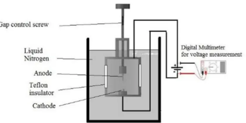
The CuO nanoparticles have been produced by the lab made arc discharge apparatus immersed in de-ionized water (Figure 1). Two Cu electrodes were used inthe instrumentas anode and cathode. The arc discharge was initialized in de-ionized water by contacting the anode to the cathode and at that rate the openness among the electrodes were inspected by measurement discharge voltage to retain resolute about arc during the experiment. When arc discharge is performed in aqueous solution, H2O decomposes into H+, O-2 and OH- ions and then Cu atoms react with O to form CuO nanoparticles.A direct current (DC) welding power source provided the arc current and it was fixed to 50 A since 50 A is the optimum amperage value for the production of large amounts and effective nanoparticle (Eskizeybek et al., 2011). The voltage remains constant throughout the arc process and is significant for the reproducibility of the nanoparticles synthesis. Arc discharge was sustained until the anode electrode consumed. The resulted particles were withheld in the room temperature to consummate decantation at the fag end of the reaction vessel for 24 h. The stagnant particles were picked up and washed with de-ionized water. After all, the particles were desiccated at 40 °C under vacuum for 24 h (Eskizeybek et al., 2012).
Figure 1. The schematic view of the arc discharge instrument.
Shimadzu XRD-6000 performed X-Ray Diffraction (XRD) analysis. X-ray diffractometer exploiting Cu Kα radiation (λ=0.15418 nm) the functioning conjuncture were 40 kV and 30 mA in the scanning interval from 10°-80° at a ratio of 2°/min. The morphology of the synthesized outputs were implemented exploiting a JEOL/JSM-6335F-EDS SEM. The Transmission Electron Microscope (TEM) images of the CuO nanoparticles were carried out exploiting JEOL-2100 HRTEM at 300kV. The Diffuse Reflectance (FTIR) spectra of all materials were inscribed by a Perkin Elmer 1725 spectrophotometer.
RESULTS AND DISCUSSIONS
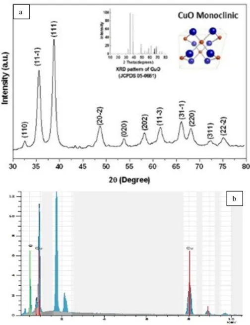
XRD pattern of the synthesized products by arc-discharge is designated in Figure 2a. The low weak intensities and wider peak widths indicate the low crystalline quality of the sample. All diffraction peaks in the pattern can be indexed to a pristine monoclinic crystalline of CuO with lattice parameters a=4.68370 Å, b=3.42260 Å, c=5.12880 Å and β=99.540 (JCPDSNo. 45-0937; Abaker et al., 2011). Two dominated peaks which appears at 2θ=35.6◦ and 38.8◦ are indexed as (111)–(002) and (111)–(200) planes and regarded in the surveyed XRD pattern. These peaks are the characteristic peaks for pristine monoclinic CuO crystalline phase and enumerate that they are privileged crystalline planes of the nanoplates (Bayansal et al., 2011). Importantly, no definite peaks of impureness are seen in this pattern. The average particle size is computed to be 37.5 and 40.6 nm using Scherrer formula for the (002) and (111) planes, alternately. In addition to XRD analysis, EDS analysis was also fulfilled to monitor the composition and pureness of the synthesized outputs (Figure 2b). The EDS spectrum indicates the explicit peaks related to copper and oxygen confirming the contamination with the presence of Si attributable resulting from the Si wafer, which was at the furthest of the detection limit of EDS.
Figure 2. (a) XRD pattern of the prepared nanoparticles presenting monoclinic CuO (b) The EDS spectrum of nanoparticles showing copper and oxygen
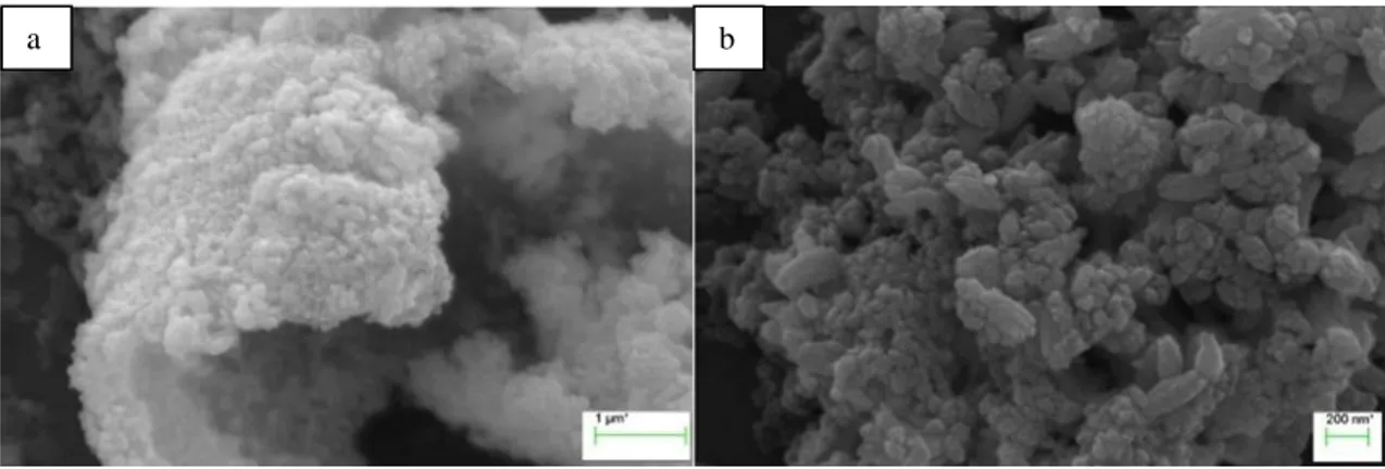
The morphologies of CuO nanostructures were characterized by SEM and TEM. Figure 3a indicates the low-magnification SEM image of as-grown CuO structures. The as-synthesized CuO nanoparticles consist of the conglomerated particles. It is important to note that no specific morphology is observed (Figure 3a). The magnified image shows that the nanostructure has a stack formation with 10–40 nm average particle size (Figure 3b).
Figure 3. SEM images of CuO nanoparticles
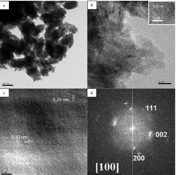
The CuO nanostructures were also characterized by TEM and high-resolution TEM (HRTEM). Figure 4a indicates the low-resolution TEM image of CuO clusters. The average size of CuO clusters is measured as of 100–150 nm (Figure 4a) and showing that the structure is associated from various numbers of nanoparticles. Figure 4b manifests the HRTEM image of the ersatz CuO nanostructure. The micrograph reveals the formation of irregularly shapely nanoparticles. The dimension of the nanoparticles aline amongst 10 and 50 nm which is in decent compliance with the centreing particle sizes assessed by Scherrer’s equation. The smooth and parallel lattice fringes of the swelled structures exposed that the synthesised nanoparticles exhibit semi-crystalline nature as seen upper inset in Figure 4b. The weighed spacing of the crystallographic plane is about 0.27 nm, ersatz to the [110] lattice fringes of the monoclinic CuO. No dislocations in the lattice fringes are beheld which predicates that the synthesised structures are pristine without any impureness or structural imperfections (Abaker et al., 2011). Figure 4c proves the structural details of high-resolution lattice fringes from another agglomerated CuO nanostructures. The interplanar spacings for the two sides are 0.252 and 0.232 nm, which are analogue the spacings for [002] or [-111] and [111] planes in monoclinic CuO (Zou et al., 2011). This further validates the multi-crystallinity of the as-prepared nanoparticles. Extended defects such as twins are not found in HRTEM observations. More thorough structural data on the CuO nanocrystal is supplied by selected area electron diffraction (SAED) analysis. The conforming SAED pattern of the CuO nanocrystal is indicated in the Figure 4d. The SAED pattern proves ordinate dot alignment and the diffraction dots can be appointed to the [200], [002], [111] crystal planes of monoclinic CuO along the [100] axis (Liu et al., 2006; Zhang et al.,2005; Xu et al., 2007).
Figure 4. TEM images of (a), (b) CuO nanoparticles, (c) HR-TEM image of CuO nanoparticles, (d) SAED pattern. The scale bars are 100 nm, 10 nm and 2 nm for a, b and c respectively
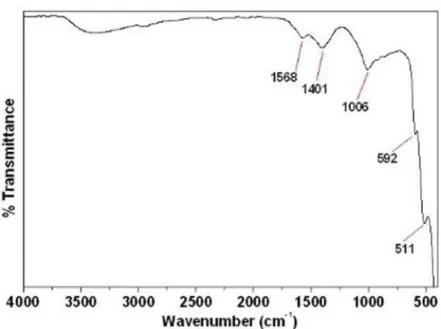
The grade and composition of as-synthesized of CuO nanostructures were characterized further by FTIR spectroscopy and the indicated in Figure 5. The availability of absorption bands at 511 and 592 cm−1 are in view of ν(Cu–O) modes which approve the composition of monoclinic CuO crystals (Zhang et al.,2016). The absorption band nascence at 1401 cm−1 was arisen based on the existence of (CO3)-2(Abaker et al., 2011). The band broadly transpires in the FTIR spectrum, if the specimens are synthesized in the presence of the oxygen atmosphere (Singh et al., 2009; Hai et al., 2010). The absorption peak at 1568 cm−1 is attributed for bending vibration of absorbed water and surface hydroxyl. A wide peak in the interval ∼ 3000–3500 cm−1 was by the reason of the O–H stretching mode (Abaker et al., 2011; Vaseem et al., 2008).
Figure 5. FTIR spectra of CuO nanoparticles CONCLUSIONS
In summary, CuO nanoparticles have been fabricated via arc discharge method in de-ionized water as a simple, low temperature owing to liquid medium and cost-effective solution method. The crystal structure and morphology of copper oxide nanoparticles were characterized by XRD, SEM, TEM and FTIR spectroscopy. XRD shows that CuO nanoparticles have monoclinic crystal system as a single phase without impurities. The as-synthesized CuO nanoparticles consist of irregularly shaped nanoparticles with diameters from through 10 to 50 nm. HRTEM survey revealed that the copper oxide nanoparticles represent multi-crystal formation and these values are consistent with the literature.
REFERENCES
Abaker, M., Umar, A., Baskoutas, S., Kim, S. H., Hwang, S. W., 2011, “Structural and optical properties of CuO layered hexagonal discs synthesized by a low-temperature hydrothermal process”, Journal of Physics D: Applied Physics, 44(15), 155405.
Ashkarran, A. A., Mahdavi, S. M., Ahadian, M. M., 2010, “Photocatalytic activity of ZnO nanoparticles prepared via submerged arc discharge method”, Applied Physics A, 100(4), 1097-1102.
Bayansal, F., Kahraman, S., Çankaya, G., Çetinkara, H. A., Güder, H. S., Çakmak, H. M., 2011, “Growth of homogenous CuO nano-structured thin films by a simple solution method”, Journal of Alloys and Compounds, 509(5), 2094-2098.
Chen, J., Wang, K., Hartman, L., Zhou, W., 2008, “H2S detection by vertically aligned CuO nanowire array sensors”, The Journal of Physical Chemistry C, 112(41), 16017-16021.
Eskizeybek, V., Sarı, F., Gülce, H., Gülce, A., Avcı, A., 2012, “Preparation of the new polyaniline/ZnO nanocomposite and its photocatalytic activity for degradation of methylene blue and malachite green dyes under UV and natural sun lights irradiations”, Applied Catalysis B: Environmental, 119, 197-206.
Eskizeybek, V., Demir, O., Avci, A., Chhowalla, M., 2011, “Synthesis and characterization of cadmium hydroxide nanowires by arc discharge method in de-ionized water”, Journal of Nanoparticle Research, 13(10), 4673-4680.
Fang, F., Futter, J., Markwitz, A., Kennedy, J., 2009, “UV and humidity sensing properties of ZnO nanorods prepared by the arc discharge method”, Nanotechnology, 20(24), 245502.
Hai, Z., Zhu, C., Huang, J., Liu, H., Chen, J., 2010, “Controllable Synthesis of CuO Nanowires and Cu2O Crystals with Shape Evolution via γ-Irradiation”, Inorganic chemistry, 49(16), 7217-7219.
carbon”, Nature, 347(6291), 354.
Lanje, A. S., Sharma, S. J., Pode, R. B., & Ningthoujam, R. S., 2010, “Synthesis and optical characterization of copper oxide nanoparticles”, Adv Appl Sci Res, 1(2), 36-40.
Lee, Y. I., Goo, Y. S., Chang, C. H., Lee, K. J., Myung, N. V., Choa, Y. H., 2011, “Tunable Synthesis of Cuprous and Cupric Oxide Nanotubes from Electrodeposited Copper Nanowires”, Journal of nanoscience and nanotechnology, 11(2), 1455-1458.
Liu, C., Cong, H. T., Li, F., Tan, P. H., Cheng, H. M., Lu, K., Zhou, B. L., 1999, “Semi-continuous synthesis of single-walled carbon nanotubes by a hydrogen arc discharge method”, Carbon, 37(11), 1865-1868.
Liu, J., Huang, X., Li, Y., Sulieman, K. M., He, X., Sun, F., 2006, “Self-assembled CuO monocrystalline nanoarchitectures with controlled dimensionality and morphology”, Crystal growth & design, 6(7), 1690-1696.
Liu, Q., Ren, W., Li, F., Cong, H., Cheng, H. M., 2007, “Synthesis and high thermal stability of double-walled carbon nanotubes using nickel formate dihydrate as catalyst precursor”, The Journal of Physical Chemistry C, 111(13), 5006-5013.
Panuthai, N., Savanglaa, R., Praserthdam, P., Kheawhom, S., 2014, “Characterization of copper–zinc nanoparticles synthesized via submerged arc discharge with successive reduction process”, Japanese Journal of Applied Physics, 53(5S3), 05HA11.
Qiu, G., Dharmarathna, S., Zhang, Y., Opembe, N., Huang, H., Suib, S. L., 2011, “Facile microwave-assisted hydrothermal synthesis of CuO nanomaterials and their catalytic and electrochemical properties”, The Journal of Physical Chemistry C, 116(1), 468-477.
Qin, Y., Zhang, F., Chen, Y., Zhou, Y., Li, J., Zhu, A., Yang, J., 2012 “Hierarchically porous CuO hollow spheres fabricated via a one-pot template-free method for high-performance gas sensors”, The Journal of Physical Chemistry C, 116(22), 11994-12000.
Sano, N., Wang, H., Alexandrou, I., Chhowalla, M., Teo, K. B. K., Amaratunga, G. A. J., Iimura, K., 2002, “Properties of carbon onions produced by an arc discharge in water”, Journal of Applied Physics, 92(5), 2783-2788.
Sano, N., Wang, H., Chhowalla, M., Alexandrou, I., Amaratunga, G. A. J., 2001, “Nanotechnology: Synthesis of carbon'onions' in water”, Nature, 414(6863), 506.
Shi, W., Chopra, N., 2011, “Surfactant-free synthesis of novel copper oxide (CuO) nanowire–cobalt oxide (Co 3 O 4) nanoparticle heterostructures and their morphological control”, Journal of Nanoparticle Research, 13(2), 851-868.
Shrestha, K. M., Sorensen, C. M., Klabunde, K. J., 2010, “Synthesis of CuO nanorods, reduction of CuO into Cu nanorods, and diffuse reflectance measurements of CuO and Cu nanomaterials in the near infrared region”, The Journal of Physical Chemistry C, 114(34), 14368-14376.
Singh, D. P., Ojha, A. K., Srivastava, O. N., 2009, “Synthesis of different Cu (OH) 2 and CuO (nanowires, rectangles, seed-, belt-, and sheetlike) nanostructures by simple wet chemical route”, The Journal of Physical Chemistry C, 113(9), 3409-3418.
Tai, Y. L., Yang, Z. G., 2012, “Preparation of stable aqueous conductive ink with silver nanoflakes and its application on paper‐based flexible electronics”, Surface and Interface Analysis, 44(5), 529-534.
Tepanov, A. A., Krutyakov, Y. A., Lisichkin, G. V., 2014, “Electric discharge in liquids as technique to obtain high-dispersed materials based on metals of IB group”, Russian Journal of General Chemistry, 84(5), 986-997.
Vaseem, M., Umar, A., Kim, S. H., Hahn, Y. B., 2008, “Low-temperature synthesis of flower-shaped CuO nanostructures by solution process: formation mechanism and structural properties”, The Journal of Physical Chemistry C, 112(15), 5729-5735.
Wang, S., Huang, Q., Wen, X., Li, X. Y., Yang, S., 2002, “Thermal oxidation of Cu 2 S nanowires: a template method for the fabrication of mesoscopic Cu x O (x= 1, 2) wires”, Physical Chemistry Chemical Physics, 4(14), 3425-3429.
Wu, R., Ma, Z., Gu, Z., Yang, Y., 2010, “Preparation and characterization of CuO nanoparticles with different morphology through a simple quick-precipitation method in DMAC–water mixed solvent”, Journal of Alloys and Compounds, 504(1), 45-49.
Xu, C., Xu, G., Liu, Y., Wang, G., 2002, “A simple and novel route for the preparation of ZnO nanorods”, Solid State Communications, 122(3-4), 175-179.
Xu, H., Wang, W., Zhu, W., Zhou, L., Ruan, M., 2007, “Hierarchical-oriented attachment: from one-dimensional Cu (OH) 2 nanowires to two-one-dimensional CuO nanoleaves”, Crystal Growth and Design, 7(12), 2720-2724.
Yao, W. T., Yu, S. H., Zhou, Y., Jiang, J., Wu, Q. S., Zhang, L., & Jiang, J., 2005, “Formation of uniform CuO nanorods by spontaneous aggregation: Selective synthesis of CuO, Cu2O, and Cu nanoparticles by a solid− liquid phase arc discharge process”, The Journal of Physical Chemistry B, 109(29), 14011-14016.
Zhang, K., Zhang, N., Cai, H., Wang, C., 2012, “A novel non-enzyme hydrogen peroxide sensor based on an electrode modified with carbon nanotube-wired CuO nanoflowers”, Microchimica Acta, 176(1-2), 137-142.
Zhang, M., Tu, X., Wang, J., Fang, T., Wang, Y., Xu, X., Chen, Y., 2016, “Hydrothermal syntheses of CuO, CuO/Cu2O, Cu2O, Cu2O/Cu and Cu microcrystals using ionic liquids”, Chemical Research in Chinese Universities, 32(4), 530-533.
Zhang, X., Zhang, D., Ni, X., Song, J., Zheng, H., 2008, “Synthesis and electrochemical properties of different sizes of the CuO particles”, Journal of Nanoparticle Research, 10(5), 839-844.
Zhang, Z. P., Sun, H. P., Shao, X. Q., Li, D., Yu, H., Han, M., 2005, “Three‐Dimensionally Oriented Aggregation of a Few Hundred Nanoparticles into Monocrystalline Architectures”, Advanced Materials, 17(1), 42-47.
Zhao, X., Ohkohchi, M., Wang, M., Iijima, S., Ichihashi, T., Ando, Y., 1997, “Preparation of high-grade carbon nanotubes by hydrogen arc discharge”, Carbon, 35(6), 775-781.
Zou, Y., Li, Y., Zhang, N., Liu, X., 2011, “Flower-like CuO synthesized by CTAB-assisted hydrothermal method”, Bulletin of Materials Science, 34(4), 967.