BIOMACROMOLECULES, MOLECULES AND FUNCTIONAL
NANOPARTICLES FOR THERAPEUTIC AND DIAGNOSTIC
APPLICATIONS
A DISSERTATION SUBMITTED TO
THE GRADUATE SCHOOL OF ENGINEERING AND SCIENCE OF BILKENT UNIVERSITY
IN PARTIAL FULFILLMENT OF THE REQUIREMENTS FOR THE DEGREE OF
DOCTOR OF PHILOSOPHY IN
MATERIALS SCIENCE AND NANOTECHNOLOGY
By Ayşe Özdemir
BIOMACROMOLECULES, MOLECULES AND FUNCTIONAL
NANOPARTICLES FOR THERAPEUTIC AND DIAGNOSTIC
APPLICATIONS
By Ayşe Özdemir March 2016
We certify that we have read this thesis and that in our opinion it is fully adequate, in scope and in quality, as a thesis for the degree of Doctor of Philosophy.
Ayşe Begüm Tekinay (Advisor)
Turgay Tekinay (Co-advisor)
Mustafa Özgür Güler
Bahri Aydın
Hatice Kader Karlı Oğuz
Approved for the Graduate School of Engineering and Science:
Levent Onural
i ABSTRACT
BIOMACROMOLECULES, MOLECULES AND FUNCTIONAL NANOPARTICLES FOR THERAPEUTIC AND DIAGNOSTIC
APPLICATIONS
Ayşe Özdemir
PhD in Materials Science and Nanotechnology Advisor: Ayşe Begüm Tekinay
Co-Advisor: Turgay Tekinay March, 2016
Cancer is one of the most important global health problem. In the last decade, researchers have focused on the development of novel sensitive diagnostic agents and potential therapeutic molecules to further contribute to the success of cancer treatment and increase survival rates of cancer patients. Magnetic resonance imaging (MRI) is a powerful diagnostic tool and used in clinics for cancer imaging. Superparamagnetic iron oxide nanoparticles (SPIONs) are used as a negative contrast agent to increase sensitivity of MRI. SPIONs can be coated with biocompatible natural or synthetic materials to maintain stability and improve their blood distribution profile. SPIONs can also be non-covalently functionalized with peptide amphiphile (PA) molecules through hydrophobic interactions to render them water soluble and biocompatible. In addition, several efforts have been made to improve specificity and sensitivity of SPIONs by attaching cancer targeting agents such as peptides. For cancer therapy, metal based drugs have attracted attention because of their biological and pharmaceutical properties over the past decades. The understanding of interactions between potential agents and biomolecules is important for designing novel anticancer
ii
drugs against tumors to overcome the toxicity of currently used chemotherapeutic drugs and achieve more precision. Herein, I investigated the potential of proline-rich PA coated SPIONs as a negative contrast agent for cancer diagnosis by MRI. To achieve water solubility and cancer targeting, positively charged K and LPPR peptide sequences were presented on the PA micelles. PA functionalization provided a water-dispersible hybrid system. Biocompatibility and efficient uptake of the SPIONs were found to be improved with PA coating. This hybrid system provided enhancement in the MR imaging of tumor tissue in chemically induced breast cancer model. In addition, in vivo experiments and histological examinations revealed the biodistribution and bioelimination profile of the nanoparticles. These SPION/PA system can potentially be used as a contrast agent in cancer diagnosis by MRI. In addition, I analyzed the interactions between metal based molecules that can be used as cancer therapeutics and calf thymus DNA or human serum albumin (HSA) by spectroscopic and calorimetric methods which showed the binding modes, affinities and the effects on the structure of these biomacromolecules. Although similar structures demonstrated similar binding characteristics, each molecule has different association with DNA or HSA. The obtained results are promising for the development of metal or half metal based anticancer agents targeting DNA and carried by HSA.
Keywords: Magnetic Resonance Imaging, Superparamagnetic Iron Oxide
iii ÖZET
TERAPÖTİK VE DİAGNOSTİK UYGULAMALAR İÇİN
BİYOMAKROMOLEKÜLLER, MOLEKÜLLER VE FONKSİYONEL NANOPARÇACIKLAR
Ayşe Özdemir
Malzeme Bilimi ve Nanoteknoloji, Doktora Tez Danışmanı: Ayşe Begüm Tekinay
Eş Danışman: Turgay Tekinay Mart, 2016
Kanser dünya genelinde en önemli sağlık sorunlarından biridir. Geçtiğimiz yüzyıl da araştırmacılar kanser tedavisinin başarısını arttırmaya katkıda bulunacak ve kanser hastalarının yaşama oranını yükseltecek yeni, hassas teşhis ajanları ve potansiyel tedavi edici moleküllerin geliştirilmesi üzerinde yoğunlaşmıştır. Manyetik rezonans görüntüleme etkili bir teşhis aracıdır ve klinikte kanser görüntülemede kullanılmaktadır. Süperparamanyetik demir oksit nanoparçacıklar (SPION) MRI hassasiyetini arttırmak için negatif zıtlık ajanı olarak kullanılmaktadır. Kararlı halin sürdürülebilmesi ve kan dağılım profillerinin iyileştirilebilmesi için SPION’lar biyouyumlu, doğal veya sentetik malzemelerle kaplanabilir. Yeni bir yaklaşım olarak, SPION’lar peptit amfifil (PA) moleküllerle hidrofobik etkileşimler yoluyla suda çözünebilir ve biyouyumlu hale getirmek için fonksiyonlaştırılabilir. Ayrıca SPION’lara kanseri hedefleyen antibadi ve peptit gibi ajanlar eklenerek özgünlük ve hassasiyeti arttırmak için pek çok çaba sarfedilmektedir. Geçtiğimiz yıllarda metal içerikli ilaçlar gösterdikleri biyolojik ve farmakolojik özelliklerden dolayı antikanser ajanı olarak dikkatleri çekmiştir. Kullanılmakta olan sisplatin gibi kemoterapi
iv
ilaçlarının toksisitesini ortadan kaldırmak ve daha hassas tedavi sağlamak için biyomoleküllerle potansiyel ajanlar arasındaki etkileşimlerin anlaşılması, yeni antikanser ilaçlarının dizaynı için önemlidir. Bu tez çalışmasında, prolince zengin peptit amfifillerle kaplanmış SPIONların MRI ile tümör görüntülemede negatif zıtlık ajanı olarak kullanılabilirliği araştırılmıştır. Suda çözünürlüğü sağlamak ve kanseri hedeflemek için pozitif yüklü K ve LPPR amino asit sekansları peptit amfifiller üzerinde sunulmuştur. PA fonksiyonlaştırması suda çözünebilir hibrit bir sistem sağlamıştır. PA kaplamasının biyouyumluluğu ve hücreye alımı arttırdığı bulunmuştur. Bu hibrit system, kimyasal yolla indüklenmiş meme kanseri modelindeki tümörün MR ile görüntülemede zıtlığı arttırmaktadır. Ayrıca in vivo deneyler ve histolojik incelemeler nanoparçacıların biyolojk dağılımlarını ve biyoatılım profilerini ortaya çıkarmıştır. Bu SPION/PA sistemi potansiyel olarak MRI ile kanser teşhisinde negatif zıtlık ajanı olarak kullanılabilir. Ayrıca, kanser tedavisinde kullanılabilecek metal bazlı moleküller ile sığır timus DNA’sı veya insan serum albumin (HSA) arasındaki etkileleşimler, bağlanma modları, ilgiler ve ilgili moleküllerin biyomakromoleküllerin yapısı üzerindeki etkileri spektroskopik ve kalorimetrik metotlarla ortaya çıkarılmıştır. Benzer yapılar benzer bağlanma karakteri gösterse de her molekül DNA veya HSA ile farklı bir ilişkiye sahiptir. Bu sonuçlar DNA’yı hedefleyen ve HSA tarafından taşınan metal yada yarı metal içerikli antikanser ajanlarının geliştirilmesi için umut vaadetmektedir.
Anahtar kelimeler: Manyetik Rezonans Görüntüleme, Süperparamanyetik Demir
v
Acknowledgement
I would like to express my gratitude to my advisors, Prof. Ayşe Begüm Tekinay and Prof. Turgay Tekinay for their guidance and support of my research, for their patience, motivation, knowledge and kindness. Their guidance helped me in not only all the time of my Ph.D. but also having a good personality as a scientist.
I would like to thank Prof. Mustafa Özgür Güler for giving me the opportunity to study with his group and for devoting his valuable time to review and comments on this thesis. My Ph.D studies could not be completed without Prof. Güler’s contributions. I would like to thank Prof. Dr. Bahri Aydın, who helped me to develop my ideas, knowledge and understanding as a thesis committee member. I want to thank to the members of my thesis jury, Prof. Aykutlu Dana and Prof. Kader Karlı Oğuz for their valuable comments.
I would like to thank UNAM (National Nanotechnology Research Center) for technical and equipment support. I would like to acknowledge the PhD fellowship from TÜBİTAK (The Scientific and Research Council of Turkey) BIDEB 2211-A. I would also like to thank the National Boron Research Institute of Turkey (Ulusal Bor Araştırma Enstitüsü (BOREN), Ankara, Turkey) (Grant No. 2012.ç0356) and COST (European Cooperation in Science and Technology) action-112S047 funded parts of the research discussed in this dissertation.
I acknowledge Dr. Alper Dilli for the many valuable discussions that helped me understand my research area better and Mr. Erdeniz Yurdakul, a fine technician who
vi
kept me in technical assistance during my study in Chapter 2. In particular, I am grateful to the hospital staff, especially to Mr. İbrahim Ulusoy during in vivo studies. I would like to express my special thanks to Melis Şardan Ekiz for her valuable contributions in the synthesis and characterization of the SPIONs and PAs used in study in Chapter 2. She helped me to improve my view of science especially during the experimental design and academic writing. I am also grateful to Ömer Faruk Sarıoğlu and Refiye Tekiner for their fruitful collaboration and contributions to my studies in Chapter 3 and 4.
I thank my fellow NBT and BML lab mates for the stimulating discussions, interchanging ideas, and for all the fun we have had at UNAM. I would also like to thank Gözde, Göksu, Seren, Gökhan, Nuray and Melike for exchanges of knowledge and skills, which helped enrich the experience and improve my knowledge during my graduate program. A very special thanks goes out to Merve for her geniality and supports during the problems I faced with. For moral support, I thank to Büşra, Seyran, Hacer, Adile and Fatma who are my best friends.
I would like to thank Prof. İhsan Doğramacı, the founder of Bilkent University for giving opportunity to study in an international unique environment to conduct high impact research.
I would like to thank to my parents, my sister Gülsüm, my brother Hasan Hüseyin and my husband Salih who always supported and encouraged me with their best wishes. Finally, this dissertation is dedicated to my son, who has been not only my source of love but also the motivating force throughout my dissertation.
vii
Contents
ABSTRACT ... i ÖZET... iii Acknowledgement... v Contents ... viiList of Figures ... xiii
List of Tables... xvi
Abbreviations ... xvii
Chapter 1 ... 1
INTRODUCTION ... 1
1.1Cancer ... 2
1.1.1 Cancer Diagnosis and Therapy ... 4
1.1.2 Importance of Material - Biomacromolecule Interactions ... 24
Chapter 2 ... 26
BIOACTIVE PEPTIDE AMPHIPHILE FUNCTIONALIZED SUPERPARAMAGNETIC IRON OXIDE NANOPARTICLES (SPIONs) FOR IN VIVO TUMOR IMAGIG ... 26
viii
2.2 RESULTS and DISCUSSION ... 31
2.2.1 Synthesis and Characterization of the Nanoparticles ... 31
2.2.2 In vitro Cytotoxicity and Uptake Studies ... 35
2.2.3 In vivo Cancer Model... 42
2.2.4 In vivo Magnetic Resonance Imaging (MRI) Studies... 44
2.2.5 Histological Examination and Tissue Distribution of SPION/K-PA ... 50
2.3 CONCLUSION ... 56
2.4 EXPERIMENTAL SECTION ... 57
2.4.1 Materials ... 57
2.4.2 Synthesis of superparamagnetic iron oxide particles... 57
2.4.3 Peptide amphiphile (PA) synthesis ... 58
2.4.4 Surface coating of SPIONs ... 59
2.4.5 Morphological characterization of SPIO nanoparticles ... 59
2.4.6 Particle size and zeta potential analysis ... 59
2.4.7 Iron content determination ... 60
2.4.8 PA content determination ... 61
2.4.9 In vitro studies ... 61
2.4.9.1 Cell viability analyses ... 62
2.4.9.2 SPION accumulation analyses in cells ... 62
ix
2.4.10 Animal breast cancer model ... 63
2.4.11 MRI analyses ... 64
2.4.12 Histological analyses ... 66
2.4.13 Statistical analysis ... 66
Chapter 3 ... 67
INTERACTIONS BETWEEN METAL IONS AND BIOMACROMOLECULES ... 67
3.1 INTRODUCTION ... 68
3.2 RESULTS and DISCUSSION ... 71
3.2.1 Binding of DNA with Metal Ions ... 71
3.2.1.1 Characterization of the Binding of Metal Ions to DNA by UV-Visible (UV-Vis) Absorbance Spectroscopy ... 71
3.2.1.2 Competitive Binding of Au(III) and Ga(III) Ions with EtBr-DNA Mixture ... 73
3.2.1.3 Analysis of Conformational Changes ... 74
3.2.1.4 Thermodynamics of DNA Binding ... 76
3.2.1.5 Investigation of Metal Ion Binding by Vibrational Spectroscopy ... 79
3.2.2 HSA Studies ... 83
3.2.2.1 Analysis of Electronic Spectra to HSA ... 83
3.2.2.2 Analyses of Effects of Binding of Metal Ions to Fluorescence Emission of HSA ... 85
x
3.2.2.3 Calorimetric Investigation of the HSA-Metal Ion Binding ... 87
3.2.2.4 Analysis of Effects of Metal Ions on the FT-IR Spectra of HSA ... 89
3.2.2.5 Antitumor Activities of Au(III) and Ga(III) Ions ... 91
3.3 CONCLUSION ... 92
3.4 EXPERIMENTAL SECTION ... 94
3.4.1 Materials ... 94
3.4.2 UV-Vis absorbance spectroscopy ... 94
3.4.3 EtBr displacement assay ... 95
3.4.4 CD spectroscopy ... 95
3.4.5 ITC ... 96
3.4.6 FT-IR spectroscopy ... 96
3.4.7 Fluorescence spectroscopy ... 97
3.4.8 Cell culture and viability assay ... 97
Chapter 4 ... 98
INTERACTIONS BETWEEN BIOMACROMOLECULES AND BORIC ACID OR BORON COMPOUNDS ... 98
4.1 INTRODUCTION ... 99
4.2 RESULTS and DISCUSSION ... 102
4.2.1 DNA Binding Characteristics of BA ... 102
xi
4.2.1.2 Investigation of Conformational Transition of DNA ... 104
4.2.1.3 Morphological Investigation of DNA-BA Interactions ... 106
4.2.1.4 Effects of BA Treatment on FT-IR Spectra of DNA ... 107
4.2.1.5 Analysis of Thermodynamics of BA-DNA Binding ... 110
4.2.2 DNA Binding Characteristics of Boron Compounds ... 112
4.2.2.1 Spectroscopic Confirmation of the Interaction of Borates with DNA 112 4.2.2.2 Competitive DNA Binding Between EtBr and Borates ... 116
4.2.2.3 Investigation of Effects of Borate on DNA Damage ... 118
4.2.2.4 Analysis of Effects of Borates on DNA Conformation ... 120
4.2.2.5 Structural Analysis of DNA-Borate Binding ... 122
4.2.3 HSA Binding Characteristics of Boron Compounds ... 129
4.2.3.1 Binding Studies Using UV-Vis Spectra ... 129
4.2.3.2 Fluorescence Quenching of HSA by Boron Compounds ... 131
4.2.3.3 Analysis of Protein Conformation ... 133
4.2.3.4 Structural Analysis of HSA Binding of Borates ... 135
4.2.3.5 Cytotoxicity of Boron Compounds on HUVECs ... 138
4.3 CONCLUSION ... 139
4.4 EXPERIMENTAL SECTION ... 141
4.4.1 Materials and solutions ... 141
xii
4.4.3 EtBr displacement assay ... 142
4.4.4 TEM ... 142
4.4.5 ITC ... 143
4.4.6 DNA cleavage and mobility experiments ... 143
4.4.7 Fluorescence measurements ... 143
4.4.8 CD spectroscopy ... 144
4.4.9 FT-IR spectroscopy ... 145
4.4.10 Cytotoxicity assay ... 146
Chapter 5 ... 147
CONCLUSION and PROSPECTS ... 147
Bibliography ... 152
xiii
List of Figures
Figure 1.1 Medical imaging techniques. ... 6
Figure 1.2 Schematic illustration of physicochemical mechanisms to alter MRI contrast ... 8
Figure 1.3 Schematic illustration of relaxations of magnetic particles. ... 9
Figure 1.4 Schematic illustration of hysteresis loop of superparamagnetic materials. ... 12
Figure 1.5 Active and passive targeting strategies. ... 19
Figure 1.6 Cellular uptake and specificity of gold nanoparticles. ... 23
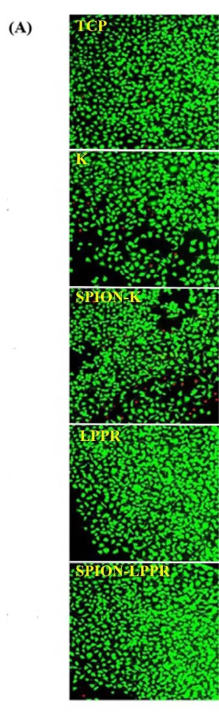
Figure 2.1 Images of hydrophobic iron oxide nanoparticles in the absence and presence of magnetic field and chemical structures of PA molecules. ... 33
Figure 2.2 DLS analysis to determine the dispersion stability of K-PA coated SPIONs and LPPR coated SPIONs. ... 34
Figure 2.3 Dose-dependent cytotoxicity profiles of PA coated SPIONs and free PAs on different cell lines ... 38
Figure 2.4 Viability of HUVECs. ... 39
Figure 2.5 Prussian blue assay of HUVECs... 40
Figure 2.6 Tube formation on Matrigel... 41
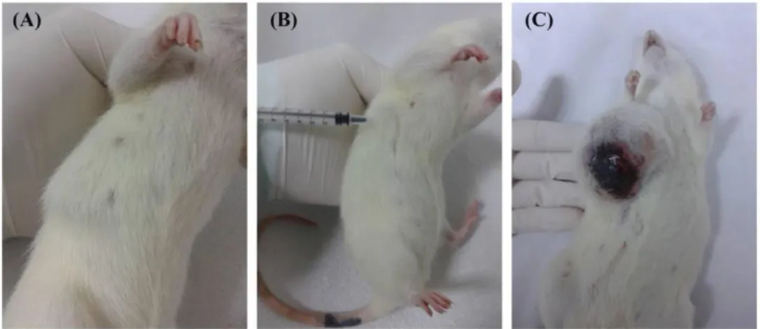
Figure 2.7 Chemically induced breast cancer progression in female Spraque-Dawley rats. ... 43
Figure 2.8 In vivo MRI ... 46
Figure 2.9 MR images from healthy rats. ... 47
xiv
Figure 2.11 Quantification of signal intensity changes. ... 49
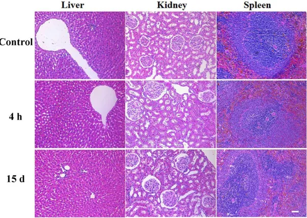
Figure 2.12 H&E staining for analyzing the tissue morphology of the kidney, liver and spleen of healthy rats. ... 52
Figure 2.13 H&E staining for analyzing the tissue morphology of the kidney, liver and spleen of tumor bearing rats. ... 53
Figure 2.14 Prussian blue staining for investigating iron accumulation in the tumor of rats. ... 54
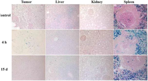
Figure 2.15 Prussian blue staining for analyzing iron accumulation in the liver, kidney and spleen of healthy rats ... 54
Figure 2.16 Prussian blue staining for investigating iron accumulation in the tumor, kidney, liver and spleen of tumor baring rats. ... 55
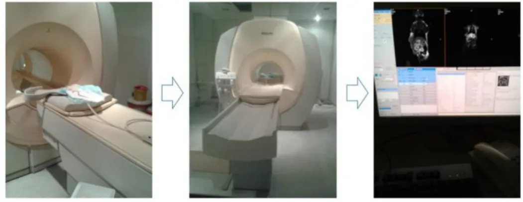
Figure 2. 17 MRI procedure. ... 65
Figure 3.1 UV-Vis absorbance spectra of CT DNA-metal system ... 72
Figure 3.2 CD spectra of CT DNA titrated with metal ions ... 75
Figure 3.3 ITC final thermograms of Au(III)-DNA and Ga(III)-DNA bindings. ... 78
Figure 3.4 FT-IR spectra of CT DNA in the presence of Au(III), and Ga(III) ions .. 81
Figure 3.5 Absorption spectra of HSA with various amount of metal ions. ... 84
Figure 3.6 Fluorescence spectra of HSA in the presence of metal ions.. ... 86
Figure 3.7 ITC final thermograms of Au(III)-HSA, and Ga(III)-HSA bindings. ... 88
Figure 3.8 Representative spectrum of curve-fitted amide I region of HSA. ... 90
Figure 3.9 Alamar blue cytotoxicity assay. ... 91
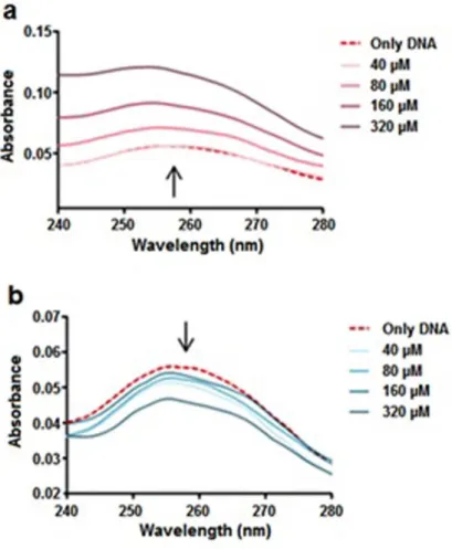
Figure 4.1 UV–Vis spectra of CT DNA in the presence and absence of BA. ... 103
Figure 4.2 CD spectra of free DNA and BA- DNA mixtures BA ... 105
xv
Figure 4.4 FT-IR spectra of DNA-BA mixtures. ... 108
Figure 4.5 ITC data for the titration of BA into CT DNA ... 111
Figure 4.6 Chemical structures of ule and col... 113
Figure 4.7 UV-Vis spectra of CT DNA in the presence and absence of boron compounds. ... 114
Figure 4.8 UV-Vis spectra of boron compounds in the presence and absence of different concentrations of CT DNA... 115
Figure 4.9 Gel electrophoresis ... 119
Figure 4.10 CD spectra of CT DNA in the presence of borates. ... 121
Figure 4.11 FT-IR spectra of CT DNA-ule mixtures. ... 125
Figure 4.12 FT-IR spectra of CT DNA-col mixtures ... 126
Figure 4.13 FT-IR difference spectra of CT DNA-ule mixtures and CT DNA-col mixtures ... 127
Figure 4.14 FT-IR second-derivative spectra of CT DNA-borates... 128
Figure 4.15 UV-Vis spectra of free HSA and different molar ratios of HSA-boron compound complexes ... 130
Figure 4.16 Fluorescence emission spectra of HSA-borate complexes. ... 132
Figure 4.17 CD spectra of HSA-ule and HSA-col interactions ... 134
Figure 4.18 FT-IR spectra and difference spectra of HSA-ule and HSA-col ... 136
xvi
List of Tables
Table 3.1 Relative fluorescence ratios of EtBr-CT DNA mixture after incubation with metal ions ... 73 Table 3.2 Thermodynamic parameters obtained by ITC ... 78 Table 3.3 Peak positions and spectral shifts of the metal ions-DNA complexes ... 82 Table 3.4 Thermodynamic parameters obtained by ITC analysis for the binding of HSA with Au(III) or Ga(III) ions. ... 88 Table 3.5 Secondary structure of HSA upon treated with Au(III) or Ga(III) ions... 90 Table 4.1 The relative fluorescence intensity ratios with respect to varying concentrations of BA... 103 Table 4.2 FT-IR of the free CT DNA and CT DNA-BA complexes ... 109 Table 4.3 EtBr displacement assay ... 117 Table 4.4 Curve fitting analysis of free HSA and protein incubated with borates .. 137
xvii
Abbreviations
ANOVA : Analysis of variance
Au : Gold
BA : Boric acid
BNCT : Boron neutron capture therapy CCD : Charge-coupled device
CD : Circular dichroism Co : Cobalt
Col : Colemanite
CT : Computed tomography
CT DNA : Calf thymus deoxyribonucleic acid DLS : Dynamic light scattering
DMBA : 7,12-Dimethylbenz(a) anthracene DMEM : Dulbecco’s modified Eagle’s medium DMF : Dimethylformamide
DNA : Deoxyribonucleic acid
EPR : Enhanced permeability and retention EtBr : Ethidium Bromide
xviii
FDA : Food and drug administration FT-IR : Fourier transform infrared Ga : Gallium
Gd : Gadolinium
HBTU : Hexafluorophosphate H&E : Hematoxylin-eosin
HPLC : High-performance liquid chromatography HSA : Human serum albumin
HUVEC : Human umbilical vein endothelial cell IR : Infrared
MCF-7 : Human breast adenocarcinoma cell MDA-MB-453 : Human metastatic breast carcinoma cell Mn : Manganese
MRI : Magnetic resonance imaging MSN : Microporous silica nanoparticles NRP-1 : Neuropilin-1
PA : Peptide amphiphile PBS : Phosphate buffered saline PET : Positron emission tomography Pt : Platinum
xix PVA : Polyvinyl alcohol QDs : Quantum dots
RES : Reticuloendothelial system RF : Radiofrequency
ROI : Region of interest ROS : Reactive oxygen species Ru : Ruthenium
SEM : Standard error of the mean
SPECT : Single-photon emission computed tomography SPION : Superparamagnetic iron oxide nanoparticle TCP : Tissue culture plate
TEM : Transmission electron microscopy TFA : Trifluoroacetic acid
TIS : Triisoproplysilane Trp : Tryptophan Ule : Ulexite US : Ultrasound UV-Vis : UV visible
VEGF : Vascular endothelial growth factor
1
Chapter 1
2
1.1 Cancer
Cancer is a complex disease characterized by the uncontrolled tissue growth and abnormal cell spreading. The accumulation of spontaneous or inherited genetic mutations1 and several molecular alterations are the main reasons of cancer2. Other
factors that result in cancer are external factors, such as tobacco3 or excessive sun exposure4, 5, infectious organisms such as human papillomavirus 6, 7, hepatitis B8 and
C virus9, human immunodeficiency virus, or Helicobacter pylori10, unhealthy diet11, obesity12, physical inactivity13, as well as internal factors, such as hormones14, and immune conditions1, 15. Generally, the presence of one or more of these factors results in a detectable cancer within ten or more years.
In the US, cancer is the second most common disease16. It is reported that approximately 14.5 million cancer patients survived until 2014 and it is expected that this value will increase nearly 1.6 million in 201517. On the other hand, there is a significant increase in the incidence of cancer cases in our country. Each year thousands of cancer cases are diagnosed in Turkey and the most commonly diagnosed cancer type is the lung cancer among men, while breast cancer is the most frequent cancer type among women according to the results of the Ministry of Health of Turkey18.
Although screening offers an opportunity for the diagnosis of cancer in early stages, it is not enough to provide extensive treatment and better outcomes. The early prognosis and treatment of cancer, before symptoms appear, remain poor with current imaging and therapeutic modalities in oncology clinic. Unfortunately, more than half of cancer patients have metastasized cancer cells that invade other organs of the body when
3
cancer is detected, which reduce the effectiveness of treatment and increase mortality rate for cancer of the breast, colon, rectum, cervix, and lung19.
Some of the cancer biomarkers alter their expressions according to biological conditions. They are genes, microRNAs20, proteins, lipids, carbohydrates, and small metabolite molecules19. They can be used as early cancer indicators which are often over-expressed in tumor cells rather than healthy cells such as gastrin-releasing peptide receptor for breast, colon, lung, and especially prostate cancer21, L-ferritin receptors for breast cancer22, KIT protein for gastrointestinal stromal tumors23, MMR enzyme loss for Lynch syndrome which is associated with malignant colon24, rectum, endometrium, stomach and small bowel cancer23, prostate-specific membrane antigen for prostate cancer, and VEGF/VEGFRs for tumor angiogenesis25. By using these
biomarkers as targets, not only could we predict tumor characteristics such as metastasis, hormone dependence, induction of angiogenesis, but also it could facilitate decision making for diagnosis and treatment of cancer accurately. As an example, Neurophilin-1 (NRP-1) is a transmembrane glycoprotein co-receptor of vascular endothelial growth factor (VEGF) and is highly expressed in cancer cells to promote angiogenesis of tumor tissue. Depending on this molecular mechanism, human monoclonal anti-NRP-1 antibody MNRP1685A was developed for the treatment of advanced solid tumors and further modification was done by combining anticancer drugs such as bevacizumab and paclitaxel to increase efficiency of therapy26, 27. In addition to early diagnosis of cancer, we also need to improve quality of the life of cancer survivors with further advancements in diagnosis and subsequently monitoring tumor progression, metastasis, and then effectiveness of the therapy with high
4
specificity21. Also, in order to develop novel imaging agents and new anticancer agents, the exact mechanism of action of these agents and their effects in biological systems need to be investigated.
1.1.1 Cancer Diagnosis and Therapy
Multidisciplinary approaches have led to biomedical imaging modalities which are not only highly sensitive, but also have good resolution properties. These techniques have assisted us in understanding the biological phenomena and diagnose several diseases over the last few decades. Currently, detection of diseases are based on several chemical or biological clinical tests, histopathological analysis of tissue specimen, as well as imaging techniques. In order to detect tumor at early stage, managing disease course in patients, and decrease mortality rate of cancer patients, diagnostic screening methods and cancer therapy agents should urgently be improved.
Imaging techniques are important for the diagnosis of cancer in early stages. They can be used for presenting accurate location of cancer cells, tumor morphology and cancer metabolism28. As imaging modalities, Positron emission tomography (PET), single-photon emission computed tomography (SPECT), computed tomography (CT), ultrasound (US), optical fluorescence imaging, and magnetic resonance imaging (MRI) are widely used. Their potential benefits and drawbacks are illustrated in Figure 1.1.
PET and SPECT are nuclear imaging methods which give information about the functional processes and metabolic activity, based on the used biologically active material. These conventional imaging techniques differ in terms of sensitivity, spatial resolution, used radioactive contrast agents and cost29.
5
CT uses X-ray for the detection of different tissues. Even though CT is non-invasive and has high resolution, radiation exposure during clinical examination may result in dangerous outcomes, such as induction of cancer30. Recent developments in
nanotechnology lead to new alternative materials that perform better than aromatic iodinated contrast agents, such as gold nanorods, fullerenes and carbon nanotubes which can be functionalized by conjugation with several targeting agents in order to increase retention rate and tissue specific accumulation31.
Besides being an inexpensive and flexible imaging modality, US has widespread use for several applications. With the advances in technology, 3D imaging systems have been produced to get higher diagnostic accuracy of pathology32. Microbubbles have been used to increase sensitivity of US imaging. For selective assessment and treatment of inflammation33, ischemia–reperfusion injury, angiogenesis, and thrombosis, these microbubbles have been modified by a variety of targeting ligands and combined with drugs34 over the last ten years. Furthermore, contrast-enhanced US method is capable of detecting early tumor angiogenesis by using microbubbles functionalized with echistatin35.
6 Figure 1.1 Medical imaging techniques.
Optical or fluorescence imaging is based on photon detection by a charge-coupled device (CCD) camera. It is used for visualization of fluorescently labeled specific cells
in vitro and in vivo. There are advantages and disadvantages of fluorescence imaging
technique. It is relatively cost effective, portable and provides real time imaging. On the other hand, autofluorescence and limited light penetration depth may be problematic when you need high spatial sampling of photons propagating through animal tissues36. The most commonly used florophores are polymethines-organic dyes suh as Cy5.5, Cy737and indocyanine green which is FDA approved for clinical use38. Activatable fluorescent probes which do not emit light as unbound but give fluorescence by enzymatic activation if bound to its target inside the cell have newly
7
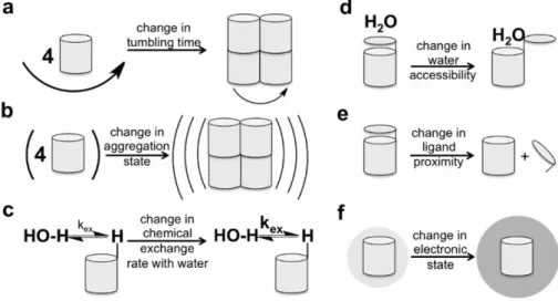
emerged38.It is possible to detect tumor early with highly sensitive fluorochromes that conjugate to overexpressed growth factor receptors on the cancer cells. For example, tumor specific fluorescence imaging was demonstrated in an ovarian cancer patient through the folate conjugated FITC in a plot study39. For the diagnosis of breast cancer, Pham W. et al. synthesized Cy7 conjugated with AREPPTRTFAYWG peptide sequence that provides uMUC-1 antigen specific binding of the dye40. With the advances in nanotechnology new materials have been evaluated for in vivo molecular cancer imaging applications such as dye-containing silica or lipoprotein or calcium phosphate nanoparticles, quantum dots (QDs), inorganic fluorescent semiconductor nanocrystals, upconversion nanoparticles, Au nanoclusters and Ag nanoclusters28. Radiology is accepted as “gold standard” for understanding the effectiveness of treatment and response of body to the applied treatment23. Up to now, the most widely used clinical and preclinical radiology imaging technique to detect physiological processes, anatomical details and disease state is MRI. The main reasons for the superiority of MRI are high spatial resolution at submillimeter scale, high contrast between soft tissues, being a noninvasive method and the absence of ionizing radiation during imaging. There are several parameters that affect signal intensity and distinct contrast in MR imaging. Some of them are proton relaxation rates, water proton densities, tumbling rates or time, field strength, acquisition sequence41, accessibility of water to the agent, electron spin state, MR frequency and (super)paramagnetism of the contrast agent42. These physicochemical mechanisms can be used to produce responsive contrast agents as well42 (Figure 1.2).
8
Classically, the principle behind MRI can be explained as alignment of protons of water molecules in the presence of an external magnetic field (B0). Longitudinal
magnetization is the alignment of proton nuclear spins in the same direction of B0 when
radiofrequency (RF) pulse is applied.
In contrast, transverse magnetization occurs when the proton nuclear spins are aligned antiparallel with B0. During transverse magnetization, longitudinal magnetization is
reduced. Under a magnetic field, nuclear spin transition is called “spin flip” and its quantum state is expressed with the following formula:
ω0 = γB0
where ω0 is Larmor frequency, γ is gyromagetic ratio43.
Figure 1.2 Schematic illustration of physicochemical mechanisms to alter MRI contrast. Reproduced by permission of Wiley42.
After removal of the RF pulse, the excited water protons relax to the ground state by emitting the energy gained from the RF pulse. There are two different relaxation processes. Longitudinal (spin-lattice) relaxation which is known as T1, derives from
9
the longitudinal magnetization recovery. On the other hand, reduction of transverse magnetization resulting from the loss of phase coherence and dephasing between the proton nuclear spins produces transverse (spin-spin) relaxation known as T2 (Figure 1.3). Upon reconstruction of these signals with Fourier transform, gray scale images are obtained. Thus, images are sorted into two groups: T1-weighted images and T2-weighted images. The inverse of relaxations is called as relaxation rate “R1 or R2” (s
-1). A brighter contrast is obtained if T1 relaxation rate (R1) is faster. Darker contrast
is observed in the presence of a faster T2 relaxation rate (R2)43.
Figure 1.3 Schematic illustration of relaxations of magnetic particles.
MRI gives more reliable results for clinicians in the diagnosis diseases related to invasion of structures when compare to CT and US 44. If the contrast between diseased region and the healthy region is uncertain, it is sometimes very hard to detect the region of interest (ROI)43. Although low expression levels of some biomarkers and relatively low sensitivity (for example, false-positive rate of 10% for breast cancer, limited detection of small tumors or subtle anatomical changes43) of MRI limit its application
10
limitations. The basic function of contrast agents is to accelerate magnetic relaxation to create much more contrast in RIO, which gives opportunity for discrimination between the pathological lesions and the healthy tissues. In 2010, approximately 35% of all clinical MRI in the USA was performed with the help of contrast agents45. There are two types of contrast agents: positive contrast agents result in brighter images, while negative contrast agents produce a darker image. Although they have strong contrast enhancement effects, their tissue specificity and selectivity need to be improved.
Contrast agents that are designed to enhance MRI signal comprise various materials, such as metals (gold, silver, and cobalt) and metal oxides (Fe3O4, TiO2, and SiO2)46.
Generally, T1 contrast agents demonstrate paramagnetic properties. The inorganic materials that have been applied clinically are mainly gadolinium (Gd3+) chelates which have unpaired electrons, a large magnetic moment and relaxivity (r1 and r2)
values around 4–6 mM−1⋅s−1 47. Food and drug administration (FDA) and European agencies have approved Gd based contrast agents for clinical use48. In 1988, the first approved contrast agent was GdDTPA (gadopentetate dimeglumine, Magnevist®) for imaging of pathologies in liver and gastrointestinal system49. For a standard MRI scan, the minimum concentration of positive contrast agent is approximately 10 to 500 μM, depending on the imaging system properties42. It is known that free Gd is very toxic. The exchange of Gd with other metal species, called transmetallation, and chelate dissociation due to the protonation of the ligands at acidic environment are the main factors for adverse effects of Gd chelates in the body50. The determined lethal dosage for mouse is 0.2 mmol kg−1 Gd3+. It causes well-known toxicity observed as nephrogenic systemic fibrosis, which is a serious syndrome, on human patients with
11
severe kidney failure. Rui et al. investigated toxicity of several T1 contrast agents using a mouse model. They observed that Gd accumulates in the liver, spleen, and especially kidneys48. In vivo studies also showed deposition of Gd in bone49. In a study
performed by Kanazawa et al. commercially available gadolinium-diethylenetriamine pentaacetic acid (GdDTPA BMA, Omniscan®), was administered intravenously to the patients with bladder cancer. They reported that use of this T1 contrast agent gives opportunity for quantification and discrimination of bladder tumors and the normal bladder wall51. Gd based nanoparticles with minimum toxicity and prolonged
circulation time have been evaluated after surface modifications by scientists. Li et al. reported potassium/gadolinium iron hexacyanoferrate nanoparticles can be used as positive MRI contrast agents and measured a relaxivity of 12.3 mM−1 s−1. Under infrared (IR) radiation, these nanoparticles convert IR energy to thermal energy which gives opportunity for cancer therapy52.
Due to their high magnetization properties, MnO containing materials have also been tested as T1 contrast agents. When compared to Gd(III) chelates, MnO contrast agents have more contrast enhancement potential in MRI. However their high toxicity often diminishes their clinical application. Potential severe effects of MnO have been studied by several researchers in vitro and in vivo 48, 53-55. Chen et al. modified the surface of
MnO nanoparticles with N-(trimethoxysilylpropyl) ethylene diamine triacetic acid (TETT) silane which increases water solubility and biocompatibility. In order to achieve glioma specific binding, these nanoparticle were further decorated with folic acid, which showed that these nanoparticles can be used for glioma diagnosis by MRI56. In another study, manganese oxide (MnO) nanocrystals with the size of 15-20
12
contrast when taken by low pH compartments in the cell such as lysosome57. Huang et
al. synthesized and characterized a new T1 contrast agent for brain tumor imaging
which was coated with HSA that provides long term stability and monodispersity of MnO nanoparticles in biological media. Unlike phospholipid-coated MnO nanoparticles (0.21 s-1mM-1), this modification does not cause a significant reduction in the relaxivity of material58.
For T2 weighted sequences, superparamagnetic iron oxide nanoparticles (SPION) have been clinically applied. Very small and single domain ferromagnetic particles demonstrate superparamagnetism where the energy barriers are relatively low with respect to thermal energy, and therefore the magnetization occurs with thermal excitations. In other words, superparamagnetic materials can be magnetized as soon as an external magnetic field is applied and their average magnetization is zero without a magnetic field like paramagnetic material but their magnetic susceptibility is larger than paramagnetic materials (Figure 1.4).
Figure 1.4 Schematic illustration of hysteresis loop of superparamagnetic materials.
There are two sources for SPIONs: one of them is magnetite (Fe3O4), other is
13
SPIONs with different coating agents and hydrodynamic sizes (ranging from 5 nm to 3500 nm) have been approved60 as T2 contrast agents by the FDA or are in clinical development61. Some of the commercial ones are called Feridex = Endorem, Clariscan,
Resovist = Cliavist, Sinerem and Abdoscan®.
In clinical study, researchers investigated MRI contrast enhancement of Resovist®, which is a reticuloendothelial system (RES) specific dextran coated SPION, with 1.5-T MR scanner. For liver lesions larger than 2 cm, these nanoparticles could be used for detection accurately (95.2%)62.
Yang et al. synthesized PEG coated SPIONs and functionalized with folate as tumor targeting ligand. They reported that these nanoparticles selectively accumulates in cancer cells both in vitro and in vivo. MRI of a mouse model revealed that the dark contrast generated from folate conjugated PEG coated SPIONs is significantly high at the tumor site rather than healthy tissue63. As an alternative strategy, hydrophobic
SPIONs with 9.9 nm diameter were encapsulated into polymeric micelles and were functionalized with the attachment of RGD peptide for tumor ανβ3 integrin imaging by
MRI64.
In the literature, there are several studies about SPIONs prepared with different sizes and compositions by using different strategies and comparison of their contrast effects in MRI65-67. Combination of Co and Mn with iron oxide nanoparticles (CoFe2O4 and
MnFe2O4) also leads to more prominent T2 contrast enhancement. However, there are
some considerations about the toxicity of these nanoparticles due to the production of free metal ions that may result in dangerous outcomes in nervous system68. Though iron oxide nanoparticles are typically considered negative contrast agents, SPIONs
14
with mean size of 5 nm or less demonstrate T1 MRI contrast effects. Some groups have developed hybrid systems by combining polymers and SPIONs to obtain dual T1/T2 responsive probe for tumor detection69-71.
SPIONs can be used for imaging of apoptosis, angiogenesis, β-amyloid accumulation, inflammation and gene expression via MRI72. On the other hand, biological applications of SPIONs are not restricted to MRI contrast enhancement. SPIONs can be used for tissue repair, immunoassay, detoxification of biological fluids, hyperthermia, drug delivery73, and cell separation purposes as well74.
Although SPIONs have several advantages such as biocompatibility and long blood circulation time, we have to take disturbance in iron homeostasis, oxidative stress, and altered cellular responses into consideration in clinical use73.
Thanks to the improvements in synthesis methodologies, there are important developments in the preparation of high quality magnetic nanoparticles. Synthesis methods of magnetic nanoparticles can be categorized into four groups: co-precipitation, thermal decomposition, microemulsion, and hydrothermal synthesis techniques. By using these methods, nanoparticles with different compositions and phases can be synthesized such as iron oxides, pure metals, alloys and ferromagnets. During nanoparticle production, the main purpose is to obtain highly stable, monodisperse, uniform, as well as size and shape controlled magnetic nanoparticles75. Co-precipitation is one of the most common method for magnetic materials’ synthesis, especially iron oxides. This process occurs under elevated temperatures or room temperature in the presence of base and inert atmosphere76. The physicochemical properties of the final products such as composition and size are mostly determined by
15
the content of the initially used salts, and the chemical properties of precipitation medium77. Although this method is very simple and fast, the nanoparticles synthesized with co-precipitation technique are very polydisperse.
The best and most common method for the synthesis of size and shape controlled magnetic nanoparticles is thermal decomposition technique which is performed at high temperatures (ranging from 240 to 320 °C) and needs organic solvents with stabilizing surfactants. Generally, metal acetylacetonates, metal cupferronates and carbonyls are used as precursors. Surfactants used as stabilizing agent include hexadecylamine, oleic acid or fatty acids such as decanoic acid, lauric acid, myristic acid, palmitic acid, oleic acid and stearic acid78. By changing the ratio of these two materials and hydrocarbon solvents, size, shape and composition of the product can be modified75. Nanoparticles
with very narrow size distribution and very small size can be synthesized by this method. However, it is a little bit more complicated and time consuming technique than other methods. Nanoparticles synthesized with thermal decomposition method are generally not water soluble, thus they need a surface capping agent such as poly(ethylene glycol) (PEG) or dextran for biomedical applications. It is also noteworthy that water soluble nanoparticles can be obtained with this method if monocarboxyl-terminated PEG79 or R,ω-dicarboxyl-terminated PEG are used as
surface capping agent80.
Microemulsion method is also used to prepare magnetic nanoparticles at ambient conditions. This method depends on stabilization of two different thermodynamically stable isotropic dispersion of immiscible liquids by an interfacial film of surfactant molecules81. The most important disadvantage of microemulsion method is that
16
generally controlling shape, size and dispersity is very hard. Moreover, it is not an efficient method for magnetic nanoparticle synthesis, because you have to use large amounts of solvents for a little amount of product.
Another method for magnetic nanoparticle synthesis is hydrothermal method. Although this synthesis method is simple, high temperature (above 200 °C) and pressure conditions (higher than 2000 psi) are required for the production of size and morphology controlled materials80. Basic mechanism behind the hydrothermal method is the separation and phase transfer at the interfaces of the liquid, solid, and solution phases. However the detail of mechanism of hydrothermal method is unclear. In order to get monodispersed and uniform metal and metal oxide nanoparticles, a mixture of high-boiling-point reducing agent, electrostatic stabilizer and surfactant are required. Nanoparticles obtained by methods mentioned before are generally unstable under physiological conditions. Therefore, they are stabilized with biocompatible materials which are monomeric stabilizers such as carboxylates or phosphates, inorganic materials such as gold and silica, or polymers such as dextran, PEG, polyvinyl alcohol (PVA), poly(D,L-lactide-coglycolide (PDL)61, alginate, starch or chitosan80 to prevent agglomeration or precipitation, and to improve colloidal stability in biological media. Up to now, the most powerful and common coating agent has been dextran, which is found on the surface of some FDA approved contrast agents for MRI82. In vitro and
in vivo investigations revealed that dextran coating decreases toxicity of nanoparticles
and it is biodegradable and improves their blood distribution profile83. Furthermore; dextran coated SPIONs give opportunity for attachment of targeting moieties on the surface of agent due to the tunable surface chemistry.
17
As an alternative to covalent attachment methods for surface coating of nanoparticles, hydrophobic interactions between alkyl chains of peptide amphiphile (PA) and lauric acid or laurylamine or octyl groups on the surface of nanoparticles can be used for the functionalization purposes84, 85. PAs are molecules containing a hydrophobic tail (usually double-chain lipids or single chain fatty acids) and a hydrophilic biologically active peptide headgroup (capable of mimicking functions of several biomolecules such as receptors) which are conjugated through a spacer86. Above the critical micelle concentration, PAs form soluble micelles. Incorporating a variety of targeting amino acid sequences to the PA micelles allows not only targeting but also imaging of several diseases like cancer87, 88.
Recently, significant progress has been made on the development of unique contrast agents with tumor targeting properties. These designs are generally based on conjugation of contrast agent with tumor specific ligands such as DNA, siRNA, aptamers, small molecules, peptides, proteins and antibodies89 which are capable of binding selectively to cancer biomarkers or receptors for not only tumor targeted imaging but also non-invasive monitoring of molecular processes, and targeted drug delivery82. For instance, cyclic(RGDyK)-4-methylcatechol labeled monodispersed Fe3O4 nanoparticles were used as integrin targeted T2 contrast agents and these
ultrasmall nanoparticles have 60 mM-1s-1 morerelaxivity value than clinically used Feridex® nanoparticles90. Radermacher et al. designed a new SPION agent coated with PEG and functionalized with TLVSSL peptide sequence, which is called phosphatidylserine-targeted hexapeptide. This new contrast agent was tested in terms of necrotic tissue targeting after cancer treatment in an animal liver tumor model.
18
When compared to non-targeted nanoparticles, coupling of targeting ligand to the SPION induced a 3-fold higher accumulation in tumor site91.
Actually, the effective diagnosis and treatment of solid tumors depend on true understanding of cancer targeting for proper material design92. The well-known differences between tumor and healthy tissue are leaky blood vessels (due to the disordered tight junctions93) and impaired lymphatic system of the tumor tissue,
namely enhanced permeability and retention (EPR) effect which could be used for accumulation of particles and drugs via active or passive targeting strategies (Figure 1.5). Size and shape are the most important factors for nonspecific accumulation of particles. Additionally, increasing blood concentration of a material enhances passive tumor targeting chance by preventing aggregation, precipitation and serum protein binding of nanoparticles. On the other hand, active targeting is achieved by coating of surfaces with tumor specific binding ligands. Chemical and physical properties of nanoparticles affect their uptake and interactions with biological substances as well92. In one study, scientists compared the effect of size, coating and addition of a targeting ligand on the tumor accumulation of iron oxide nanoparticles in vitro and in vivo94. According to their observations, 30 nm nanoparticles accumulate more than particles with 100 nm size. Coating of nanoparticles with PEG did not result in significant differences between in vitro and in vivo uptake of nanoparticles. Furthermore, in vivo imaging of mice tumor tissue has been achieved by a monovalent Fab’ fragment of Tmab (FDA approved monoclonal antibody for breast cancer targeting) moiety coupled to iron oxide nanoparticles. Antibody targeted nanoparticles with smaller size have greater tumor concentration than passively targeted nanoparticles because of high
19
washing out rate of nonspecific particles from neoplastic tissues. Several studies have explored that upon intravenous administration, materials with a molecular weight above the renal threshold (40 kDa) and diameter ranging from 10 to 150-200 nm tend to accumulate preferentially in tumor93.
Figure 1.5 Active and passive targeting strategies. Reprinted from reference [89] with permission89.
Currently used cancer management methods are surgery, radiation, chemotherapy, endocrinotherapy, immune therapy, and molecular targeted therapy. However, these
20
methods have limited advantages due to the poor effectiveness especially for metastatic disease with acquired resistance at late stage.
Nowadays, one important class of potential therapeutics are peptides that have high specificity and low toxicity for the treatment of diseases, which depend on angiogenesis. In the last decade, several promising amino acid sequences have been identified for the inhibition of cancer progression through preventing vessel formation. The advantages of peptides for cancer targeting are their small size which leads to good tissue penetration, functionalization capability which gives opportunity for encapsulation or surface coating of nanoparticles, and being a component of combination therapies95.
Actually, inorganic chemistry in medicine can be categorized into two different approaches: one of them utilizes ligands (metal ions in different forms; either free or bound to a biomacromolecule such as protein) as drugs, other uses metals as imaging agents previously discussed in detail96.
Herein, inorganic chemotherapeutics used in cancer treatment will be discussed in detail. The main mechanism of conventional cancer drugs is eliminating malignant cells without considering the presence of healthy cells in the environment. Although they usually manage their duty efficiently, their precision is insufficient and they have several adverse side effects such as nephrotoxicity97, ototoxicity, and allergy98. Research on chemotherapeutics for cancer is important for designing novel anti-cancer drugs against tumors to overcome the toxicity of these drugs and achieve more precision.
21
Over the past decade, metal based drugs have attracted attention because of their biological and pharmaceutical properties as anticancer agents. Metallopharmaceuticals such as BBR3464, satraplatin and picoplatin contain platinum (Pt), which binds to DNA and interferes with its normal function, halting DNA replication, and thereby leading to cell death. The most commonly used Pt containing potent anticancer drugs are cisplatin, carboplatin, oxaliplatin and KP1537. However acquired resistance after repeated administrations limits their medical use99. By mimicking superoxide dismutase (SOD) enzyme, Mn chelates can be used for cancer therapy. NAMI-A and KP1339, which are chemotherapeutic agents, include ruthenium (Ru) compound. Besides having strong complex forming ability with various ligands100, greater resistance to hydrolysis provide advantages to Ru compounds as anticancer drugs96. Other examples of use of inorganic compounds are Arsenix trioxide, namely Trisenox® for the treatment of acute promyelocytic leukaemia, gallium (KP46) for melanoma101, and lanthanum (KP772) for multi-drug resistant cancer102.
Several researchers have investigated the use of new metal (iron, copper, cobalt, zinc, cadmium and mercury) compounds for their antineoplastic potential103-107. One interesting study demonstrated that successful Gd neutron capture therapy was achieved by incorporation of Gd(III) chelates into calcium phosphate micelles without therapeutic drugs. This finding is important because this system can effectively accumulate in tumor tissue and disrupted cancer cells through γ-rays or electron emission from the Gd nuclides. Furthermore, being an MRI guided cancer targeting system facilitates imaging of tumor lesions and minimizing toxicity on healthy tissue108.
22
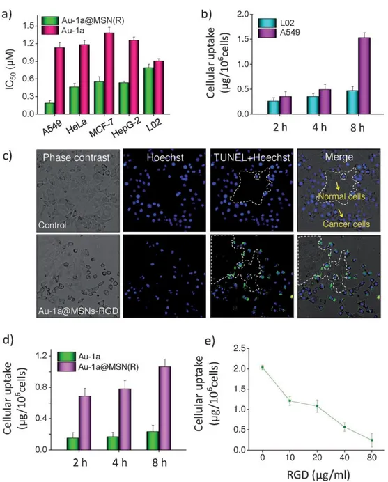
He et al. reported the anticancer properties of a gold(III) porphyrin complex (Au-1a) loaded microporous silica nanoparticles (MSN) denoted with RGD peptide which binds integrins overexpressed on cancer cells109. Cancer cell specific uptake of these
nanoparticles by various cell lines and in vitro selective induction of apoptosis by gold encapsulated silica nanoparticles in a co-culture model were demonstrated (Figure 1.6). Time dependent and dose dependent accumulation of this silica-gold drug delivery system in cancer cells revealed the importance of targeting ligand for efficient therapy.
23
Figure 1.6 Cellular uptake and specificity of Au-1a@MSN(R). a) Cytotoxicity of gold nanoparticles on different cell lines. b) Quantification of cellular uptake of nanoparticles in L02 normal human liver cells and A549 lung carcinoma cell lines. c) TUNEL-Hoechst 33342 co-staining of A549/L02 co-culture cells after treatment with gold nanoparticles. d) Quantification of time dependent cellular uptake of nanoparticles. e) Dose-dependent effects of RGD peptide on the cellular uptake of Au-1a@MSN(R). Reproduced by permission of Wiley109.
24
1.1.2 Importance of Material - Biomacromolecule Interactions
Conventional cancer treatment is based on chemotherapy and radiotherapy. The discovery and characterization of new drug candidates have received a great deal of attention110 to improve the quality of life of patients for those undergoing treatments.
Up to now, although several different kinds of materials have been developed in order to increase effectiveness of cancer treatment, we still need to improve capabilities of these materials29. Significant improvements in both cancer diagnosis and treatment can be achieved through detailed understanding of the interactions between materials and biomolecules which provides important information about the effects of material on the target of interest in terms of thermodynamic and kinetic property changes toward biological molecules such as receptors and deoxyribonucleic acid (DNA), the metabolic fate in blood and pharmacological activity of material.
One of the most important application of inorganic compounds in medicine is cancer chemotherapy. There are extensive studies focusing on use and increasing effectiveness of these metal based antitumor drugs with fewer side effects. The detailed examination of the chemistry of the material, as well as the kinetics and affinity of material's interaction with its potential tumor related molecular targets can yield important insights into material design99.
Currently used chemotherapeutic agents generally demonstrate their function through DNA damage by binding to DNA and thereby inhibiting replication and transcription processes. For example; DNA is accepted to be the cellular target of a well-known and commonly used anticancer drug: cisplatin. The intercalation, groove binding and covalent binding are the main binding modes for an association between an agent and
25
DNA111. The extensive understanding of binding modes is important to assess the biological consequences of these DNA adducts. In literature, enormous efforts have been done to determine physical interaction of a compound with DNA by using several spectroscopic, calorimetric and molecular modeling methods111-114.
On the other hand, analyses of interaction between inorganic compounds with serum proteins are crucial to understand fate of them in blood stream and their pharmacokinetic behavior. Although efficient transfer of an agent to the target often requires serum protein binding as a drug delivery system, unwanted or random protein-metal ion interactions may result in toxicity. Human serum albumin (HSA) binding contributes significantly to the discovery of novel potential materials because it is capable of transporting various kinds of materials such as drugs115, metabolites,
endogenous ligands, and metal ions116, 117. HSA is a globular protein structurally dominated by α-helices. It has three homologous domains (I, II and III) and each contain two separate subdomains (A and B)118. Although they are very similar in terms of their structures, these domains demonstrate different binding properties. Most HSA-binding drugs bind to the IIA and IIIA domains, while metals utilize other HSA-binding sites; such as sites A and B, N-terminal binding sites, the Cys34 binding site and others119. In the presence of external small molecules, proteins undergo modifications
in their secondary structure, which may affect their stability and binding properties120. Thanks to the physical and biophysical characterization techniques, it is possible to determine binding sites of several potential inorganic drug candidates and enhance cytotoxicity of compounds in cancer cells118, 121. Thus, HSA based carriers, hybrid systems and metal based drugs could be designed and developed rationally for biomedical applications.
26
Chapter 2
BIOACTIVE PEPTIDE AMPHIPHILE FUNCTIONALIZED
SUPERPARAMAGNETIC IRON OXIDE NANOPARTICLES
27 2.1 INTRODUCTION
Breast cancer is the second most commonly diagnosed cancer type among women worldwide. According to the estimation from the National Cancer Institute, about 232,670 new cases were diagnosed in 2014, with 40,000 associated deaths due to the breast cancer in the US17. Mortality rate from breast cancer has been increasing each year all over the world. Breast cancer risk is two times higher if a woman has one or more affected first degree relatives diagnosed with breast cancer due to the inherited mutations in cancer susceptibility genes such as BRCA1 and BRCA2122. Aging is
another factor for the development of breast cancer as well123.
Over recent years, enormous studies have focused on different diagnostic and therapeutic approaches to cancer. The most promising strategy has been based on tumor targeting with overexpressed specific receptors. The conjugation of these targeting molecules with contrast agents or drugs improves the selective uptake by tumor cells124. Judah Folkman proposed anti-angiogenic therapy in 1971125. Although angiogenesis is a physiological process by which new blood vessels are formed, it is also crucial for the tumor growth and metastatic spread of cancer cells by supplying nutrients and oxygen, as well as removing waste products126. The role of angiogenesis in supporting solid tumor growth is well documented. Most tumor endothelial cells produce high levels of VEGF, and VEGF receptors (VEGFRs). VEGF expression is up-regulated in response to hypoxia and often increases in regions of necrotic tumor. Neuropilin-1 (NRP-1) is a co-receptor for VEGF and its expression mediates angiogenesis and invasion127 of several tumor cells, such as breast, prostate, neuroblastomas, astrocytomas, lung, pancreas, bile duct, gastric, colon cancers and melanoma cells128, 129. Each breast carcinoma cell has nearly 1–2 x 10⁵ NRP-1
28
molecules on its surface. There is a strong affinity between NRP-1 and VEGF165 but not VEGF121, because the NRP-1 binding site in VEGF165 is encoded by VEGF exon 7, which is absent in VEGF121127.
Recently, the short peptide sequence ATWLPPR, was identified by Starzec et al. through peptide libraries displayed on bacteriophage130. This peptide prevents the binding of VEGF165 to NRP-1 which plays role as an anti-angiogenic and anticancer agent. Positive MRI contrast enhancement of a multifunctional nanoparticle including silica nanoparticles with gadolinium chelate and ATWLPPR peptide as tumor targeting ligand was tested in a rat intracranial U87 glioblastoma model131. The intratumoral retention of ATWLPPR-targeted nanoparticles was higher than the non-targeted nanoparticles which demonstrates tumor cell selectivity of this peptide sequence. It has reported that the shortest amino acid sequence interacting with NRP-1 coreceptor is LPPR. In this study, I used LPPR as an antiangiogenic tumor vascularate targeting peptide to hinder the interaction between VEGF and NRP-1 required for angiogenesis of cancer tissue.
Breast exam and mammography are often used to detect breast cancer at an early stage. Although mammography can detect tumor and contribute to treatment of patients, this technique is insufficient if patient is young or has dense breast tissue. MRI is a powerful medical imaging modality for high resolution visualization of the cancer non-invasively. Tissue anatomic structures can be monitored through the detection of magnetization of water protons in the presence of a strong magnetic field. Normal and pathological tissues may have different intrinsic relaxation times depending on their physiological environment, specific contrast can be observed between the site of
29
interest and the surrounding regions at advanced stages of disease132. The sensitivity of MRI can be greatly enhanced by using contrast agents such as SPIONs which increase the signal-to-noise ratio. SPIONs are already used in clinic as contrast agents in MRI (Feridex®, Endorem®, Gastromark®, Sinerem®) for the diagnosis of liver or lymph node pathologies and diseases of the bowel wall or mesentery.
Recent advances in the development of surface modified nanoparticles have increased use of MRI for cancer diagnosis133. Several groups have reported that tumor specific MRI T2 imaging agents are biocompatible, biodegradable and have long blood circulation time, which provide high accumulation in the tumor tissue134, 135. As contrast agents, iron oxide nanoparticles which are known to be strong enhancers of proton spin–spin relaxation, provide several opportunities in the accurate diagnosis of cancer135. The size and coating of nanoparticles strongly influence their contrast enhancement and tumor targeting properties. There are currently several efforts under way to reduce hydrophobic interactions between SPIONs and biological molecules by coating the surface of nanoparticles with different synthetic and natural chemical moieties that also give opportunity for the functionalization of nanoparticles for target specific interaction74. Attachment of a biomarker associated with tumor metastasis and/or progression on the surface of nanoparticles is an effective approach towards the early detection of cancer136 which is called active targeting. Also, nanoparticles can passively accumulate at tumor site due to their small size as discussed in previous chapter. For example, iron oxide nanocubes encapsulated with polyethylene glycol-phospholipid have been shown to increase sensitivity in tumor imaging compared to Feridex137. Lee and coworkers have developed branched PEI and PEG coated SPIONs
30
that are able to bind to negatively charged plasmid DNA due to the positive charge on their surface and are detectable at the tumor site by MRI138.
Our group previously reported that lauric acid and laurylamine capped hydrophobic SPIONs can be noncovalently functionalized with peptide amphiphile (PA) molecules through hydrophobic interactions to render them water soluble and biocompatible84. We hypothesized that LPPR coated SPIONs could actively reach the cancerous tissue following systemic administration in vivo. On the other hand, K-PA coated SPIONs without a tumor targeting moiety were used as a control. We further hypothesized that the MRI negative contrast enhancement of PA functionalized SPIONs could be utilized to monitor breast tumors noninvasively and to evaluate the effect of SPIONs on the time course of distribution and elimination from tumor and different organs. In this chapter, I demonstrated noncovalent coating of SPIONs with proline-rich PAs, and investigated the efficiency of this superparamagnetic, water-dispersible hybrid system in enhancing the MR imaging of tumor tissue in rats. The potentials of PA functionalized SPIONs as an MRI contrast agent were assessed in Sprague-Dawley rats with mammary gland tumors. In particular, the cellular viability and uptake behavior of the SPION/PA system were investigated using different healthy and cancer cell lines, while the biodistribution and bioelimination of the nanoparticles were investigated by MRI and histological examination in animal tumor model.
![Figure 1.5 Active and passive targeting strategies. Reprinted from reference [89] with permission 89](https://thumb-eu.123doks.com/thumbv2/9libnet/5615200.111054/41.892.171.785.341.813/figure-active-passive-targeting-strategies-reprinted-reference-permission.webp)