CONTRIBUTION OF MESENCHYMAL STEM CELLS IN CELL BASED THERAPIES
A THESIS SUBMITTED TO
THE DEPARTMENT OF MOLECULAR BIOLOGY AND GENETICS AND THE INSTITUTE OF ENGINEERING AND SCIENCE OF
BİLKENT UNIVERSITY
IN PARTIAL FULFILLMENT OF THE REQUIREMENTS FOR THE DEGREE OF DOCTOR OF PHILOSOPHY
BY
ZEYNEP TOKCAER KESKİN
I certify that I have read this thesis and that in my opinion it is fully adequate, in scope and in quality, as a thesis for the degree of Doctor of Philosophy
Assist. Prof. Dr. K. Can Akçalı
I certify that I have read this thesis and that in my opinion it is fully adequate, in scope and in quality, as a thesis for the degree of Doctor of Philosophy.
Assoc. Prof. Dr. İhsan Gürsel
I certify that I have read this thesis and that in my opinion it is fully adequate, in scope and in quality, as a thesis for the degree of Doctor of Philosophy.
Assist. Prof. Dr. H. Uygar Tazebay
I certify that I have read this thesis and that in my opinion it is fully adequate, in scope and in quality, as a thesis for the degree of Doctor of Philosophy.
Prof. Dr. Belma Turan
I certify that I have read this thesis and that in my opinion it is fully adequate, in scope and in quality, as a thesis for the degree of Doctor of Philosophy.
Assoc. Prof. Dr. Z. Günnur Dikmen
Approved for the Institute of Engineering and Science
Director of Institute of Engineering and Science Prof. Dr. Levent Onural
ABSTRACT
CONTRIBUTION OF MESENCHYMAL STEM CELLS IN CELL BASED THERAPIES
Zeynep TOKCAER KESKİN Ph.D. in Molecular Biology and Genetics Supervisor: Assist. Prof. Dr. K. Can Akçalı
August 2010, 110 Pages
Stem cell research evolved as a new hope and has gained tremendous interest in the last two decades to develop new strategies for many of debilitating diseases. Mesenchymal Stem Cells (MSCs) are multipotent cells capable of self-renewal and differentiating into multiple lineages such as osteocytes, adipocytes, chondrocytes, myoblasts, and hepatocytes. MSCs can migrate to the injured tissue and have immunomodulatory effects. Due to these features, MSCs have high therapeutic value in tissue engineering and regenerative medicine. In this thesis, our aim was to investigate the further contribution of the MSCs in different cellular therapies. We used two approaches to accomplish our aim. First, we investigated the possibility of obtaining functional cardiomyocytes from rat MSC within a shorter time period by determining the induction timing of cardiomyocyte differentiation of MSCs. Our data revealed that it is possible to get functional cardiomyocytes from in vitro MSC culture in a shorter time period than previously achieved. This reduction in time may provide emergency cases with access to cell-based therapies that may have previously been unavailable. In the
second part of this thesis, we examined in vivo and in vitro effects of a telomerase antagonist, imetelstat (GRN163L) on MSCs. Telomerase activity is essential for the continued growth and survival of malignant cells, therefore inhibition of this activity presents an attractive target for anti-cancer therapy. MSCs also show telomerase activity in maintaining their self-renewal; therefore the effects of telomerase inhibitors on MSCs may be an issue of concern. Our results showed that inhibiting the telomerase activity does not interfere with the self-renewal and differentiation of MSCs under short term in vitro culture conditions.
ÖZ
MEZENKİMAL KÖK HÜCRELERİN HÜCRE TEMELLİ TERAPİLERE KATKISI
Zeynep TOKCAER KESKİN Moleküler Biyoloji ve Genetik Doktorası
Tez Yöneticisi: Doç. Dr. K. Can Akçalı Ağustos 2010, 110 Sayfa
Kök hücre araştırmaları tedavisi olmayan hastalıklar için yeni bir umut olmuş ve son 20 yılda hastalıklara tedavi geliştirmek için yeni bir strateji olarak büyük ilgi görmüştür. Mezenkimal kök hücreler (MKH) kendini yenileme kapasitesine sahip, osteosit, adiposit, kondrosit, miyoblast ve hepatosit gibi değişik hücre kökenlerine farklılaşabilen, hasarlı dokuya ulaşabilen ve bağışıklık sistemini baskılayıcı etkiye sahip hücrelerdir. Bu özelliklerinden ötürü MKHlerin doku mühendisliği ve rejeneratif tıptaki terapötik önemi yüksektir. Bu tezde, amacımız MKHlerin hücre terapilerine katkısını incelemekti. Amacımıza ulaşmak için iki yöntem kullandık. İlk olarak, rat MKHlerinden kardiyomiyosit farklılaşmasının daha kısa sürede elde edilebilirliğini, MKHlerden kardiyomiyosit farklılaşmasını indükleme süresini belirleyerek araştırdık. Verilerimiz in vitro MKH kültüründen daha kısa sürede fonksiyonel kardiyomiyosit elde edilebildiğini göstermiştir. Süredeki bu kısalma acil vakaların daha önce erişimleri sağlayamadıkları hücre temelli terapilere erişimini sağlayabilecektir. Tezin ikinci bölümünde telomeraz antagonisti olan imetelstat’ın (GRN163L) MKHler üzerindeki in vivo and in vitro etkilerini araştırdık. Telomeraz aktivitesi malign hücrelerin büyüme ve hayatta kalmaları için gereklidir, bu nedenle bu aktivitenin engellenmesi kanser tedavisi için çekici bir hedef oluşturmaktadır. MKHler de kendilerini yenilemek için telomeraz aktivitesine sahiptirler ve telomeraz inhibitörünün MKHler üzerindeki etkisi kesin olarak bilinmemektedir. Sonuçlarımız, telomeraz aktivitesinin engellenmesinin kısa süreli in vitro kültür koşullarında MKHlerin kendini yenileme ve farklılaşma potansiyellerini etkilemediğini göstermiştir.
ACKNOWLEDGEMENT
I would like to express my gratitude to my supervisor Assoc. Prof. Dr. Can Akçalı for his personal and academic guidance and for his trust in me. He always treated like a father to me. I hope I didn’t let him down, in any part of this study. It was an honor for me to work with him.
I would like to thank the present and former members of Akçalı Group, Fatma Ayaloğlu Bütün, Verda Bitirim, Sumru Bayın, Sinan Gültekin, Hande Koçak and Ece Terzioğlu Kara for their friendship, support and patience during my PhD. I couldn’t accomplish this without their help.
I would like to thank to Assoc. Prof. Dr. İhsan Gürsel and Prof. Dr. Wayne Criss for their supervision and support whenever I needed.
I would like to thank to MBG faculty for providing me the necessary background during my studies and all MBG grad members for providing me a very peaceful and enjoyable work place with their friendship since 2005.
I would like to thank to Gizem Tinçer, Chigdem Aydın Mustafa, Hande Erünlü, Ceren Sucularlı, Emre Onat, Bala Gür Dedeoğlu, Sevgi Bağışlar, Elif Uz, Tolga Acun and Fuat Yağcı for their support, friendship and help whenever I needed.
I would like to thank to Tokcaer and Keskin families for their endless love, support, guidance and understanding. I couldn’t manage without them.
I would like to dedicate this thesis, as we started and completed this work together, to my husband Volkan Keskin who always supported, encouraged and guided me with his endless patience and love.
TABLE OF CONTENTS
ABSTRACT ... iii
ÖZ ... iv
ACKNOWLEDGEMENT ... v
TABLE OF CONTENTS ... vi
LIST OF TABLES ... viii
LIST OF FIGURES ... ix
ABBREVIATIONS ... xi
CHAPTER 1 ... 1
INTRODUCTION ... 1
1.1 Stem cells ... 2
1.1.1 Embryonic Stem Cells ... 3
1.1.2 Induced Pluripotent Stem Cells ... 5
1.1.3 Adult Stem Cells ... 7
1.1.4 Mesenchymal Stem Cells ... 10
1.1.5 Mesenchymal Stem Cells in Cellular Therapies ... 14
1.1.6 Mesenchymal Stem Cells in Cardiac repair ... 18
1.2 Telomeres and Telomerase ... 21
1.2.1 Telomeres, Telomerase and Cancer ... 23
1.2.2 Telomerase and Stem Cells ... 25
1.2.3 Telomerase Antagonist GRN163L (Imetelstat) ... 26
CHAPTER 2 ... 29
AIM OF THE STUDY ... 29
CHAPTER 3 ... 31
MATERIALS AND METHODS ... 31
3.1 Animals ... 31
3.2 Isolation of the Cells from Rat Bone Marrow ... 31
3.3 Culturing of Mesenchymal Stem Cells (MSCs) ... 32
3.4 Treatment of the MSCs with Telomerase Template Antagonist GRN163L (GRN163L) and the Experimental Groups ... 33
3.5 Colony Forming Unit (CFU) Assay ... 33
3.6 Total RNA Isolation from Rat MSC ... 33
3.7 cDNA Synthesis ... 34
3.8 Primer Design ... 34
3.9 RT-PCR ... 36
3.10 Agarose Gel Electrophoresis ... 37
3.11 Semiquantitative Analysis of RT-PCR ... 37
3.12 Q-RT-PCR ... 38
3.14 Total Protein Isolation from MSCs ... 39
3.15 Western Blotting ... 39
3.15.1 Sodium Dodecyl Sulphate – Polyacrylamide Gel Electrophoresis (SDS-PAGE) ... 39
3.15.2 Semi-dry transfer of proteins to a PVDF membrane ... 40
3.15.3 Immunological detection of Immobilized Proteins ... 40
3.16 Immunocytochemistry ... 41
3.17 Differentiation of MSCs ... 42
3.17.1 Differentiation of MSCs into Cardiomyocytes ... 42
3.17.2 Differentiation of MSCs into Adipocytes ... 42
3.17.3 Differentiation of MSCs into Osteocytes ... 43
3.18 Intracellular Ca+2 measurement ... 43
3.19 Statistical Analysis ... 44
3.20 Buffers and Solutions ... 44
CHAPTER 4 ... 45
RESULTS ... 45
4.1 Timing of Induction of Cardiomyocyte Differentiation for in vitro Cultured Mesenchymal Stem Cells ... 45
4.1.1 Characterization of Mesenchymal Stem Cells (MSCs) on 9th and 14th Days of Cell Culture ... 46
4.1.2 Validation of the Cardiomyocyte Differentiation ... 49
4.2 Effects of GRN163L on MSCs ... 52
4.2.1 Phenotypic Effects of GRN163L on MSCs ... 53
4.2.2 Effects of GRN163L on the Differentiation capacities of MSCs ... 56
4.2.3 Effects of GRN163L on Telomerase of MSCs ... 58
4.2.4 Effects of GRN163L on the Cell Cycle of MSCs ... 59
CHAPTER 5 ... 64 DISCUSSION ... 64 CHAPTER 6 ... 71 FUTURE PERSPECTIVES ... 71 REFERENCES ... 73 APPENDIX ... 107
LIST OF TABLES
Table 3.8 List of Primers and the product sizes they amplify………35
Table 3.9 RT-PCR Conditions………..………...36
Table 3.12 Q-RT-PCR conditions………38
LIST OF FIGURES
Figure 1.1.1 1 Characteristics of ESCs ... 5
Figure 1.1.3 1 Adult Stem Cells ... 8
Figure 1.1.4 1 MSC multilineage differentiation potential ... 12
Figure 1.2 1 Elongation of telomeres by the enzyme telomerase ... 22
Figure 1.2.3 1 Action mechanism of GRN163L on telomerase ... 27
Figure 4.1.1 1 Characterization of MSCs on the 9th day of cell culture ... 47
Figure 4.1.1 2 Characterization of MSCs on the 14th day of cell culture ... 47
Figure 4.1.1 3 Cytoplasmic calcium levels of MSCs in response to KCl and caffeine measured with fura-2AM on the 9th day of the culture ... 48
Figure 4.1.1 4 Cytoplasmic calcium levels of MSCs in response to caffeine measured with fura-2AM on the 14th day of the culture ... 48
Figure 4.1.2 1 Cytoplasmic calcium levels of cardiomyocytes, differentiated from the MSCs on 9th day of the culture, in response to KCl and caffeine measured with fura-2AM. ... 50
Figure 4.1.2 2 Cytoplasmic calcium levels of cardiomyocytes, differentiated from the MSCs on 14th day of the culture, in response to KCl and caffeine measured with fura-2AM. ... 50
Figure 4.1.2 3 CD90 immunocytochemistry after induction of differentiation (A) on 9th day (B) on 14th day (magnification 20x) ... 51
Figure 4.1.2 4 Expression profile of cardiac-specific markers in 5-azacytidine-treated MSCs on the (A) 9th and (B) 14th day of culture (* indicates p<0.05). ... 52
Figure 4.2.1 1 Effect of GRN163L (a-d: phenotypic effects, A. untreated MSCs (control), B. mismatch oligonucleotide treated MSCs (mismatch) C. 1µM GRN163L treated MSCs (163L) D. MSCs treated with GRN163L and then left for recovery (163LR) E. expression of CD markers of MSCs. ... 55
Figure 4.2.2 1 Adipogenic differentiation of MSCs determined by Oil Red O staining ... 57 Figure 4.2.2 2 Osteogenic differentiation of MSCs determined by Alizarin Red staining ... 58 Figure 4.2.3 1 TRAP assay of MSCs to determine telomerase activity A. normal samples B. heat inactivated samples ... 59 Figure 4.2.4 1 mRNA expressions of cell cycle genes in MSCs from control, 163L, 163LR and mismatch groups, determined with Q-RT-PCR. * indicates
p<0.05. ... 61
Figure 4.2.4 2 Protein expressions of cell cycle genes in MSCs from control, 163L and 163LR groups determined with Western blotting ... 63
ABBREVIATIONS
ALT Alternative Lengthening of Telomeres ASC Adult Stem Cells
BM Bone Marrow
bp Base Pair
BSA Bovine Serum Albumin BrDU Bromodeoxyuridine CCR Cytokine Receptor CD Cluster of Differentiation
cDNA Complementary Deoxyribonucleic Acid CFU-F Colony Forming Unit Fibroblasts CXCR Chemokine receptor
ddH2O Double distilled water DEPC Diethylpyrocarbonate
DMEM Dulbecco’s Modified Eagle Medium DNA Deoxyribonucleic Acid
DNase Deoxyribonuclease
dNTP deoxy Nucleotide Triphosphate ESC Embryonic Stem Cells
FBS Fatal Bovine Serum FGF Fibroblast Growth Factor GFP Green Fluorescent Protein GVDH Graft versus Host Disease HIF Hypoxia-Inducible Factor HLA Human Leukocytes Antigen HRP Horse Raddish Peroxidase
HS Hot Start
HSCs Hematopoietic Stem Cells ICM Inner Cell Mass
IL Interleukin
IP3 Inositol-3-Phosphate ISCs Intestinal Stem Cells
iPS Induced Pluripotent Stem Cells kDa Kilo Dalton
kg kilogram
LG-DMEM Low Glucose Dulbecco’s Modified Eagle Medium LIF Leukemia Inhibitory Factor
M Molar
MEF Mouse Embryonic Fibroblast MeOH Methyl Alcohol
mg milligram
MHC Major Histocompatibility Complex mL Milliliter
mM milliMolar
MSC Mesenchymal Stem Cells MRI Nagnetic Resonans Imaging NK Natural Killer cell
nM nanoMolar
NSCs Neural Stem Cells
PBS Phosphate Buffered Saline PD Population Doubling PCR Polymerase Chain Reaction PFA Paraformaldehyde
PVDF Polyvinylidenedifluoride
Q-RT-PCR Quantitative Reverse-Transcriptase Polymerase Chain Reaction RNA ribonucleic acid
RNA Ribonucleic acid Rpm revolution per minute RT Room Temperature
RT-PCR Reverse-Transcriptase Polymerase Chain Reaction rtTA Reverse Tetracyclin Transactivator
SDS Sodium Dodecyl Sulfate TAE Tris Acetate EDTA TBS Tris Buffered Saline
TERC Telomerase RNA Component TERT Telomerase Reverse Transcriptase Tet Tetracyclin
TGF Transforming Growth Factor Th Helper T-cell
TNF Tumor Necrosis Factor
TRAP Telomeric Repeat Amplification Protocol VEGF Vascular Endothelial Growth Factor
μg Microgram
μl Microliter μL Microliter
CHAPTER 1
INTRODUCTION
Radiometric age dating indicates that the world is 4.5 billion years old and the origins of first human beings date back to 200.000 years ago. Throughout these 200.000 years, human beings have been facing with many problems. Majority of these problems have been caused by the humans themselves in the past and present such as pollution, war and feminine. In the last 50 years or so, humans have been facing another problem, aging. Aging is the result of the increased life quality due to the technological improvement to diagnose and treat diseases. According to World Health Organization’s 2008 health report 85% of the world population is supposed to be over the age of 60 by 2050 while today the mean life expectancy in the world is over 75 years of age (http://www.who.int/whr/2008/whr08_en.pdf). In this chamber of globalization, urbanization and aging the incidence of chronic and non-infectious diseases now are the main health concerns of the population, like cancer, chronic heart diseases and diabetes. Moreover, together with the increase at life span more than 50% of the people above age 70 have more than one chronic disease. Thus, treatment of these chronic diseases is very important for a healthy future.
Stem cell research evolved as a new hope and has gained tremendous interest in the last two decades to develop new strategies for many of these
diseases. Applications involving the use of stem cells in humans that might have been considered “science fiction” fewer than 20 years ago are now being utilized with a great success rate (Akar et al 2006). In this thesis, our aim was to investigate the contribution of the mesenchymal stem cells (MSCs) in cellular therapies. We used two approaches to accomplish our aim:
i) directly by determining the induction timing of cardiomyocyte differentiation of mesenchymal stem cells and;
ii) indirectly by examining the in vitro effects of a telomerase antagonist, imetelstat (GRN163L) on mesenchymal stem cells.
Introduction section starts with a brief explanation of the stem cells. Embryonic stem cells, induced pluripotent stem cells and adult stem cells will be introduced. Among the adult stem cells, mesenchymal stem cells will be discussed in detail including their use in cellular therapy. Finally, the structural and functional features of telomerase enzyme will be analyzed.
1.1
Stem cells
Stem cells are group of cells that are able to self-renew and differentiate into several lineages to form specialized somatic cells (Morrison et al 1997, Weissman 2000; Till and McCulloch 1961). Stem cells are able to accomplish these tasks by undergoing symmetric and/or asymmetric cell division (Preston et al 2003). Symmetric division yields two daughter stem cells having the same properties of stemness which are undifferentiated and have the capacity to differentiate into other lineages. If there is a requirement for differentiated somatic cells, then a stem cell undergoes asymmetric division to yield a progenitor cell
and an undifferentiated copy of itself to maintain the stem cell pools of the body. Stem cells can be categorized based on their time of onset. Embryonic stem cells are present on in the very early part of the embryonic development, but germ line and adult stem cells become active later in the development and post nataly respectively. Better categorization of the stem cells can be made based on their potencies. Embryonic stem cells are pluripotent, which can form all the cell types from all body lineages where as adult stem cells are multipotent and able to produce multiple lineages.
1.1.1 Embryonic Stem Cells
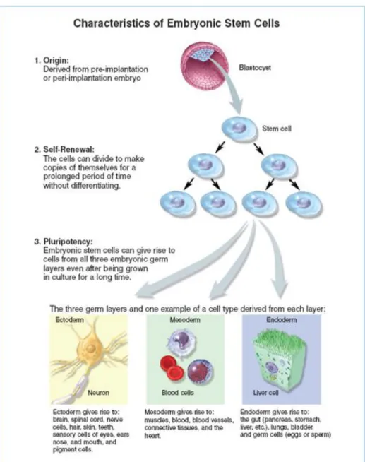
After fertilization, zygote undergoes serial divisions during the embryonic development. At the blastula stage, cluster of cells form inner cell mass (ICM) where embryonic stem cells are obtained (Evans and Kaufman, 1981, Martin 1981, Thomson et al, 1998, Reubinoff et al 2000). Embryonic stem cells are pluripotent and can differentiate into any of the cells belonging to three germ layers, endoderm, mesoderm and ectoderm. (Figure 1.1.1.1). Furthermore, they can maintain their pluripotency for many passages without losing their genetic integrity (Burdon et al 2002, Smith 2001, Keller 2005).
ESCs form teratomas in vivo, which contains cell types from all germ layers (Wobus and Boheler 2005). X chromosome is active and ESCs do not undergo X inactivation until they are differentiated (Yang et al 2007). ESCs require a feeder layer composed of irradiated human or mouse embryonic fibroblast (MEF) cells. Feeder layers not only provide better attachment but also secrete growth factors for their maintanence (Amit et al 2003). In addition, mouse ESCs require leukemia inhibitory factor (LIF) to maintain their undifferentiated
state in vitro while human ESCs need FGF-2 (Odorico et al 2001, Pera and Trounson 2004, Keller 2005). When ESCs are removed from the feeder layers, some cells start differentiating spontaneously and some of them remain undifferentiated. These cells form clusters called embryoid bodies which are unique features of ESCs in suspension cultures. ESCs express transcription factors such as Oct4, Sox2 and Nanog which are important to keep them undifferentiated and pluripotent (Mitsui et al 2003, Strumpf et al 2005, Kuroda et al 2005, Rodda et al 2005). Human ESCs also express SSEA3 and 4 on their surfaces as markers of their undifferentiated state. They have active telomerase enzyme (Semb 2005, Hiyama and Hiyama 2007) that is rapidly reduced upon the differentiation (Armstrong et al 2005).
ESCs are important tools for studying embryogenesis, understanding the mechanisms of genetic diseases and for cellular therapies. There are severe ethical problems when human ESCs are considered despite the fact that these embryos are leftover embryos obtained from IVF clinics. In addition, the risk of tumor formation and immune rejection also the problems that need to be solved.
Figure 1.1.1 1 Characteristics of ESCs ©2006 Terese Winslow (http://stemcells.nih.gov/info/2006report/2006Chapter1.htm)
1.1.2 Induced Pluripotent Stem Cells
The problems that are associated with ESCs prompted the researchers to investigate new sources of stem cells that have the features of ESCs. Attempts
finally reached to a success in 2006 when two Japanese scientists, Takahashi and Yamanaka announced the induction of pluripotent cell from normal somatic cell. Their study revealed that, when mouse fibroblast cells were transfected with lentiviral vectors carrying Oct-4, Sox-2, Klf-4 and c-myc, the cells became pluripotent and this process was named as “Induced Pluripotent Cells (iPS)”. These four factors are called as “Yamanaka Factors”. In addition, iPS cells form embryoid bodies, regain their telomerase activity, elongate their telomeres and form teratomas in vivo, but more importantly they are capable of restarting the mouse developmental process after being injected into a blastocyst. When results of their study were first published, it opened a new era and raised hopes to obtain patient specific pluripotent stem cells. Afterwards studies towards better understanding the induced pluripotent stem (iPS) cells and their uses have gained tremendous interest. In 2008 iPS cells were developed by using human fibroblasts as a source (Park et al 2008, Takahashi et al 2008, Yu et al 2008). Several different cell types like pancreatic beta cells, dermal fibroblasts and keratinocytes (Lowry et al 2008, Stadtfeld et al 2008, Aasen et al 2008) were also used as a source with different transfection efficiencies. There are also some drawbacks in iPS cells. All these studies were employing retroviral vectors which might have mutagenic effects. In addition, introducing c-myc raised considerations against generation of tumors. As the main aim of the induced pluripotency was to generate patient specific cells for cell therapy, to improve the methods of iPS cell generation have become one of the fasted evolving areas of stem cell research. To tackle these problems drug induced systems, addition of N-myc instead of c-myc and using strategies without viral vectors (Wernig et al 2008, Nakagawa et al 2008, Okita et al 2008) have been utilized. A successful system employed piggyBac transposase to generate iPS cells. In this system, Yamanaka factors were inserted by a single cassette controlled by tet/rtTA. By this way the expression of the transgenes were conditionally controlled. After a successful
induction was generated, the genes inserted at the first transfection were removed with a second transposase transfection (Woltjen et al 2009). However at the end of all protocols the morphological, genetically and functional properties of iPS cells should be checked according to their similarities with ESCs. Recent studies also revealed that the cells which are reprogrammed into iPS cells retain their initial epigenetic codes that may affect their differentiation properties (Polo et al 2010, Kim et al 2010). Polo et al (2010) showed that continuous passaging reduced the differences between differentiation capacities into different lineages and the transcript identities between ESC and iPS cells derived from muscle, blood and fibroblast origins.
Although there are some drawbacks, using iPS cells in cellular therapies is one of the most promising options, at least in animal models. Alpio et al (2010) achieved to reduce high glucose levels in two different mouse models for diabetes mellitus by using differentiated stem cells obtained from iPS cells. In a mouse spinal cord injury model iPS cells were also used to induce neural differentiation and contributed to remyelination, formed functional neurons and astrocytes (Tsuji et al 2010). In addition, there are several studies that employ iPS cells successfully in the models of cardiovascular regeneration (Nelson et al 2009, Narazaki et al 2008, Zhang et al 2009) as well as Parkinson’s disease (Wernig et al 2008).
1.1.3 Adult Stem Cells
Adult stem cells (ASCs), also known as somatic stem cells, are found in organs and tissues and maintain homeostasis. Compared to ESCs, they are more differentiated and their potential is more limited (multipotent vs pluripotent).
There are ASCs unique for almost all tissue types such as blood, bone, intestine, brain, liver and heart (Figure 1.1.3.1). Although tissue specific stem cells have their own specific markers, but they are not universal markers for all ASC types. Since, ASCs are quiescent cells, retention of DNA labeling dyes like BrdU or histone-GFP is used to overcome such identification problems (Tumbar et al 2004, Kiel et al 2007).
Figure 1.1.3 1 Adult Stem Cells (Raghunath et al 2005)
The first discovery of ASCs leads back to 1950s when irradiation studies were performed to find a cure after atomic bomb sent to Hiroshima and Nagasaki. Scientists discovered that, when mice were severely irradiated to deplete their bone marrow cells, they showed the same symptoms with the people survived after atomic bomb. If they were injected again with bone marrow of another mouse on the other hand, the whole blood system was regenerated (Lorenz et al 1951, Ford et al 1956, Nowell et al 1956, Gengozian et al 1957). These initial
studies lead to the discovery and characterization of the hematopoetic stem cells (HSCs) (Wu et al 1963, Sprangrude et al 1988).
HSCs are the most widely studied ASC type. They express CD34, CD117, Sca-1 as the surface markers (Kim et al 1998, Negrin et al 2000). They are able to differentiate into several cell types other than blood cells such as muscle cells, cardiomyocytes, neurons and hepatocytes (Ferrari et al 1998, Peterson et al 1999, Brazelton et al 2000, Jackson et al 2001). One of the main aims of bone marrow (BM) transplantation is actually the transplantation of HSCs. BM transplantation is considered as an effective treatment in many different hematopoetic diseases including malignancies. However HSC transplantation still has some draw backs mainly due to the immune rejection manifested as graft versus host disease (GVDH).
There are also tremendous amount of cell removal in the intestines. Therefore intestines are also another source of stem cells. Intestinal stem cells (ISCs) are localized in the crypts (Marshman et al 2002) and responsible for the maintenance of different cell types of the intestines. Their localization is believed to be at the +4 position according to the Paneth cells at the bottom of the crypt as shown by Potten (1974) and Marshman et al (2002). Several molecules were identified a intestinal stem cell marker like musashi-1, sFRP5, Dcamkl1, prominin1/CD133 and Lgr5 (Kayahara et al 2003, Asai et l 2005, Gregorieff et al 2005, Giannakis et al 2006, Barker et al 2007, Snippert et al 2009, Sato 2009). Among these markers Lgr5, which is an orphan G protein coupled receptor, is widely accepted as an intestinal stem cell marker since 2007 (Barker et al 2007).
Neural stem cells (NSCs) are found in the neural tissue generating neurons, astrocytes and oligodendrocytes and can be isolated from sub-ventricular
zone of the brain (Kokavay et al 2008). Although the clues about their presence were discovered in 1960s, the idea of brain can not regenerate was dominant until 1992. First clues about the functionality of NSCs were gathered when Reynods and Weiss (1992) discovered that the cells taken from adult mice brain could be stimulated in vitro with epidermal growth factor and they were able to proliferate and form neurons and astrocytes. Six years after, the proliferation of the cells in hippocampus and dentate gyrus of adult humans were demonstrated from the autopsy samples taken from the ex-patients treated with BrdU (Eriksson et al 1998). The mostly known markers of neural stem cells are Sox2, GFAP, Pax6, LeX and nestin (Kokavay et al 2008, Conti and Cattaneo 2010). Studies performed in several animal models of neurological diseases including Parkinson’s disease, Huntington’s disease, Alzheimer’s disease, multiple sclerosis as well as spinal cord injury models revealed the success in cellular therapy models by using NSCs indicating their functional role in maintenance (Ronaghi et al 2010, Kim and Vellis 2009 and Taupin 2008).
In addition to ISCs, NSCs and HSCs there are several somatic stem cells as well and one of them is mesenchymal stem cells (MSCs) which are also localized mainly in BM like HSCs. In the next section MSCs will be described in detail.
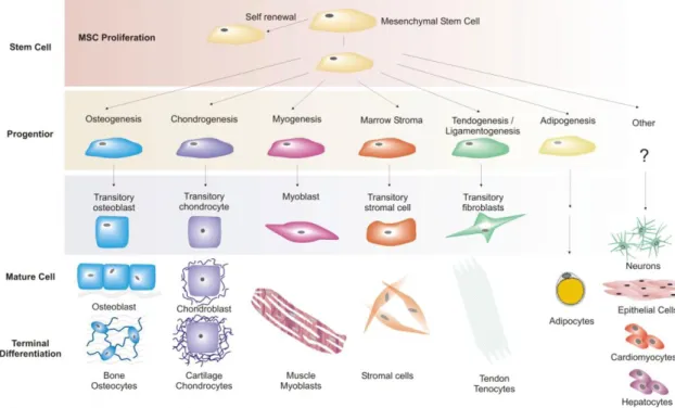
1.1.4 Mesenchymal Stem Cells
Mesenchymal stem cells (MSCs), also known as bone marrow stromal cells or mesenchymal stromal cells were discovered by the pioneering studies of Friedenstein in 1968. During the in vitro culturing of HSCs, it was discovered that there was a different type of cell in the bone marrow other than HSCs with
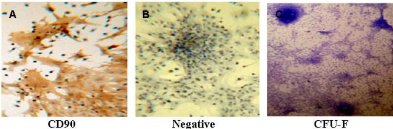
forming colonies which were afterwards named as colony forming unit fibroblasts, CFU-F (Lanotte et al 1981). Moreover, they were also able to differentiate into bone, adipose, cartilage and muscle tissue (Figure 1.1.4.1). Later on, these adherent, fibroblastic and colony forming cells were first named by Owen as stromal stem cells (1988) which were then finally called as mesenchymal stem cells by Caplan (1991). First characterization study of MSCs was performed by Pittenger et al in 1999 obtained from human bone marrow aspirates. The BM aspirate was first separated according to the density gradient and plated afterwards. Attached cells were counted according to their colony formation capacities and when proportioned to the total cell number, only 0.001 to 0.01% of the nucleated cells formed colonies. Moreover, they have shown that MSCs were positive for CD29, CD90, CD71 and CD106 and negative for CD45, CD14 and CD34. Furthermore these cells were able to undergo 40 population doublings (PD) in vitro within 10 weeks.
Figure 1.1.4 1 MSC multilineage differentiation potential (Caplan and Bruder 2001)
Other than bone marrow, MSCs were also isolated from many other sources such as adipose tissue, umbilical cord blood, placenta and even from dental pulp. However, studies revealed that MSCs from these sources had different CD marker expressions, phenotypes and PD. These results forced the scientific world to define certain criteria in identifying these cells. International Society for Cellular Therapy published a position paper by Dominici et al (2006) and set following features for MSCs. MSCs should be positive for CD73, CD90 and CD105, negative for CD19, CD34, CD45, CD11a and HLA-DR. In addition, MSCs should be isolated from fresh tissue and should attach to the plastic culture plates and differentiate into adipocytes, chondrocytes and osteocytes in vitro.
An important feature of MSCs is their homing capacity. Homing is defined as the migration of these cells to the site of injury. In the homing process of
MSCs, chemokines, cytokines and receptors on their surface were shown to be important (da Silva Meirelles et al 2008). When inflammation occurs at the site of injury, the gradient of cytokines and chemokines increase and the expression of chemokine receptors mediate the migration of MSCs to the injured tissue (Salem and Thiermann 2010). Especially CD44 was found to be important in homing of both mouse and human MSCs (Herrera et al 2004, Sackstein et al 2008). In addition, CXCR4 and VCAM-1 (CD106) had also a critical role in the migration of the MSCs (Segers et al 2006, Shi et al 2007, Hung et al 2007). It was also shown that MSCs can activate matrix metalloproteases to enter to the tissue from blood to localize the niche at the site of injury (De Becker et al 2007).
MSCs also have important antiapoptotic and immunomodulatory effects, which makes them non immunogenic. In animal models when the MSCs were injected to the scarred tissue, they were able to reduce the apoptotic rate of the surrounding cells, which were mediated by the secretion of several growth factors like VEGF, FGF2 and TGF-β especially in hypoxic conditions (Togel et al 2007, Parekkadan et al 2007, Block et al 2009). When immunomodulatory effects are considered, it was shown that the proliferation of the T-cells were inhibited during
in vitro co-culturing (Di Nicola et al 2002, Krampera et al 2003, Le Blanc et al
2003). Furthermore, not only cytotoxic and helper T cells but also natural killer cells, B-cells and immature dendritic cells were found to be affected by MSCs. The immunomodulatory effects of MSCs on T-cells and NK cells were associated via secretion of molecules like TGF-β, PGE2 and IL10. Whereas their effect on B-cells was found to be indirect, via the modulation of plasma cells by leading the inhibition of immunoglobulin secretion (Sotiropoulou et al 2006, Nasef et al 2007, Rafei et al 2008, Nemeth et al 2009, da Silva Meirelles 2009).
MSCs are important source for cellular therapies for the following reasons: i) Their well defined markers enable to obtain with high purity, ii) They can easily be expanded in vitro with high numbers without
losing their properties,
iii) They can differentiate into different cell types, iv) They can migrate to the injured site in vivo,
v) They have immunosuppressive effects that make their use possible in allogeneic grafting and off-shelf use.
These special features of MSCs make them good candidates in regenerative medicine and cellular therapies.
1.1.5 Mesenchymal Stem Cells in Cellular Therapies
In the literature there are many studies investigating the regenerative capacities of MSCs in different disease models generated by employing different non-human animal species. Cardiac regeneration, liver regeneration, kidney regeneration, autoimmune diseases and graft versus host disease (GvHD), neurological diseases, pulmonary diseases, osteogenic diseases, and cartilage repair are the most widely studied conditions. Moreover MSCs are also being investigated extensively by clinical trials, mostly in United States, Europe and East Asia. Some of the clinical trials are studying their use in neurological, liver, bone, heart diseases, GvHD and some autoimmune diseases like diabetes and Chron’s disease. In the following section, the application of MSCs in different cell based therapies will be discussed and a particular attention will be given to their roles in heart diseases.
1.1.5.1 MSCs in renal injuries
Kidney injuries if left untreated can progress to acute kidney failures followed by chronic kidney failures. At the end stage of renal disease dialysis or kidney transplantation is needed. Renal injuries could be due to immunological problems or to ischemia (Salem and Thiemermann 2010) suggesting the importance of restoring the microvasculature (Bussolati et al 2008). Treatment of kidney injuries requires either an immunomodulatory effect or replacement of the scared tissue. Therefore, MSCs are good candidates in curing kidney diseases. In the sheep model of ischemia reperfusion injury, it has been shown that the intraarterially injected MSCs differentiated into tubular and glomerular cells (Behr et al 2007). In a previous study performed with a rat model of ischemia reperfusion injury, the restoration of renal function was also observed after MSCs were introduced by intracarotid administration. However, this improvement was found to be a result of immunomodulatory action of MSCs as no differentiation was reported (Tögel et al 2005, Lange et al 2005). In recent studies performed with human umbilical cord derived MSCs similar result was obtained indicating the improvement of renal failure via immunomodulatory effects of MSCs (Chen
et al 2010, Cao et al 2010). In this immunomodulatory activity homing of the
MSCs to the injury site was found to be important. It has also been shown that the way of the administration of MSCs is also important for their immunomodulatory effect. In a rat model of injury better immunomodulatory effects were obtained when MSCs were introduced intraarterially than intravenously (Zonta et al 2010). Also in the homing of MSCs in renal injuries CXCR4 and CD44 were found to be important (Ji et al 2004, Herrera et al 2007).
1.1.5.2 MSCs in liver injuries
Although liver is able to regenerate upon injury, in the case of end stage liver injuries also require transplantation which may take very long time to find a donor. Therefore, cellular therapies are also important in liver injuries to replace the need for transplantation. Since isolation and culture of hepatocytes at efficient amounts is not possible in vitro (Serralta et al 2003, Serralta et al 2005), cellular therapies involving stem cells have an important role in curing liver diseases. Among the stem cells, MSCs receive special attention since their differentiation into hepatocyte like cells were reported (Chamberlain et al 2007, Sato et al 2005, Lee et al 2004).
Differentiation of MSCs into hepatocytes was reported both in vitro and in
vivo. These hepatocytes were positive for the hepatocyte markers and found to be
functional evidenced by their secretion of albumin and storing glycogen (Chamberlain et al 2007, Lee et al 2004, Schwartz et al 2002). Immunomodulatory effects of MSCs were also found to be important during liver regeneration. It was shown that engraftment of the MSCs into the liver, but not differentiation, stimulated proliferative and regenerative properties of the liver (Banas et al 2008, Parekkadan et al 2007, Caplan and Dennis 2006). In the murine injury models of carbontetrachloride treatment or partial hepatectomy the effect of MSCs on hepatic stellate cells were demonstrated, reducing fibrosis (Cavalho et al 2008, Abdel Aziz et al 2007, Zhao et al 2005, Sakaida et al 2004). Chamberlain et
al (2007) also demonstrated that when the MSCs were administered by
intrahepatic injection they formed hepatocytes more efficiently compared to intraperitoneal administration.
factor (HGF) were found to be important in the differentiation of MSCs into hepatocytes (Dong et al 2010, Parekkadan et al 2007) while TNF-α and IL-6 were found to be important in antifibrotic effects (Pulavendran et al 2010, Parekkadan
et al 2007). The expression of matrix metalloproteinases by MSCs was also found
to be important in the reduction of fibrosis (Fang et al 2004, Sakaida et al 2004, Oyagi et al 2006). During the homing of MSCs to the injured liver it was demonstrated that CXCR4 and CCR9 were important molecules (Chen et al 2009).
1.1.5.3 MSCs in GvHD
GvHD is an inflammatory reaction caused by the T-cells of the host against the donor tissue HLAs after the transplantation which results in the rejection of the transplant (Flomenberg et al 2004, Goulmy et al 1996). Interestingly, MSCs were found to be very effective in suppressing the immune reaction during GvHD (Koç et al 2000, Frassoni et al 2002, Le Blanc et al 2004).
The inhibitory role of MSCs against GvHD depends on the effects of MSCs on dendritic cells, T cells, B cells and NK cells. The main inhibitory effect was found to be via dendritic cells, thereby preventing the antigen presentation (Reddy et al 2005). The interaction between antigen presenting cells and T-cells as well as the signaling mediated via co-stimulatory factors like CD28, ICOS and CD40 cause the initiation of immunological response against the graft (Blazar et
al 2001, Blazar et al 1997). MSCs lacking co-stimulatory molecules like CD40
were not able to contact with the host immune cells (Tse et al 2003). When T-cells and MSCs were cultured together it was demonstrated that the release of IFN-γ from inflammatory Th1 cells decreased while IL-4 secretion from anti-inflammatory Th2 cells increased. Moreover, when MSCs were cocultured with activated T cells, PGE-2 secretion by MSCs was found to be increased and T-cell
inhibition was observerved. The same effect couldn’t be observed when the MSCs were cocultured using PGE-2 inhibitors suggesting the importance of PGE-2 synthesis by MSCs (Aggarwal and Pittenger 2005).
In a baboon model of skin transplantation, it was shown that the MSCs suppress T-cells and enabled graft survival with a single intravenous administration (Bartholomew et al 2002). At murine heart transplantation model,
i.v. injected MSCs prolonged the survival of the transplanted heart which was
shown to be due to the regulatory effects of MSCs on Th1 and Th2 (Zhou et al 2006). Treg cells were shown to be induced by MSCs at another study using rat heart transplantation model mediating the graft survival (Casiraghi et al 2008).
The immunomodulatory effect of MSCs on immune system was also demonstrated in human cancer patients and GvDH patients after bone marrow transplant (Koç et al 2000, Frassoni et al 2002, Le Blanc et al 2004). Le Blanc et
al (2004) also reported that the MSCs administered to the patient could be of any
type of HLA antigens. MSCs were administered twice after transplantation and fully recovered within a year. A recent study reported the use of MSCs with the bone marrow transplant patients irresponsive to immunosuppressors and they have also demonstrated the regression of the disease after injection of MSCs twice (Lim et al 2010).
1.1.6 Mesenchymal Stem Cells in Cardiac repair
According to the World Health Statistics Report published by WHO in 2008, ischemic heart diseases have the highest mortality rate among all diseases today and 20 years later (http://www.who.int). Therefore, the treatment of heart
diseases is very important to provide a better living to the patients. Ischemic heart diseases are characterized by a shortage in the blood supply to the different regions of the heart. Cardiomyocytes in these regions go under necrosis and apoptosis which is called infarction. Although heart tissue renews itself very slowly by the help of cardiac stem cells, the regeneration rate is very slow to replace the damaged cardiac muscle with the healthy new cardiomyocytes (Oh et
al 2003, Beltrami et al 2003). If left untreated at its end stage heart transplantation
is the only thereupethic option.
Taylor and Jones (1979) reported the generation of cells, having mesenchymal phenotypes after several weeks, when 3T3 and 10T ½ cells were treated with 5-azacytidine which incorporates and leads the methylation of DNA reverting the cells to a more pluripotent state. Based on their findings in 1995, Wakitani et al published the first paper about the generation of cardiomyocytes in
vitro from rat BM derived MSCs. Later on several studies were perfomed which
reported the successful differentiation of MSCs into cardiomyocytes (Makino et al 1999, Bittira et al 2002, Xu et al 2004).
In vivo studies with different animal models were performed to reveal the
effect of MSCs in cardiovascular diseases. Shake et al (2002) showed the differentiation of MSCs expanded from swine BM, into functional cardiomyocytes when injected into the infracted swine myocardium. In a canine model it was observed that the intracardialy injected MSCs were differentiated into smooth muscle cells and endothelial cells rather than cardiomyocytes however they have observed a better functionality at the infracted area (Silva et al 2005). In a myocardial infarct model it was demonstrated that when the rats were treated with MSCs, the infracted area get significantly smaller 4 weeks after the treatment and the MSCs that were labeled prior to the treatment were expressing
cardiac markers such as cardiac troponin and smooth muscle actin (Tang et al 2006). In a similar mouse model the in vivo effects of human MSCs obtained from the patient underwent ischemic heart disease were tested in which the infarct size and heart functions were assessed with MRI. According to the results of the study, MSCs were able to home to the scar tissue and improve the function of the left ventricule by differentiating into smooth muscle and endothelial cells as well as cardiomyocytes (Robert et al 2007). Administration ways and times were also investigated. Introducing MSCs by transendocardial electromechanical-guided delivery was more efficient than intracoronary delivery (Perin et al 2007) and it was demonstrated that if the MSCs were delivered to the heart 1 week after infarction better results were obtained in cardiac function and formation of blood vessels when compared to 1 hour and 2 weeks delivery (Jiang et al 2008). Moreover, there are studies which claim the regenerative effect of MSCs to their paracrine effects. MSCs that were genetically modified for the overexpression of Akt, exerted a better curing effect on the left ventricles of animal myocard infarction models (Lim et al 2006, Mangi et al 2003). Also, according to another study, the secretion of HGF from MSCs were triggered the migration of cardiac stem cells to the site of infarction and regeneration (Urbanek et al 2005).
Besides animal models, transplantation of MSCs had been performed to patients with myocardial infarction, who showed improved myocardial activity after the transplant (Katritsis et al 2005). The results of a clinical trial revealed that the function of left ventricle in myocardial infarction patient treated with autologous MSCs, was improved (Wollert et al 2004, Chen et al 2004). There are clinical trials going on that employ MSCs in the treatment of myocardial infarction. A recent trial to be carried on in France is still recruiting patients to investigate the administration of the MSCs intracardially (Phase1/2 study), yet another trial is investigating the intravenous administration (Phase 2 study)
(http://clinicaltrials.gov). Today there are 21 clinical trials either completed or recruiting patients that investigate the treatment capacities of MSCs in heart diseases. By the help of these in vivo and in vitro studies and clinical trials, the theurapeutic potential of MSCs in cardiac repair will be better understood.
1.2 Telomeres and Telomerase
Although the understanding of telomeres and telomerase goes back to 1880s, the first real evidence was discovered by Elizabeth Blackburn in 1978, who was awarded with Nobel Prize in 2009 with her colleagues for their studies on telomeres and telomerase.
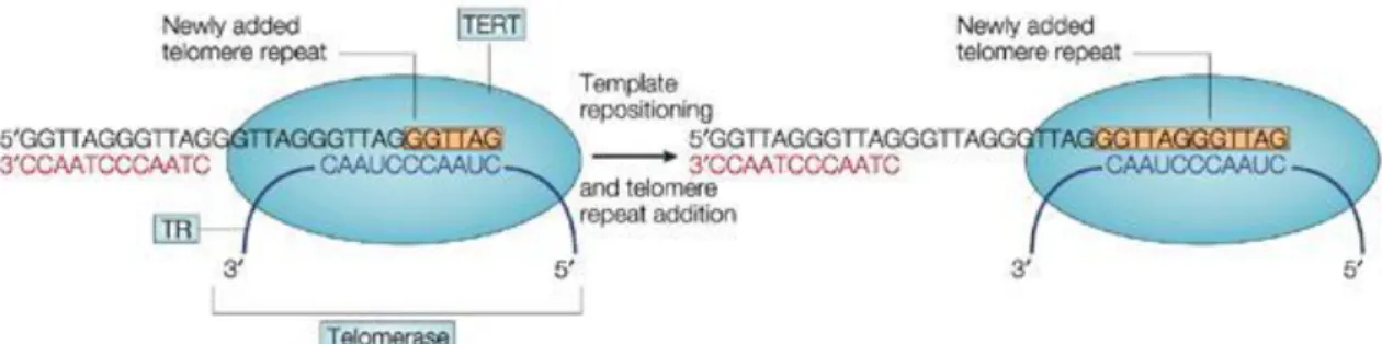
Telomeres were identified in Tetrahymena thermophila with their unique sequence of TTGGGG (Blackburn and Gall 1978) which were present at the ends of the chromosomes and protect them from degradation. Later on, the telomeric sequence of Tetrahymena was ligated to the end of a budding yeast plasmid and it was observed that the sequence was replicated and also protected the plasmid ends. When the duplicated sequences were separated from the Tetrahymena sequences it was discovered that they were the yeast telomeric fragments (Szostak and Blackburn 1982). Further studies revealed the presence of a reverse transcriptase enzyme named telomerase which was composed of RNA and a protein subunit (Greider and Blackburn 1985, Greider and Blackburn 1987, Greider and Blackburn 1989) (Figure 1.2.1).
Figure 1.2 1 Elongation of telomeres by the enzyme telomerase (Manthon and Lloyd, 2001)
It is now known that mammals have the conserved telomeric sequence of TTAGGG (Moyzis et al 1988) and shortening of telomeres are associated with replicative senescence and aging of somatic cells (Lundblad and Szostak 1989, Harley et al 1990, Allsopp et al 1992) and replicative senescence can be overcame due to genomic instability and reexpression of telomerase enzyme (Hayflick 1965, Counter et al 1992, Kim et al 1994). The telomeres can be upto 15 kb in humans and at the end of the telomeric double stranded repeats a G-rich sequence is present which forms the T-loop that packages the chromosome ends and protects from 5’- 3’exonucleases activity together with the shelterin complex (Sullivan and Karlseder 2010, Gilson and Ségal-Bendirdjian 2010, Verdun and Karlseder 2007, de Lange 2005).
Shelterin complex that protects the chromosome ends is composed of six subunits (de Lange 2005). These factors are TRF1, TRF2, POT1, TIN2, TPP1 and Rap1 (Chong et al 1995, Bilaud et al 1996, Broccoli et al 1997, Kim et al 1999, Li et al 2000, Baumann and Cech 2001, Houghtaling et al 2004). These proteins
function together to form the T-loop and prevent telomerase from binding after adequate elongation of the telomeres (de Lange 2005).
Like shelterin complex that protects the telomeres, telomerase enzyme that elongates them is also a complex. Besides from its well known protein subunit telomerase reverse transcriptase (TERT) and telomerase RNA (TERC), it contains an RNA binding protein dyskerin (Cohen et al 2007). The three subunits of telomerase enzyme have to be packaged in the Cajal bodies which are then carried to the telomeres by telomerase cajal body protein1 (TCBP1) (Cristofari et al 2007, Venteicher et al 2009). At telomeric end binding of the proteins reptin and pontin provides the correct structure to the telomerase complex which then initiates the elongation of the telomeres (Venteicher et al 2008).
The elongation of telomeres is important for the maintenance of the cell. Therefore, to gain the telomerase enzyme activity provides a survival advantage to the cell. Stem cells undergo division and proliferation whenever their residing tissue requires regeneration. Therefore the presence of telomerase holoenzyme is also important for the maintenance of the stem cells pools and tissue regeneration.
1.2.1 Telomeres, Telomerase and Cancer
Hayflick et al (1961) showed that the somatic cells were able to replicate only for a limited number of passaging in vitro, which is called replicative senescence (Hayflick 1965). It was also shown that replicative senescence was due to the loss of telomeres which provide the genomic stability to the chromosomes. Today, it is known that somatic cells do not express telomerase which is essential for the maintenance of the telomeres and serves as a tumor
suppressor to prevent the proliferation of the cells having defects in their genomes (Li et al 2005). However when telomerase was introduced to senescent somatic cells it was observed that they could overcome replicative senescence and start dividing unlimitedly (Bodnar et al 1998). Moreover, in vivo studies performed with mouse models demonstrated that when telomerase was overexpressed, the mice formed more tumors compared to the wild types. If the mice were resistant to tumor formation, prolonged life span and increased stem cell activity were reported (Tomas-Loba et al 2009, Sarin et al 2005, Gonzalez-Suarez et al 2005, Canela et al 2004).
In many cancer types, the expression of telomerase is regained to maintain the telomere length (Harley 2008). However some cancer types maintain their telomere lengths by using the pathway called alternative lengthening of telomeres (ALT) (Bryan et al 1997, Bryan et al 1995). ALT is observed mostly in the tumorigenesis of the mesenchymal cell types (Cesare and Reddel 2010). ALT pathway employs homolog recombination for the elongation of telomeres (Lubland and Blackburn 1993, Bryan et al 1995, Bryan et al 1997). Different from the telomerase extended telomeres, the chromosome end of the cells contains mostly C-rich strands (Henson et al 2009) and it is proposed that the choice towards ALT can be due to the reduction in shelterin complex concentrations (Cesare and Reddel 2010).
Recent finding revealed that the epigenetic modifications of the subtelomeric repeats, which are highly methylated in normal cells, towards hypomethylation increased tumorigenesis and caused telomere elongation (Vera et
al 2008). Moreover, telomeric repeat containing RNAs (TERRAs) were
discovered which are transcribed from the telomeric repeats that are normally highly methylated. TERRAs are shown to be telomerase inhibitors and have low
TERRA transcript concentrations were observed in cancer (Azzalin et al 2007, Schoeftner and Blasco 2008, Luke and Lingner 2009, Redon et al 2010).
1.2.2 Telomerase and Stem Cells
Presence of telomerase enzyme was demonstrated in many stem cell types including ESC, HSCs and MSCs (Hiyama and Hiyama 2007). Among them, ESCs has the highest level of telomerase activity (Brumendov and Balabanov 2006, Izadpanah et al 2006). Embryonic stem cells which have limitless potential to proliferate, conserved the activity of telomerase enzyme after extensive passaging, that is more than 120 (Xie et al 2010). Moreover their differentiation potentials were demonstrated to depend on telomerase activity, the concentration of which is diminished after differentiation (Armstrong et al 2000).
In HSCs, it has been shown that the telomerase activity was not sufficient to maintain telomere length which was further proved by the overexpression studies of TERT (Allsopp et al 2001, Allsopp et al 2003). The regenerative capacities of the cells were also found to be determined by their niches (Conboy et
al 2005). HSCs were shown to express more telomerase when they were cultured
with cytokines in vitro and the enzyme levels were also found to be decreased after differentiation and extensive proliferation resulting in aging (Chiu et al 1996) although it prevents sudden telomere length decrease during regeneration which requires rapid proliferation (Allsopp et al 2003).
There are conflicting data in the presence of telomerase enzyme in MSCs. According to a study human MSCs in vitro could undergo 50 population doublings and have telomerase activity (da Silva and Nardi 2003), in contrast
another one indicates that MSCs do not express TERT and have life spans as long as somatic cells (Fehrer and Lepperdinger). Moreover another study states that without the expression of TERT, MSCs could maintain their telomere lengths (Yanada et al 2006). There are suggestions that maintenance of the telomeres in MSCs could be by ALT pathway (Serakinci et al 2008). It was demonstrated in
vitro that when MSCs were isolated from telomerase knockout mice, they lost
their potential to differentiate and proliferate in contrast with the wild type mice (Liu et al 2004).
1.2.3 Telomerase Antagonist GRN163L (Imetelstat)
During transformation of somatic cells into cancer cells telomerase is activated which make the cancer cells immortal, for this reason telomerase is a good target in the treatment of cancer as normal somatic cells do not express telomerase. There are several methods proposed to cure cancer by targeting telomerase. One of the approaches depends on the inhibition of telomerase by the use of small molecules to promote their efficient binding on the active site of the telomerase or telomerase RNA. Another approach employs the disruption of the telomeres and prevent the binding of telomerase enzyme. Furthermore there are strategies developed to inhibit the expression of the telomerase genes or for gene therapy and immunotherapy (Harley 2008).
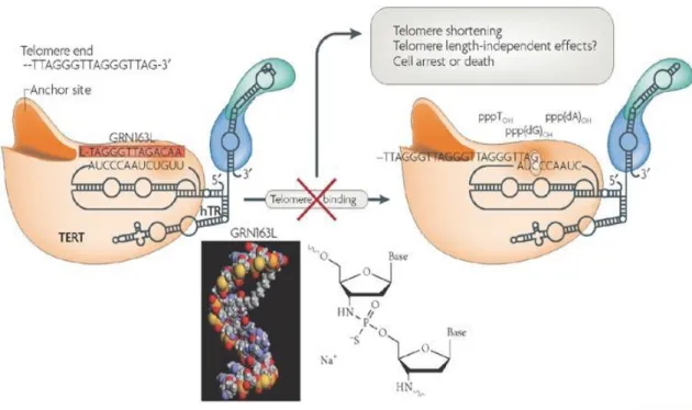
GRN163L, Imetelstat, is an oligonucleotide that inhibits the activity of telomerase. It is a 13-mer oligonucleotide having the sequence of TAGGGTTAGACAA containing a N3' P5'-thio-phosphoramidate lipid conjugate which is palmitoyl. The palmitoyl serves in the entry of the GRN163L into the cell while thio-phosphoramidate provides the backbone a better
interaction with proteins and pH stability. When these properties are combined GRN163L can easily enter the cell without any requirements to liposomes and specifically binds to telomerase RNA forming a double strand that is durable to RNAse H degradation (Pongracz and Gryaznov 1999, Gryaznov et al 2001, Herbert et al 2005) (Figure 1.2.3.1).
Figure 1.2.3 1 Action mechanism of GRN163L on telomerase (Harley 2008)
To date in vivo and in vitro properties of GRN163L is widely tested in animal models and several cancer cell lines. Dikmen et al (2005) demonstrated the effects of GRN163L on A549 luciferase (A549-Luc) cells in vitro, resulting in shortened telomeres, loose and less colonies. Moreover they successfully showed that immunodeficient mice have less metastasis when the A549-Luc cells were previously treated with GRN163L and intraperitoneal administration of GRN163L prevented the tumorigenesis of these cells in vivo. Djojosubroto et al (2005)
showed the effect of GRN163L on liver cancer cell lines Hep3B and Huh7. These cells became more sensitive to chemotherapeutics when previously treated with GRN163L and formation of tumors was prevented with intraperitoneal injections in nude mice. Similar results were also obtained when breast cancer cells were treated with GRN163L both in vitro and in vivo (Gellert et al 2006, Hoechreiter et
al 2006). Moreover a telomerase independent effect of GRN163L was revealed by in vivo and in vitro studies performed with A549-Luc cells. The morphology of
the cells were becoming round and they were detached from the plate surface loosing their adherent properties (Jackson et al 2007). There are also studies reporting the successful inhibition of tumorigenesis in glioblastomas, prostate cancer, breast cancer, bladder cancer and myelomas (Marian et al 2010, Calin et
al 2009, Goldblatt et al 2009, Goldblatt et al 2009, Dikmen et al 2008, Shammas et al 2008, Gomez-Millan et al 2007, Gellert et al 2006, Hochreiter et al 2006).
GRN163L has ben in clinical trials since 2005 and today there are total of 7 clinical trials (http://clinicaltrials.gov/ct2/results?term=GRN163L). The aim of these trials is to reveal the effects of GRN163L in the treatment of cancer alone or as a combinatory treatment in lung cancer, myelomas and breast cancer. Although
in vivo and in vitro effects of GRN163L is well described in cancer, its effects on
the quiescent residers of the body, adult stem cells are not known as they also maintain telomerase.
CHAPTER 2
AIM OF THE STUDY
Mesenchymal stem cells are bone marrow derived stem cells that are able to differentiate into adipocytes, osteocytes, chondrocytes, cardiomyocytes and they are easy to isolate and manipulate. They can home to the injured tissue and have immunomodulatory effects. For these reasons they are important candidates in cellular therapies. MSCs can also be frozen to preserve them, and when they are thawed they can function apparently normally, thus allowing for future "off-the-shelf" therapy approaches. However, optimal timing of stem-cell delivery to acutely injured tissues or organs can be a major handicap. This issue is of paramount importance in cardiovascular medicine (Bartunek et al 2006). The assessment of donor MSC functional activity at the cellular level, which is the focus of several research groups, is also critically important. There is no available data from experimental studies or clinical trials that shows the exact timing of the induction of cardiomyocyte differentiation prior to MSC delivery to an injured heart or ischemic limb. In the first part of the study, we aimed to demonstrate the best timing to induce the differentiation of cardiomyocytes from rat bone marrow derived MSCs.
The presence of the telomerase activity in many different kinds of human tumors, but not in normal somatic cells has been known for years. Thus, inhibition
of telomerase activity is now an attractive tool on targeted cancer therapy. Telomerase inhibitor, GRN163L has been shown to have established effects on the treatments of the cancers. However this inhibition may lead to side effects since germline cells, proliferating stem and progenitor cells also exhibit telomerase activity. The effects of telomerase-targeting therapies must be well defined on MSCs. Therefore, we aimed to investigate in vitro effects of GRN163L on the self-renewal and differentiation processes of MSCs at the second part of this thesis.
CHAPTER 3
MATERIALS AND METHODS
3.1 Animals
For all the experiments, adult female 9-week-old, 280–300 g Spraque Dawley rats were used. The animals were kept in the animal holding facility of the Department of Molecular Biology and Genetics at Bilkent University under controlled conditions at 22o C with 12 hour light and 12 hour dark cycles. They were provided with unlimited access of food and water. The experimental procedures have been approved by Bilkent University Local Ethical Committee (BILHADYEK).
3.2 Isolation of the Cells from Rat Bone Marrow
After the rats were sacrificed by cervical dislocation, heterogeneous cell population was collected from the femurs and tibias by flushing with a 5 mL syringe containing 10% FBS (HyClone, Logan, USA) and 1% penicillin/streptomycin solution (Hyclone) in DMEM (HyClone). The mixed suspension was then centrifuged at 3000 rpm for 3 min. After the supernatant was removed, the cells were washed with 10 mL 1X PBS buffer once. The cells suspended in 1X PBS were centrifuged at 2500 rpm for 3 min and then the buffer was removed and cells were re-suspended in 10 mL 1X PBS buffer again. The
mixture was centrifuged at 2000 rpm for 3 min and then the supernatant was removed.10mL 1X PBS buffer was added to the cell pellet and the cells were re-suspended once more. The cell suspension was centrifuged at 1500 rpm for 3 min to get rid of the remaining impurities like fat, connective tissue and hair and then after the removal of supernatant, cell pellet was re-suspended in MesenCult medium (StemCell Technologies, Vancouver, Canada) with a 20% supplement (StemCell Technologies) and a 1% penicillin–streptomycin solution (HyClone) to prepare for tissue culturing.
3.3 Culturing of Mesenchymal Stem Cells (MSCs)
Cells were counted with hemocytometer and the cell number was calculated according to the below formula
Number of cells in the mixture = number of cells counted x 104 x dilution factor After the cells were seeded to the plastic culture plates at equal numbers in MesenCult medium (StemCell Technologies, Vancouver, Canada) with a 20% supplement (StemCell Technologies) and a 1% penicillin–streptomycin solution (HyClone), they were cultured in a 5% CO2 incubator at 37°C. The next day, the
media of the tissue culture plates were changed and the nonadherent cells were removed. The media of the cells were changed every 3 days, after washing with sterile 1X PBS. The adherent cells on the 14th day of the cell culture were mostly MSCs.
3.4 Treatment of the MSCs with Telomerase Template Antagonist
GRN163L (GRN163L) and the Experimental Groups
After 14 days of culture, the cells were trypsinized (HyClone) and transferred to culture dishes. One day after the transfer, GRN163L (Geron Co., CA, USA) and its mismatch control oligonucleotide were added at a concentration of 1 μM to the cell plates. The media were changed every 3 days with the fresh GRN163L
(hereafter stated as 163L group) and mismatch control oligonucleotide (hereafter stated as mismatch group) together with control MSCs with no treatment
(hereafter stated as control group). 163L and mismatch cultures continued for 1 week. Finally, GRN163L was removed and MSCs were left for recovery for 1 week (hereafter stated as 163LR (recovery) group).
3.5 Colony Forming Unit (CFU) Assay
MSCs were washed with 1X PBS and were air dried. They were fixed by ice cold methanol (MeOH) for 5 minutes. After fixation MeOH was removed and the cells were washed with 1X PBS buffer. The fixed cells were then treated with giemsa staining reagent (Carlo Erba) for 5 min. To stop the reaction of giemsa staining, tap water was added and the cells were washed with tap water trice. The colonies stained in purple were counted under bright field light microscope.
3.6 Total RNA Isolation from Rat MSC
Total RNA from the MSCs were isolated at the 14th day of the cell culture. The cells were first washed with 1X PBS buffer twice and were trypsinized at 37oC for 3 min. Trypsinization was ended by the addition of DMEM (Hyclone)
containing 10% Fetal Bovine Serum (Hyclone) and 1% penicillin-streptomycin solution (Hyclone). Then the cell suspension was centrifuged at 1500 rpm for 5 min. After centrifugation the media was removed and the cell pellet was washed with 1X PBS buffer and centrifuged again at 1500 rpm for 5 min. Finally, the supernatant was removed and the total mRNA was isolated from the cell pellet by using RNeasy Mini Kit (Qiagen) according to the manufacturer’s protocol.
3.7 cDNA Synthesis
The cDNAs were synthesized from the total RNA samples by using DyNAmo cDNA Synthesis Kit (Finnzymes, Finland) according to the manufacturer's protocol. 2 µg RNA was mixed with DEPC treated ddH2O to a
total volume of 10,5 µl and 1,5 µl of oligo(dT) primer was added and the mixture was centrifuged for 3 sec. The samples were incubated at 65°C for 5 minutes and then chilled on ice for 3 minutes. Then 15 µl of 2X RT buffer including dNTP mix and 10 mM MgCl2 and 3 µl of M-MuLV RT RNase H+ enzyme was added to
the mixture, after a 3 sec centrifugation they were incubated at 25ºC for 10 min, 45ºC for 45 min, and 85ºC for 5 minutes, respectively.
3.8 Primer Design
Primer designs were performed by using the combination of Primer exe, Primer Blast (NCBI) and Primer Premier. The specificity of the primers and product sizes they would amplify were checked via Ensemble and NCBI databases publicly available through the internet. Primers for all the genes amplified were listed in Table 3.9.