Hepato1og.v 2000; 33: 254-265 Printed in Denmark Ail rights reserved
Mwrksgaard Copenhagen Journal of Hepatology
ISSN 0168-8278
‘I
~53 but not ~16~~~ induces growth arrest in retinoblastomadeficient
hepatocelhilar carcinoma cells
Anne Pierre Morel”, Kezban Unsal**, Tolga Cagatay2, Frederique Ponchel’, Brian Carr” and Mehmet Ozturk’,2
NSERM U453, Centre Leon Berm-d, Lyon, France, ‘Department of Molecular Biology and Genetics, Bilkent University, Ankara, Turkey, and 3Pittsburgh Transplantution Institute, University of Pittsburgh, Pittsburgh, PA, USA
Background/Aim: Both ~16’~~” and p53 proteins are negative regulators of the cell cycle. In human hepato- cellular carcinomas @ICC), the loss of function of ~53, retinoblastoma @Rb) and ~16’~~~~ genes by dif- ferent mechanisms has been largely documented, but their hepatocellular effects are poorly known. We compared the growth-inhibitory effects of p16rNK4” and p53 proteins in Hep3B cell line-derived clones. Met/z&: Cells were transfected with inducible ~16’~~~” and p53 expression vectors, and stable clones were analyzed for transgene expression by Western blotting and immunoperoxidase staining. Ef- fects on cell growth were analyzed by in vitro growth assay, thymidine incorporation and flow cytometry. Biochemical effects of p53 were tested by Northern blotting of p21 ‘iPI transcripts and by Western blotting of p2P1, mdm-2, bax, cyclin-dependent kinase 2 and cyclin E proteins. The pRb protein was studied by Western blotting and immunoprecipitation assays.
H
EPATOCELLULAR carcinoma (HCC), which is etio- logically associated to hepatitis B virus (HBV), hepatitis C virus (HCV) and aflatoxins, is one of the most frequent cancers world-wide (1). Genetic studies have revealed so far that a dozen genes are altered in these cancers. These genetic alterations (mostly so- matic) are related to at least four different pathways, including DNA damage response (~53 gene), cell cycle regulation (retinoblastoma, p 1 61NK4a and cyclin D genes), TGF-P (M6P/IGF2R, SMAD2 and SMAD4* A. P M. and K. U. contributed equally to this work.
Received 4 October: revised 17 December; accepted 24 December 1999
Correspondence: Mehmet Ozturk, Department of Mol- ecular Biology and Genetics, Bilkent University, 06533 Bilkent Ankara, Turkey. Tel: 90 3 12 266 50 81.
Fax: 90 312 266 50 97. e-mail: [email protected]
Results: The induction of ~16’~~’ protein expression did not affect in vitro growth of cells. In contrast, p53 protein in its wild-type conformation provoked a growth arrest accompanied by transactivation of p21Cipr gene and accumulation of p21ciP’, bax and mdm-2 proteins. p53-induced growth arrest was due to a cell cycle arrest at the GUS transition, probably mediated by p21ciP’ protein, which inhibits cyclin-de- pendent kinase 2/cyclin E complexes.
Conclusions: The lack of detectable pRb protein and resistance of cells to p16rNK4” strongly suggest that p53 is able to arrest the growth of HCC cells by a mechanism independent of “p53-retinoblastoma path- way”. These findings are applicable to HCC with ab- berrations of both p53 and pRb genes, and may not represent the universal effects of p53 in hepatic cells. Key words: Cell cycle arrest; Cyclin E; Hepatoma; p16rNK4”; p21ciP’; ~53; Retinoblastoma.
genes) and wnt (B-catenin and APC genes) signaling pathways (2).
p53 was the first tumor suppressor gene found to be mutated in HCC (3). Many reports now indicate that the p53 gene, which is located at chromosome 17p, is somatically mutated in about 30% of HCCs worldwide (2). Both the frequency and the type of p.53 mutations are different depending on the geographical location and suspected etiology of these tumors. An HCC-spe- cific codon 249 mutation (AGG+AGT leading to p53- 249Ser), suspected to be induced by aflatoxins, was found in most HCCs from geographical areas with a high incidence of HCC and a high risk of exposure to aflatoxins (4,5). The frequency of all p53 mutations in HCC varies between 15% in Europe and 42% in China (2). On the other hand, physical and functional interac- tion between p53 protein and HBx viral antigen has
p53, pkW=a and growth arrest in hepatoma
been reported. Based on this experimental evidence, viral inactivation of wild-type ~53 in HCC has been proposed (68).
The ~53 protein is a transcription factor that en- hances the rate of transcription of different genes that carry out, at least in part, the p53-dependent functions in a cell. Most of the ~53 mutations in HCC, as in other cancers, occur in the central DNA binding do- main, leading to a loss of its transcriptional activity. The ~53 protein functions to integrate cellular re- sponses to stress, such as exposure to DNA damaging agents and hypoxia (9). Under normal conditions, ~53 is rapidly degraded in cells by a mechanism that in- volves p 14ARF and mdm-2 proteins. Upon cellular stress, ~53 is modified post-transcriptionally, dis- sociates from mdm-2 protein, becomes more stable, mi- grates into the nucleus and transactivates its target genes (9,lO). One of the outcomes of ~53 activation is cell cycle arrest at Gl phase mediated by p21ciP1 pro- tein, which is an inhibitor of different cyclin-dependent kinases, including those involved in the phosphoryla- tion of the retinoblastoma protein (pRb). The so-called “p53-retinoblastoma pathway” implicates an import- ant role for pRb and its two related gene products, ~107 and ~130, in p53-mediated Gl-S phase regulation (9). ~53 also plays a role in triggering apoptosis under several different physiological conditions. The apoptot- ic role of ~53 appears to be limited to certain cell types such as hematopoietic cells. There is also experimental evidence that p53-dependent apoptosis can occur as a response to the expression of a viral or cellular onco- gene or the absence of a critical tumor suppressor gene
product such as pRb (9). Studies on the response of HCC cells to ~53 are limited in number, and obser- vations from different laboratories are sometimes con- tradictory. For example, we reported that ~53 acts as a growth suppressor in Hep3B cells (11) and p53-me- diated apoptosis was reported in the same cell line in several reports (12-14). However, these observations were challenged by Friedman et al. (15), who reported that Hep3B cells were resistant to p53-mediated growth arrest and apoptosis.
The p 1 61NK4a gene, which is located at chromosome 9p, codes for two alternatively spliced transcripts (see ref. 16 for a review). One of the transcripts is for P161NK4” protein, an inhibitor of cyclin-dependent ki- nase 4 (CDK4) and CDK6, whereas the other is pl4ARF protein which regulates ~53 stability (10,17). The ~16’~~~~ status in HCC has been studied exten- sively. Both germ-line and somatic mutations of the p16rNK4” gene were found in HCC patients. It was also reported that about 50% of HCC display de mm
methylation of the p161NK4” gene, as observed in many
other cancers (reviewed in ref. 2). As an inhibitor of CDK4 whose main substrate appears to be pRb pro- tein, pl 6rNK4a protein is an antagonist of cyclin D for CDK4 activation (17). Therefore, it is not surprising that retinoblastoma gene mutations and cyclin D gene amplifications also occur in HCC, albeit at lower fre- quencies (2). Thus, the main consequence of p161NK4” gene alterations is considered to be a loss of CDK4 inhibition. As a result, the phosphorylation of pRb by CDK4 is no longer regulated, leading to an increase in “free E2F” transcription factors involved in the pro- gression of Gl phase of cell cycle and Gl/S transition. The expression of p161NK4” progressively increases as cells undergo senescence (17). This suggests that the loss of ~16’~~~~ function in HCC is related to cellular immortalisation. However, the effects of ~16’~~~~ pro- tein in hepatoma cells have not been reported yet. It is also unknown whether and how ~16’~~~~ gene alter- ations affect the cellular functions of ~14~~~ protein (the product of alternatively spliced transcripts of the same gene), whose main role appears to be the acti- vation of ~53 protein.
In this report, we compare the effects of ~53 and p1(jINK4a proteins of Hep3B and its derivative cell lines. Hep3B is a differentiated HCC cell line with inte- grated HBV DNA and deleted ~53 gene (3). Its deriva- tive Hep3B-TR is a TGF+resistant clone due to a homozygous deletions of TGF-P receptor type II (18). The status of the retinoblastoma gene in the Hep3B cell line is a matter of debate (15,19). Our main goal was to know whether p16rNK4” and ~53 proteins, known to be upstream regulators of “retinoblastoma growth control pathway”, differ in their phenotypic ef- fects in these cells. We demonstrate that ~53, but not ~16’~~~” is able to suppress the growth of these HCC cells by cell cycle arrest at Gl/S transition. We provide evidence that the resistance of cells to ~16’~~~~ protein is correlated with the lack of pRb protein. The same observation also demonstrates that p53-induced growth arrest in Hep3B-TR cells occurs independently from the retinoblastoma growth control pathway.
Materials and Methods Plasmid constructs
The plasmids pLTRp53cGvall35 and pSV2neo (ref. 20) were a gift from M. Oren (Rehovot, Israel). The plasmid pAUCT/CCW was con- structed by A. Fattaey (Charlestown, MA, USA). This plasmid con- tains a Tet Repressor-vp16 fusion cDNA under the control of CMV promoter. It also contains a multiple cloning site downstream to a promoter sequence composed of a Tet operator and CMV TATA box. The pAUCT/CCW also contains a Neoe gene allowing the selection of stable clones. A pBluescript plasmid containing human ~16’~~~~ cDNA (a gift from A. Samarut, Lyon, France) was used for cloning of p16rNK4” cDNA into pAUCT/CCW plasmid. The p16 cDNA in- sert was removed from the pBluescript plasmid by EcoRI-XhoI re- striction enzyme digestion. Purified p16rNK4” cDNA fragment was
A. P. Morel et al.
then ligated with pAUCT/CCW plasmid, which was previously linear- ized by digestion EcoRI and XhoI restriction enzymes to obtain pAUCTp 16 tNK4a plasmid. In the presence of tetracycline, the Tet re- pressor-Vpl6 fusion peptide suppresses the expression of ~16’~~~~ protein. In the absence of tetracycline, the suppressive function is abolished.
Cell lines and antibodies
Hep3B, Hep3B-TR, Mahlavu, Huh-7 and Saos-2 cell lines were grown in MEM medium containing 10% fetal calf serum, 2 mM L- glutamine, 200 units/ml penicillin, and 200 pg/ml streptomycin under 5% COz, unless otherwise described. Antibodies to pRb (clone IF8 from Santa-Cruz, clones Ab-5 and Ab-6 from Calbiochem), ~53 (clone Ab-1 from Calbiochem and HR 231; a gift from T. Soussi. Paris, France), ~21~‘~’ (Ab-1 to wafl from Calbiochem), mdm-2 (2AlO; a gift from B. Vasylyk, Strasbourg, France), bax (N-20 from Santa Cruz), p16’“K4a (Ab-1 from Calbiochem) and cyclin E (Ab-1 from Calbiochem) were used.
Generation of stable cell clones expressing p161NK4” andp53 proteins
Appropriate plasmids (lo-20 pg of total DNA) were transfected into cells by the calcium phosphate precipitation method. The plasmids pLTRp53cGvall35 and pSV2neo were used at a ratio of 1:20. The plasmid pAUCTp16tYK4” was used alone. The p53-transfected cells were selected at 39°C in the presence of G 418 (400 iLg/ml). The p16iNK4” clones were selected in the presence of tetracycline (5 &ml) and G 418 (400 &ml).
Cell growth assays
For p161NK4”, 3000 to 5000 cells were plated into 6-well plates. Start- ing with a low number of cells allowed to continue the experiment for at least 10 days without a cell death due to an overgrowth of these cells which are not contact inhibited at full confluence. Twenty-four hours after plating (time zero), the medium was changed and the cells were incubated in the absence or in the presence of tetracycline (5 pg/ ml) for up to 10 days. The number of viable cells was determined by manual counting, using a hemocytometer, at time zero and at days 2, 4, 6, 8, and 10. As an alternative assay, the same cell lines were grown as described for 7 days, and cells were visualized in situ by methyl green staining after fixation in ethanol. For ~53 experiments, cells were seeded in 60-ml Petri dishes (at three different cell densities rang- ing from 50 000 to 500 000 cells per dish) and grown overnight at 39°C. The following day (time zero), half of the plates were moved to a 32°C incubator and further incubated for 7 days. Colony forming cells were visualized as described by crystal violet staining.
In situ thymidine incorporation assay
Cells were plated and grown at the permissive temperature (39°C) for 24 h. The following day, half of the plates were kept at the same temperature and the cells in the other half were shifted to the non- permissive temperate (32°C). Cells were labeled with [“HI-thymidine for 24 h before the arrest of the experiment at 24 h or 48 h, and studied in situ for thymidine incorporation. 13H]-thymidine (2.5 &i/ ml; Amersham) was used to label 50 000 cells grown on cover slips overnight in 6-well plates. After 24 h labeling, cells were fixed in meth- anol: acetic acid (3: 1), dried at room temperature and cover slips were transferred onto glass slides and exposed to a autoradiography emul- sion (NTB2; Kodak) for several days and stained with Giemsa tech- nique. Slides were then analyzed under a light microscope and used to obtain pictures.
Flow cytometry
Cytometric analyses following BrdU labeling was performed as de- scribed previously (21). Briefly, cells were labeled with BrdU (30 FM) for 1 h or 2 h, rinsed with PBS, detached from plates by trypsin incubation, rinsed with PBS and fixed in 70% (v/v) ethanol. Cells were rehydrated in PBS, incubated in 2 ml of 2N HCl, permeabilized in PBS containing 0.5% Tween-20 and 0.5% BSA and incubated with FITC-conjugated anti-BrdU antibody (from Becton-Dickinson). Then RNAse A (50 ,ug/ml) was added, followed by propidium iodide
(25 fig/ml; from Sigma). Cells were analyzed using a flow cytometer (Facscalibur, Becton-Dickinson).
Northern blotting
Total RNAs were extracted by the guanidium isothiocyanate method. RNA samples (10 pg) were separated by electrophoresis through de- naturing formaldehyde agarose gel and transferred to nylon mem- brane (Hybond-N; Amersham). Membranes were hybridized with [‘2P]-labeled p21c’p’ cDNA. as described (21) and subjected to auto- radiography.
Western blotting
Cell protein extracts were prepared in a lysis buffer containing 50 mM Tris-HCl, 0.25 M NaCl, 1 mM CaCl,. 0.1% Triton X-100, 50 mM NaF and a cocktail of protease inhibitors, as described previously (22). Equal amounts (usually 100 /lg) of proteins were then separated by SDS-polyacrylamide gel electrophoresis in a Tris-glycine buffer and transferred to PVDF membranes (Millipore) using a semi-dry transfer apparatus (Biorad). following the manufacturer’s recommen- dations. Membranes were checked for equal amounts of protein transfer by staining with Ponceau-S, and blocked in TBS-T (O.OSY” Tween-20 in Tris-buffered saline) containing 5% non-fat dry milk for 2 h, then incubated with the appropriate antibody overnight at 4°C. The blots were then rinsed in TBS-T and incubated with a peroxidase- conjugated secondary antibody for 1 h at room temperature. After washing in TBS-T, detection was performed using the chemilumi- nescence method (ECL, Amersham), followed by exposure to X-ray film. For most experiments. the same PVDF membranes were used to test different proteins from the same SDS-PAGE gels.The HR231 monoclonal antibody which recognizes both human and mouse pS3 was used for ~53 detection (23).
Immunoprrcipitation
Subconfluent cells were grown for 2 h in methionine-free medium containing 5% dialyzed fetal calf serum. Cells were labeled with 0.2 mCi/ml [‘%I methionine (Amersham) for 4 h in IO-cm culture dishes. Cells were then washed in PBS and proteins were extracted in 1 ml lysis buffer, prepared as described for Western blotting. Samples were precleared with 80 ~11 of 50’s (v/v) protein-A-Sepharose suspension (Pharmacia) for 1 h at 4°C and 5 ~11 of anti-pRb mouse antibody was added to the supernatant. After overnight incubation on a rocker at 4°C and a 5-min centrifugation at 13 000 rpm, supernatants were added to Eppendorf tubes containing 80 /tl of 50% (v/v) protein-A- Sepharose beads. Antigen-antibody complexes were captured by a 60 min of incubation at room temperature and the beads were pelleted by centrifugation. The beads were washed 4 times in lysis buffer and the immunoprecipitates were directly resuspended in Laemmli buffer, heated at 80°C for 5 min and loaded on SDS-polyacrylamide gels (7.5%). After electrophoresis, gel was processed with Amplify solution (Amersham), dried and subjected to autoradiography.
Results
Hep3B hepatoma cells are resistant to p16’h’K4” overexpression
Randomly selected clones from pAUCTp1 61NK4a transfections into Hep3B cells were first analyzed for tetracycline-regulated expression of p 1 61NK4a protein by Western blotting. Saos-2 cell line extracts, which ex- press p161NK4”,, but not pRb protein, were used as a positive control (24,25). As shown in Fig. 1 A, five clones (clones 9, 20, 23, 33 and 34) displayed increased expression of pl6 ‘NK4a in the absence of tetracycline, as compared to the presence of 5 ,uglml tetracycline. Three clones (clones 9, 20 and 34) did not express de-
Clones
Tetracycline
~53, pl 6rNK4a and growth arrest in hepatoma
PI6
INK4aClone 9
Clone 23
Clone 33
0 2 4 6 8 10 0 2 4 6 8 10
Incubation time (days)
Fig. 1. Resistance of Hep3B-derived clones to PI&mediated growth inhibition. (A) Tetracycline-dependent expression of p16 in selected Hep3B clones. Cells were grown for 24 h in the absence (-) or in the presence (+) of 5 pglml tetracycline and cell lysates were tested for ~16’~~~~ protein by Western immunoblotting. Saos-2 cells were used as positive control. Equal protein loading was tested by Ponceau S staining. (B) The clones 9 (left), 23 ( center) and 33 (right) were grown in the presence (filled circles) or the absence (open circles) of tetracycline for 10 days in &well culture dishes and the cell number
was measured by manual counting at 2-day intervals.
tectable p 1 61NK4a protein in the presence of tetracy- cline, while clones 23 and 33 displayed weak immuno- reactivity. In all clones tested, the levels of ~16’~~~~ were induced in the absence of tetracycline. The induc- tion was strong in clones 20, 23 and 33, moderate in clone 9, but weak in clone 34. Clone 20 expressed p16rNK4” as a doublet.
Next, the effect of ~16’~~~~ overexpression on cell growth was tested using clones 9, 20, 23 and 33. For initial experiments, all four clones were grown in paral- lel in the presence or in the absence of tetracycline for 7 days and total number of cells were estimated by in situ staining. There was no apparent difference between the number of cells (data not shown), suggesting that p16rNK4” overexpression did not affect their growth rate. The lack of growth inhibition by pl 61NK4a overex- pression was confirmed by comparing growth rates of clones 9, 23 and 33 for 10 days and manual counting
of cell numbers every 2 days. As shown in Fig. lB, the growth rate of these clones was not affected by overex- pression of p16 1NK4a in the absence of tetracycline. These observations indicated that Hep3B cells are re- sistant to growth inhibitory effects of pl 61NK4a protein.
Hep3B-TR cells are sensitive to p53-induced growth inhibition
To study the effects of wild-type ~53, we used Hep3B- TR clone rather than the parental Hep3B cells, as Hep3B-TR clone is resistant to TGF-j? (ref. 18). Apoptosis studies with Hep3B cells are complicated by the fact that these cells express TGF-jI and may under- go apoptosis in the absence of wild-type p53 (22). To study ~53 effects, we used as well-known and widely- used experimental model based on a mouse tempera- ture-sensitive mutant p53-135val protein (20). Hep3B-
A. P. Morel et al.
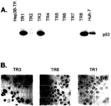
TR cells were transfected with the appropriate plasmids and eight G418-resistant clones were tested for p53 expression by Western blotting. As shown in Fig 2A, only three clones (TRl, TR3 and TR8) were positive initially. Our previous experiments indicated that certain cell lines tend to lose the expression of transfected plasmids during long-term culture (data now shown). Thus, it was important to test that the expression of p53 was stable and maintained during the in vitro growth of selected clones. TRl, TR3 and TR8 clones were grown for several weeks in the pres- ence of G-418 and the expression of ~53 was checked at different time points by immunoperoxidase staining with the anti-p53 monoclonal antibody HR 231 (ref. 23). As shown in Fig. 2B, the immunoperoxidase stain- ing studies indicated that the expression of p53 was maintained at nearly 100% in TR3 cells, only 30% of TR8 cells maintained its expression, whereas TRl cells lost p53 expression completely. Therefore, we per- formed the additional studies with TR3 and when
B.
TR3 TR8 TRI
P53
Fig. 2. Selection ofHep3B-TR clones stably expressing the mouse temperature-sensitive p.53-135val protein (A) Neo- mycin-resistant clones were selected at the permissive tem- perature (39°C) and testedfor ~53 protein by Western blot- ting. Parental HepSB-TR and Huh-7 cells were used as negative and positive controls, respectively. (B) p53-posi- tive TRI, TR3 and TR8 clones were expanded in vitro and cultured at the permissive temperature for several weeks, and thep53 expression was tested by indirect immunoperox- idase staining. The expression of ~53 was fully retained in TR3 (left), but lost partially in TR8 (center). or totally in TRI clone (right).
needed with TR8 clone, in comparison to TR4 used as negative control (see Fig. 2A). To avoid any experimen- tal error, TR3 and TR8 cells were checked regularly for the expression of ~53 by immunoperoxidase staining throughout all the experiments described here.
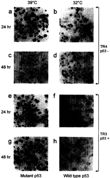
First, we compared the growth of TR3 and TR4 clones by two different techniques after incubation of cells at either 32°C (non-permissive temperature, wild- type ~53 conformation) or 39°C (permissive tempera- ture, mutant p53 conformation). Initially, the effects of wild-type p53 on DNA synthesis were tested by thymi- dine incorporation studies. Radioactive [‘HI-thymidine was added to standard culture medium at times 0 and 24 h, following temperature-shift, and cells were grown for an additional 24 h in the presence of [3H]-thymi- dine. Almost 100% of p53-negative TR4 cells were positive for thymidine incorporation at 24 h (Fig. 3a and b) as well as at 48 h (Fig. 3c and d), independent of temperature shift. TR3 cells grown at 39” also incor- porated thymidine at a rate of about 100% at both 24 h (Fig. 3e) and 48 h (Fig. 3g). In contrast to these observations, the temperature shift to 32°C caused an almost total block of thymidine incorporation into DNA of TR3 cells, as early as 24 h (Fig. 3f). Only a few TR3 cells were positive at 48 h (Fig. 3h). These observations demonstrated that the activation of wild- type p53 in Hep3B-TR cells provoked a total loss of DNA synthesis.
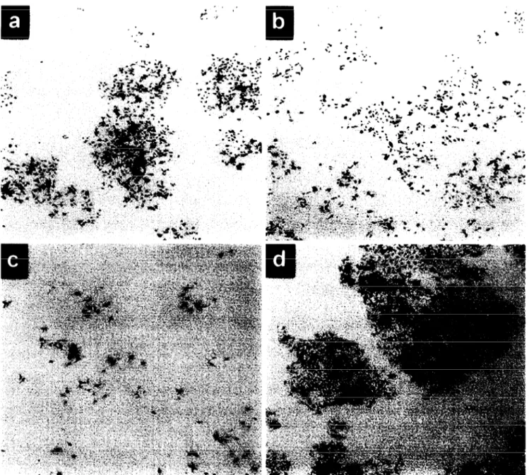
To test the long-term effects of wild-type ~53 ex- pression, Hep3B-TR clones were grown at 32°C and 39°C for 7 days at different cell densities. When forced to grow at 32°C the parental Hep3B-TR cells (Fig. 4a) as well as p53-negative TR4 clone (Fig. 4b) formed large colonies. In contrast, the p53-positive TR3 clone did not form colonies at 32”C, although single and scattered cells were still detectable after 7 days of cul- ture (Fig. 4~). The growth inhibition of TR3 cells was due to the expression of wild-type p53 at 32°C in these cells which grew well and formed colonies at the per- missive 39°C (Fig. 4d).
Taken together, these studies showed that p53 acti- vation in TR3 cells induced a total loss of DNA syn- thesis and a permanent growth arrest. These cells did not display any visible evidence of p53-dependent apoptotic cell death during these experiments. Ad- ditional apoptosis experiments using morphological analysis, in situ DNA staining by H33258 and DNA ladder tests indicated that the apoptotic cell death was minimal (<5%) in all Hep3B-TR clones, independent of p53 status and growth temperature (data not shown). Next, we further explored the effects of wild- type p53 in Hep3B-TR clones through study of the ex- pression of several known p53 target genes.
39°C 32°C a 24 hr
h
48 hr ---.-, : 4 rR4 xX3- TR3 353 +Mutant p53 Wild type ~53
Fig. 3. ~53 activation in TR3 cells at non-permissive tem- perature leads to an arrest of DNA synthesis. The p53- negative TR4 (a, b, c, d) and p53-positive TR3 (e, J g, h) cells were plated and grown at the permissive temperature (39°C) for 24 h. The following day, half of the plates (a, c, e, g) were kept at the same temperature and the cells in the other half (b, d, J h) were shifted to the non-permissive temperature (32°C). Cells were labeled with r3H]-thymi- dine for 24 h before the arrest of the experiment at 24 h (a, b, e, f) or 48 h (c, d, g, h), and studied in situ for thymidine incorporation. Note the loss of thymidine incorporation in TR3 cells at the non-permissive temperature (J h), in con- trast to TR4 cells (b, d).
p.53-dependent induction of mdm-2, p21 c@l and bax genes in Hep3B-TR clones
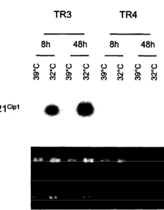
Among known p53 target genes, the expression of mdm-2, p21ciP1 and bax were studied. Northern blot analysis of p21ciP’ transcripts showed a strong induc- tion of p21ciP’ gene expression at 32°C in TR3 cells, but not in TR4 cells (Fig. 5). The p21ciP1 transcripts were nearly detectable in both clones when grown at
p53, plP= and growth arrest in hepatoma
39°C. Next, protein levels of mdm-2, p21ciP1 and bax were tested under similar conditions. The mdm-2 pro- tein was easily detectable in both parental Hep3B-TR and TR3 cells grown at 39°C (Fig. 6A). At 32”C, mdm- 2 levels did not change significantly in Hep3B-TR and TR4 cells, although a slight increase at 48 h was seen with Hep3B-TR (Fig. 6A, data not shown for TR4). In contrast, there was a clear-cut increase of mdm-2 protein levels in TR3 cells when grown at the same non-permissive temperature at both 48 h and 72 h (Fig. 6A). A p53-dependent increase of mdm-2 levels was detectable as early as 24 h in these cells (data not shown).
Accumulation of protein products of two additional p53 target genes, namely p21ciP1 and bax, was investi- gated following ~53 activation in TR3 cells (Fig. 6B). At 39”C, p21ciP1 protein was undetectable in both TR3 and TR4 cells. At 32”C, the expression of p21ciP1 was strongly induced in TR3, but not in TR4 (Fig. 6B). In contrast to p21ciP1, bax protein was present in both TR3 and TR4 clones grown at 39°C. At the non-per- missive temperature (32”C), a weak increase in bax levels was observed in TR3, but not in TR4 cells (Fig. 6B).
Taken together, these studies demonstrated that the basal expression of p21ciP1 gene in Hep3B-TR-derived clones was weak (low levels of transcripts, but no de- tectable p2 1 ciP1 protein). The activation of wild-type ~53 provoked an immediate and strong accumulation of both p21ciP’ mRNA and protein. In contrast, basal levels of bax protein in these cells were quite high and the activation of wild-type ~53 resulted in a moderate increase. Thus, we reasoned that the growth arrest in- duced by ~53 in TR3 cells was mainly due to the acti- vation of p21ciP’ gene.
p53-dependent cell cycle arrest in Hep3B-TR clones
To further analyze the growth response of TR3 cells to ~53 activation, we studied their cell cycle profiles over a period of 3 days. Both TR3 and TR4 cells were first grown at 39°C for 24 h, and then half of the plates were kept at the same temperature and the other half transferred to 32°C. Cells were labeled at 24 h, 48 h and 72 h after temperature-shift. The BrdU-labeling was done for 1 h to 2 h at different time-points tested. Cell cycle distributions at different times were shown in Table 1. TR3 cells grown at 32°C displayed a drop in S phase cells from 29% to 4%, 2325% to 924% and 20?6% to 112 5% at 24 h, 48 h and 72 h, respectively. Concomitantly, there was a moderate increase in both Gl and G2/M phase cells at all time points tested. This effect was dependent on ~53 expression. Indeed, p53- negative TR4 cells showed an increase, rather than a
A. P. Morel et al.
Fig. 4. ~53 uctivation in TR3 cells at non-permissive temperature provokes a stable loss of proliferation. Hep3B-TR (a), TR4 (b) and TR3 (c, d) cells (50 000) were plated and grown at permissive temperuture for 24 h. The following day (time 0) cells were either shifted to non-permissive temperature (a, b, c) or left at the permissive temperature (d), and cultivated for 7 days. Colony forming cells were then visualized by crystal violet staining. As compared to Hep3B-TR (a) and TR4 (b) cells, TR3 cells (c) did not form visible colonies ut the non-permissive temperature, but maintained growth at the permissive temperature (d).
decrease in S phase cells (from 18-22% to 31-34% at different time points). In addition, the ratio of Gl cells was consistently lower at 32°C in these cells, while the ratio of G2/M cells remained essentially similar to those observed at 39°C. The apparent increase in S phase cells of TR4 at 32°C is probably due to a slow- down of DNA synthesis at low temperature. Thus, the temperature shift to 32°C had an opposite effect on TR3 cells as compared to TR4, causing a strong de- crease in the fraction of S phase cells in the former.
This p53-dependent effect remained effective as long as the TR3 cells were kept at 32°C (Table l), but the frac- tion of S phase cells increased again when the cell cul- ture temperature was shifted back to 39°C (data not shown).
Lack of retinoblastoma protein in Hep3B and Hep3B- TR cells
The Hep3B cell line and its TGF-P-resistant Hep3B- TR clone were tested for pRb expression. Mahlavu and
p53, pl f.sNK4a and growth arrest in hepatoma
Saos-2 cells were used as positive and negative controls, respectively Mahlavu cells express a normal-sized pRb (ref. 19), whereas Saos-2 cells express an abnormal pRb protein of 95 kDa with a C-terminal truncation due to the deletion of exons 21-27 of the retinoblas- toma gene (24). Three anti-pRb monoclonal antibodies directed against distinct epitopes (IF8, Ab-5, and Ab- 6) were used for Western blot and Immunoprecipit- ation experiments. Western immunoblotting with Ab- 5 antibody (Fig. 7A) detected a single 1 lo-kDa poly- peptide in Mahlavu cell line, but not in Saos-2 and Hep3B-TR cells. The Ab-6 antibody provided a similar result, except that this antibody also reacted weakly with a smaller polypeptide in all three cell lines (Fig. 7B). With both antibodies, Hep3B-TR cells did not show any evidence of full-length pRb protein and re- acted similarly to Saos-2. In a third Western blot assay using the IF8 antibody, we compared Mahlavu, Hep3B-TR and Hep3B cells. As shown in Fig. 7C, 110 kDa pRb protein was detected only in Mahlavu cell line, but not in Hep3B-TR or Hep3B cells. This anti-
TR3
8h 48h 8h 48h
- - - -
Fig. 5. ~53 activation in TR3 cells at non-permissive tem- perature induces the expression of p21 ciP1 gene. The p53- positive TR3 and p53-negative TR4 cells were grown at either permissive (39°C) or non-permissive (32°C) tem- peratures for the indicated times, following overnight cul- ture at the permissive temperature. Total RNAs were ex- tracted and tested for p21 ciP’ transcripts by Northern blot analysis. Ethidium bromide staining of total RNAs (bot- tom) is also shown.
body also reacted strongly with a smaller polypeptide in all three cell lines. However, immunoprecipitation experiments with Mahlavu and Hep3B-TR allowed de- tection of a single llO-kDa band in Mahlavu, but not in Hep3B-TR cells. The smaller polypeptide was not detected by this technique (Fig. 7D). Thus, it appears that, under denaturing conditions of Western blotting assays, IF8 antibody reacts with a polypeptide unre- lated to pRb protein. Under the PAGE conditions used for these experiments (8% gel), pRb protein is not sep- arated to its differentially phosphorylated forms. Therefore, it is also highly unlikely that the smaller bands observed here represent underphosphorylated forms of pRb protein.
These findings obtained with three different anti- pRb monoclonal antibodies, together with our pre- viously published studies using a polyclonal anti-pRb
A.
Hep JB-TR TR3 48h 72h 48h 72h -- -- - mdm-2B.
Oh 24h 48h 72h 96h --- Eggg&gggg TR3 TR4 pzw bax pzw baxFig. 6. p53 activation in TR3 cells at non-permissive tem- perature induces the accumulation of mdm-2, p21c@” and bax proteins. (A) p53-positive TR3 cells were grown at per- missive (39°C) or non-permissive (32°C) temperature and testedfor mdm-2 protein by Western immunoblotting assay. Parental Hep3B-TR cells were used as a negative control for ~53 expression. (B) p53-positive TR3 (top) and p53- negative TR4 (bottom) cells were grown as in A for the indicated times and tested for p21 ‘~t and bax proteins by Western immunoblotting assay.
A. P. Morel et al.
TABLE 1
Effect of p53 activation on cell cycle distribution of Hep3B-TR-de- rived clone TR3, as compared to p53-negative control clone TR4 TR3 24 h (n= l)* 48 h (n=3)* 72 h (n=3)* Gl 32°C 52 52-t6 5427 39°C 43 4523 4823 S 32°C 4 9-c4 1125 39°C 29 2325 2026 G2iM 32°C 44 38210 3526 39°C 29 33k3 3223 TR4 24 h (n= l)* 48 h (n=3) 72 h (n=3) Gl 32°C 48 4426 4829 39°C 53 57-cl 6022 s 32°C 32 3423 3126 39°C 26 2220 18?4 G2lM 32°C 20 21k4 2224 39°C 22 22k2 22?2
* n=number of experiments, 9/o value of each fraction (mean?SD) is given.
antibody (19), clearly establish that pRb is not detect- able in Hep3B and Hep3B-TR cells. Obviously, we can- not formally exclude the possibility that the pRb is present at extremely low levels in Hep3B and Hep3B- TR cells. However, it is clear that these cells are not different from Saos-2 cells in terms of normal pRb ex- pression. Unlike us, Friedman et al. (15) were able to detect low levels of pRb protein in Hep3B cells. How- ever, these authors reported that the pRb protein in Hep3B cells protein was not functional. Thus, despite a discrepancy for the presence of pRb, the final con- clusion of both studies was that Hep3B (ref. 15 and our observations), as well as Hep3B-derived Hep3B-
A
B
TR cells (our observation) were pRb-deficient. This conclusion provided a plausible explanation for the re- sistance of Hep3B cells to p161NK4” overexpression.
Accumulation of cyclin E protein in TR3 cells indicates a cell cycle arrest at the GIIS transition
The Gl growth arrest induced by p53 in TR3 cells was accompanied by an accumulation of p21ciP1 protein due to a p53-dependent increase in p21”r gene ex- pression. This protein acts as an inhibitor of CDK2I cyclin E complexes during the late stage of the Gl phase (17). To test any effect of p21c’P accumulation on cyclin E and CDK2 protein levels, we analyzed these two proteins by Western immunoblotting at time zero, 8 h, 24 h, 48 h and 72 h in TR3 and TR4 cells grown at either 32°C or 39°C. As shown in Fig. 8, basal levels of CDK2 protein were high in TR3 cells, and they remained constant for at least 72 h, independent of growth temperature. Cyclin E protein was weakly detectable at time zero. At non-permissive temperature (32°C) there was no change at 8 h, but a strong and continued increase was observed at times 24 h, 48 h and 72 h. At permissive temperature (39°C) there was also a slight and continued accumulation of cyclin E in these cells. This was probably due to a slow increase in the number of cells at Cl phase as a result of in- creased confluency of cells. However, even at 72 h, these levels remained far below the levels observed at the non-permissive tempeature (Fig. 8). Studies with the negative control cell line TR4 were also shown in Fig. 8, for comparison. Basal levels of CDK2 were also high in this clone at early times (first 24 h), but slightly lower CDK2 levels were detected thereafter. Cyclin E
c
D
Ab-5
Western
Ab-6
Western
IF8
Western
IF8
I. P.
Fig. 7. The absence of detectable retinoblastoma protein (pRb) in Hep3B and Hep3B-TR cells. Lysates were tested for pRb protein by either Western blotting (A, B, C) or immunoprecipitation from (3SS]-methionine-labeled cells (0). pRb protein was tested with monoclonal antibodies Ab-5 (A), Ab-6 (B) and IF8 (C, 0). Mahlavu and Saos-2 cells were used as positive and negative controls, respectively.
TR3
p53, pl Pk4a and growth arrest in hepatoma
TR4
Oh
8h
24h
48h
72h
Oh 8h
24h
48h
72h
CDK2
Cyclin E
Fig. 8. Accumulation of cyclin E protein in TR3 cells following ~53 activation. TR3 and TR4 clones were grown at either non-permissive (32°C) or permissive (39°C) temperature for the indicated times and cell lysates were tested for cyclin- dependent kinase 2 (CDIL?) and cyclin E protein levels by Western immunoblotting.
levels, low at time zero remained essentially the same at 39°C but there was a slight increase at 32°C. Strong accumulation of cyclin E in TR3 cells at non-permiss- ive temperature was a consequence of p53 activation, but the delayed accumulation suggested that this change was not due to the direct transactivation of cy- clin E gene by ~53. As reported in Fig. 5, the response of p21ciP1, a direct target of ~53 was detectable as early as 8 h, whereas cyclin E accumulation started at 24 h.
Discussion
We compared the effects of p53 and ~16’~~~~ proteins in pRb protein-deficient hepatocellular carcinoma cells. The overexpression of ~16’~~~~ did not affect the cell growth rate. To our knowledge, the effects of P 1 61NK4a in hepatoma cells have not been reported pre- viously. Spillare et al. (25) reported that the number of colonies of Hep3B cells following a transfection with a ~16’~~~” expression plasmid was about 30% lower than those obtained with a control plasmid and 50% of colonies expressed the transfected p16rNK4” cDNA. Thus, both the weak effect of p16rNK4” transfection on colony formation and the in vitro growth of at least 50% of colonies despite p161NK4” overexpression also confirm our hypothesis that Hep3B cells can tolerate P 1 61NK4a overexpression. Moreover, studies with other cell types demonstrated that pRb-deficient cell lines are resistant to Gl phase growth arrest by p16rNK4” pro- tein (2629). As illustrated in Fig. 9, p161NK4” in- directly blocks cell cycle progression by inhibiting the release of “free E2F” transcription factors from the pRb/E2F complexes by CDK4-catalyzed phosphoryla- tion of pRb protein (30). In cells lacking pRb protein, the availability of “free E2F” transcription factors
(more specifically E2F-1, E2F-2 and E2F-3) is no longer controlled.
In contrast to p16 1NK4a, the overexpression of wild- type p53 in pRb-deficient hepatoma cells provoked a strong and sustained growth inhibition. This effect was consistent with our previous studies showing that in
vitro growth of these cells was not compatible with the expression of wild-type ~53, in contrast to p53-249Ser mutant (11,22). Other studies with Hep3B cell line re- ported that wild-type p53 induce apoptotic cell death
i
lxl
Fig. 9. A model highlighting the roles of cyclin D, p161NKa, pRb and ~53 gene mutations in the loss of cell cycle control
in human hepatocellular carcinoma. The numbers 1 and 2 indicate the two critical steps of cell cycle progression at the GI phase. The step 1 is controlled mainly by cyclin Dl CDK4 activity on pRb phosphorylation and E2F release. The step 2 is controlled by both E2F and cyclin-EICDIQ activity for the transition from Gl to S phase. In pRb-nega- tive cells, step I is not inhibited by p161NK4” but step 2 is inhibited by ~53 via p21c@.
A. P. Morel et al.
(12-14), with the exception of one study reporting that these cells were resistant to p53-induced apoptosis and growth arrest (15). Both the choice of cell line (TGF- p-resistant Hep3B-TR clone in our studies in contrast to TGF-P-sensitive parental Hep3B cells used in others) and different experimental conditions could ef- fect the outcome of experiments. The use of a mouse mutant p53-135val expression vector in this study con- trast with the use of human wild-type ~53 in previously reported apoptosis studies (12-14). Although the mouse mutant ~53 used here was initially used to dem- onstrate p53-mediated apoptosis (31), we cannot ex- clude the possibility that this particular protein is un- able to induce apoptosis in Hep3B-TR cells.
The p53-mediated growth arrest in Hep3B-TR clones was confirmed by three independent tests in- cluding thymidine incorporation, flow cytometry and
in vitro long-term growth assays. The mechanism by which ~53 induces growth arrest in Hep3B-TR cells is not known yet, but our observations are in favor of the following hypothesis: ~53 activation in these cells causes an accumulation of p21”‘P’ protein leading to an inhibition of kinase activity of CDK2/cyclin E com- plexes. Recent studies, as reviewed by Sherr & Roberts (17), showed that Gl phase cell cycle inhibition by p21ciP1 (as well as other members of Cip/Kip family of proteins) is concordant with specific inhibition of cy- clin E-dependent CDK2 rather than cyclin D-depend- ent CDK4 enzyme (Fig. 9). The active CDK2/cyclin E complexes have at least two different functions in cell cycle progression: (1) further phosphorylation of pRb allowing an amplification of “free E2F” release from pRb/E2F complexes; (2) phosphorylation of other pro- teins (i.e. NPAT) involved in Gl/S transition (17,30,32). It is expected that, in pRb-negative cells, the first func- tion is no longer needed and the second function be- comes rate-limiting. Indeed, cyclin E is able to induce S phase entry without activation of the retinoblastoma/ E2F pathway (33) suggesting that CDK21cyclin E-me- diated S phase entry is independent of pRb protein. In Hep3B-TR cells, p53-induced growth arrest was ac- companied by a progressive accumulation of cyclin E, as well as p2 lciP’ protein. Cyclin E gene is known to be activated by E2F factors (30). Thus, the accumu- lation of cyclin E may be taken as evidence that, in these pRb-negative cells, pRb-dependent E2F mol- ecules (E2F-1, E2F-2 or E2F-3) are “free” (30). How- ever, this accumulation of cyclin E lasted at least 72 h (Fig. 8) and was accompanied by a loss of DNA syn- thesis (Fig. 3) rather than entry into S phase, This pattern differs from the fluctuating pattern of cyclin E protein levels (and CDK2/cyclin E activity) in prolif- erating cells with peak levels at Gl/S transition fol-
lowed by a loss at early S phase (34). We did not test CDK2/cyclin E enzyme activity in our cells, but the pattern of cyclin E accumulation and DNA synthesis inhibition suggest that, following ~53 activation, cells are able to progress in the early Gl phase of cell cycle (no functional pRb), but are maintained at the Gl/ S transition because of p21ciP1-mediated inhibition of CDK2/cyclin E enzyme activity. Alternatively, cyclin E accumulation could be linked to a direct induction of cyclin E gene expression by ~53, but cyclin E gene is not a known target of ~53. As a first conclusion, our observations clearly indicate that the ~53 is able to provoke a Gl arrest independent of “retinoblastoma pathway” in hepatoma cells and that this “retinoblas- toma-independent pathway” may involve the inhibition of phosphorylation of no-pRb proteins such as NPAT by CDK2/cyclin E complexes.
With regard to genetic changes observed in HCC, it appears that, in one group of tumors, genetic changes affecting retinoblastoma, cyclin D and p16iNK4” genes lead to a loss of growth control in early Gl phase of cell proliferation. As demonstrated here with a pRb- deficient cell line, in this group of tumors, p53-me- diated growth control leading to a late Gl phase arrest remains functional. Another group of tumors displays alterations in ~53 gene. In these tumors, a functional retinoblastoma pathway may still protect cells against a severe loss of growth control. The worst situation would be with a third group of tumors displaying alter- ations in both ~53 and retinoblastoma pathway genes with almost a total loss of growth control of Gl phase (Fig. 9). HCCs displaying alterations on both ~53 and retinoblastoma genes have been described (35). This peculiar situation could explain why the prognostic value of individual genetic changes in HCC was not rewarding. A systematic study of several gene alter- ations in these tumors might provide better infor- mation for disease prognosis.
Acknowledgements
We thank A. Fattaey. M. Oren, J. Samarut, T. Soussi and B. Vasylyk for providing some of the reagents used here. This work was supported by grants to M. 0. from INSERM, TUBITAK and TWAS. K. U. was a recipi- ent of a BDP fellowsip from TUBITAK.
References
1. Strauss RM. Hepatocellular carcinoma clinical, diagnostic, and therapeutic aspects. In: Rustgi AK, editor. Gastrointestinal Can- cers: Biology, Diagnosis and Therapy. Philadelphia: Lippincott- Raven Publishers; 1995. p. 479-96.
2. Ozturk M. Genetic aspects of hepatocellular carcinogenesis. Sem- in Liver Dis 1999; 19: 23542.
p53, plP= and growth arrest in hepatoma
Ozturk M. Abnormal structure and expression of ~53 gene in human hepatocellular carcinoma. Proc Nat1 Acad Sci USA 1990; 87: 1973-7.
4. Bressac B, Kew M, Wands J, Ozturk M. Selective G to T muta- tions of p53 gene in hepatocellular carcinoma from southern Africa. Nature 1991; 350: 429-31.
5. Hsu IC, Metcalf RA, Sun T, Welsh JA, Wang NJ, Harris CC. Mutational hotspot in the p53 gene in human hepatocellular car- cinomas. Nature 1991; 350: 427-8.
6. Wang XW, Forrester K, Yeh H, Feitelson MA, Gu JR, Harris CC. Hepatitis B virus X protein inhibits ~53 sequence-specific DNA binding, transcriptional activity, and association with tran- scription factor ERCC3. Proc Nat1 Acad Sci USA 1994; 91: 223&t.
7. Ueda H, Ullrich SJ, Gangemi JD, Kappcl CA, Ngo L, Feitelson MA, et al. Functional inactivation but not structural mutation of ~53 causes liver cancer. Nat Genet 1995; 9: 41-7.
8. Ehnore LW, Hancock AR, Chang SF, Wang XW, Chang S, Callahan CP, et al. Hepatitis B virus X protein and ~53 tumor suppressor interactions in the modulation of apoptosis. Proc Nat1 Acad Sci USA 1997; 94: 14707-12.
9. Levine AJ. ~53, the cellular gatekeeper for growth and division. Cell 1997; 88: 323-31.
10. Prives C. Signalling to ~53: breaking the MDM2-~53 circuit. Cell 1998; 95: 5-8.
11. Puisieux A, Ponchel F, Ozturk M. ~53 as a growth suppressor gene in HBV-related hepatocellular carcinoma cells. Oncogene 1993; 8: 487-90.
12. Zhuang SM, Shvarts A, Jochemsen AG, van Oorschot AA, van der Eb AJ, Noteborn MH. Differential sensitivity to Ad5 ElB- 21kD and Bcl-2 proteins of apoptin-induced versus p53-induced apoptosis. Carcinogenesis 1995; 16: 2939-44.
13. Roemer K, Mueller-Lantzsch N. ~53 transactivation domain mu- tant Q22, S23 is impaired for repression of promoters and me- diation of apoptosis. Oncogene 1996; 12: 2069-79.
14. Mitry RR, Sarraf CE, Wu CG, Pignatelli M, Habib NA. Wild- type p53 induces apoptosis in Hep3B through up-regulation of bax expression. Lab Invest 1997; 77: 369-78.
15. Friedman SL, Shaulian E, Littlewood T, Resnitzky D, Oren M. Resistance to p53-mediated growth arrest and apoptosis in Hep3B hepatoma cells. Oncogene 1997; 15: 63-70.
16. Chin L, Pomerantz J, DePinho RA. The INK4a/ARF tumor sup- pressor: one gene - two products - two pathways. Trends Bio- them Sci 1998; 23: 291-6.
17. Sherr CJ, Roberts JM. CDK inhibitors: positive and negative regulators of Gl-phase progression. Genes Dev 1999; 13: 1501- 12.
18. Inagaki M, Moustakas A, Lin HY, Lodish HF, Carr BI. Growth inhibition by transforming growth factor beta (TGF-beta) type I is restored in TGF-beta-resistant hepatoma cells after expression of TGF-beta receptor type II cDNA. Proc Nat1 Acad Sci USA 1997; 90: 5359-63.
19. Puisieux A, Galvin K, Troalen F, Bressac B, Marcais C, Galun E,
et al. Retinoblastoma and ~53 tumor suppressor genes in human hepatoma cell lines. FASEB J 1993; 7: 1407-13.
20. Michalovitz D, Halevy 0, Oren M. Conditional inhibition of transformation and of cell proliferation by a temperature-sensi- tive mutant of ~53. Cell 1990; 62: 671-80.
21. Rouault J-P, Falette N, Guehenneux F, Guillot C, Rimokh R, Wang Q, et al. Identification of BTG2, an antiproliferative p53- dependent component of the DNA damage cellular response pathway. Nat Genet 1996; 14: 4826.
22. Ponchel F, Puisieux A, Tabone E, Michot J-P, Froschl G, Morel A-P, et al. Hepatocarcinoma-specific mutant p53-249ser induces mitotic activity but has no effect on transforming growth factor beta l-mediated apoptosis. Cancer Res 1994; 54: 2064-8. 23. Legros Y, Meyer A, Ory K, Soussi T. Mutations in ~53 produce
a common conformational effect that can be detected with a panel of monoclonal antibodies directed toward the central part of the ~53 protein. Oncogene 1994; 9: 3689-94.
24. Shew JY, Lin BT, Chen PL, Tseng BY, Yang-Feng TL, Lee WH. C-terminal truncation of the retinoblastoma gene product leads to functional inactivation. Proc Nat1 Acad Sci USA 1990; 87: 610. 25. Spillare EA, Okamoto A, Hagiwara K, Demetric DJ, Serrano
M, Death D, et al. Suppression of growth in vitro and tumorigen- icity in vivo of human carcinoma cells by transfected ~16’~~~~. Mol Carcinogen 1996; 16: 53360.
26. Guan K-L, Jenkins CW, Li Y, Nichols MA, Wu X, O’Keefe CL, et al. Growth suppression by ~18, a pl6INK4/MTSl- and plSINK4b/MTS2-related CDK6 inhibitor, correlates with wild- type pRb function. Genes Dev 1994; 8: 2939-52.
27. Koh J, Enders GH, Dyntacht BD, Harlow E. Tumour-derived p16 alleles encoding proteins defective in cell-cycle inhibition. Nature 1995; 375: 50610.
28. Lukas J, Parry D, Aagaard L, Mann DJ, Bartkova J, Strauss M, et al. Retinoblastoma protein-dependent cell cycle inhibition by the tumor suppressor ~16. Nature 1995; 375: 503-6.
29. Medema RH, Herrera RE, Lam F, Weinberg RA. Growth sup- pression by p16ink4 requires functional retinoblastoma protein. Proc Nat1 Acad Sci USA 1995; 92: 6289-93.
30. Dyson N. The regulation of E2F by pRB-family proteins. Genes Dev 1998; 12: 224562.
31. Yonish-Rouach E, Resnitzky D, Lotem J, Sachs L, Kimchi A, Oren M. Wild-type ~53 induces apoptosis of myeloid leukaemic cells that is inhibited by interleukin-6. Nature 1991; 352: 345-7. 32. Zhao J, Dynlacht B, Imai T, Hori T, Harlow E. Expression of
NPAT, a novel substrate of cyclin E-CDKZ, promotes S-phase entry. Genes Dev 1998; 12: 45661.
33. Lukas J, Herzinger T, Hansen K, Moroni MC, Resnitzky D, Hel- in K, et al. Cyclin E-induced S phase without activation of the pRb/E2F pathway. Genes Dev 1997; 11: 1479-92.
34. Dulic V, Lees E, Reed SI. Association of human cyclin E with a periodic Gl-S phase protein kinase. Science 1992; 257: 1958-61. 35. Murakami Y, Hayashi K, Hirohashi S, Sekiya T. Aberrations of
the tumor suppressor ~53 and retinoblastoma genes in human hepatocellular carcinomas. Cancer Res 1991; 51: 552&5.