IDENTIFICATION OF GENERAL TRANSCRIPTION
FACTOR-TWO-E AND MEDIATOR COMPLEX
INTERACTIONS IN THE CONTEXT OF PRE-INITIATION
COMPLEX FORMATION
A THESIS SUBMITTED TO
THE GRADUATE SCHOOL OF ENGINEERING AND SCIENCE
OF BILKENT UNIVERSITY
IN PARTIAL FULFILLMENT OF THE REQUIREMENTS FOR
THE DEGREE OF
MASTER OF SCIENCE
IN
MOLECULAR BIOLOGY AND GENETICS
By
Onur Rojhat Karasu
i
IDENTIFICATION OF GENERAL TRANSCRIPTION FACTOR-TWO-E AND
MEDIATOR COMPLEX INTERACTIONS IN THE CONTEXT OF PRE-INITIATION COMPLEX FORMATION
By Onur Rojhat Karasu
June 2018
We certify that we have read this thesis and in our opinion it is fully adequate, in scope and in quality, as a thesis for the degree of Master of Science.
_______________________________________ Murat Alper Cevher (Advisor)
_______________________________________ Onur Çizmecioğlu
_______________________________________ Ayşe Elif Erson-Bensan
Approved for Graduate School of Engineering and Science:
_________________________________ Ezhan Karaşan
ii
ABSTRACT
IDENTIFICATION OF GENERAL TRANSCRIPTION FACTOR-TWO-E AND MEDIATOR COMPLEX INTERACTIONS IN THE CONTEXT OF PRE-INITIATION
COMPLEX FORMATION
Onur Rojhat Karasu
M.Sc. in Molecular Biology and Genetics
Advisor: Murat Alper Cevher
June 2018
Transcription is a multistep process which requires the presence of many different proteins and protein complexes. The minimal protein components enabling transcription to happen are identified to execute the event in vitro. However, formation of the transcriptional machinery and the interactions of the proteins while forming the machinery are still not fully understood. General Transcription Factors (GTFs) are identified as the essential elements required for the transcription yet, it is still not clearly known how they are being recruited to the promoter site. Mediator Complex is known to relay the signal received from enhancer sequences to the transcription machinery which is called the Pre-Initiation Complex (PIC) right before the transcription. Here, we are aimed to characterize how Transcription Factor-Two-E is being recruited to TATA promoter by showing this recruitment is strongly corelated with Mediator complex. Besides, this recruitment of Transcription Factor-Two-E to TATA promoter may require more than one subunit of Mediator complex in addition to functional core. Also, here we confirm that Transcription Factor-Two-E co-precipitates with RNA Polymerase II (RNAP II) and TFIIH which are consistent with the existing data in literature. However, the details and the nature of these interactions are yet to be clarified. Identification of these interaction will hopefully result in insights about the sequential formation of PIC or pre-formed holoenzyme multi proteins.
Key Words: Mediator Complex, Pre-initiation Complex, General Transcription Factor-Two-E, transcription,
iii
ÖZET
GENEL TRANSKRİPSİYON FAKTÖRÜ II-E VE MEDİATOR KOMPLEKSİNİN ETKİLEŞİMİNİN BAŞLAMA ÖNCESİ KOMPLEKSİ OLUŞUMU BAĞLAMINDA TANIMLANMASI
Onur Rojhat Karasu
Moleküler Biyoloji ve Genetik Yüksek Lisans
Tez Danışmanı: Murat Alper Cevher
Haziran 2018
Transkripsiyon birçok proteinin ve protein kompleksinin gerekli olduğu çok basamaklı bir olaydır. Transkripsiyonu sağlayan proteinler, olayın yapay ortamda yapılmasına olanak sağlayabilecek kadar saptandı. Ancak, transkripsiyon sisteminin oluşumu ve bu oluşumu sırasında proteinlerin etkileşimlerinin neler olduğu hala tam olarak anlaşılmış değil. Genel Transkripsiyon Faktörleri (GTFler) transkripsiyon olması için temel elementler olarak tanımlandı ancak bu elementlerin de promoter bölgesine nasıl getirildiği tam olarak bilinmemekte. Mediator kompleksinin ise arttırıcı sekanslardan aldığı sinyali Başlama Öncesi Kompleksi denen transkripsiyon sistemine ilettiği biliniyor. Bu çalışmada, Transcription Factor-iki-E’nin TATA promoter bölgesine nasıl getirildiğini, bu olayın Mediator kompleksiyle güçlü bir ilgisi olduğunu göstererek karakterize etmeyi amaçladık. Ayrıca Transcription Factor-iki-E’nin Mediator TATA promoter bölgesine getirilmesi için fonksiyonel çekirdek alt birimlerine ek olarak birden fazla Mediator Kompleksi alt birimine ihtiyaç duyulabileceğini belirttik. Ayrıca, literatürdeki var olan bilgilere uygun olarak, Transcription Factor-iki-E’nin RNA Polimeraz II (RNAP II) ve TFIIH ile de birlikte çöktüğünü doğruladık. Her ne kadar bu etkileşimlerin detayı ve doğası henüz tam olarak aydınlatılamamış olsa da bu etkileşimlerin tanımlanmasının Başlama Öncesi Kompleksinin oluşumuna veya daha önceden oluşmuş holoenzim multi proteinlerine dair fikir vereceği ümit edilmektedir.
Anahtar Kelimeler: Mediator Kompleksi, Başlama Öncesi Kompleksi, Genel Transkripsiyon Faktörü-iki-E, transkripsiyon
iv
To my dear family and my beloved sister; I dedicate this study, for which I had to stay away from them a long time.
v
TABLE OF CONTENTS
ABSTRACT ... ii ÖZET ...iii TABLE OF CONTENTS ... v Acknowledgements ...viii List of Figures ... ix List of Tables ... xi Abbreviations ... xii CHAPTER 1……….INTRODUCTION ... 11.1. Eukaryotic Transcription System and Machinery ... 1
1.1.1. RNA Pol II Mediated Transcription and Transcriptional Steps ... 1
1.1.2. Difference of Basal and Activator Driven Transcription ... 2
1.1.3. Recruitment of RNAP II and GTFs to the Promoter Site ... 2
1.1.4. TAFs, PCs and NCs ... 5
1.2. Human Mediator Complex ... 6
1.2.1. Presence of Different Forms of Mediator Complex in Cell and The Assembly Question ... 7
1.2.2. Function, Structure and Architecture of Human Mediator Complex ... 7
1.2.3. Effects of Mediator’s Architecture and Modular Structure on Its Function and Regulations of Mediator Complex in Transcription and Pre-initiation Complex Formation and Function ... 9
1.3. General Transcription Factor IIE (GTF-TWO-E\TF-TWO-E) ... 12
1.3.1. Structure of GTF-TWO-E ... 12
vi
1.4. Baculovirus Expression System ... 16
CHAPTER 2………MATERIALS AND METHODS ... 18
2.1. MATERIALS ... 18
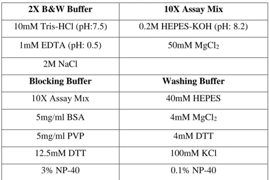
2.1.1. Tables for Required Materials, Buffers and Solutions ... 18
2.2. METHODS ... 23
2.2.1. Production and Purification of Mediator Complex Modules ... 23
2.2.1. Depletion of Mediator Complex from HeLa Nuclear Extracts ... 24
2.2.2. Immobilized Template Recruitment Assay Using Streptavidin Dynabeads 24 2.2.2.1. Production of Template... 24
2.2.2.2. Fixing the Template to the Streptavidin Dynabeads ... 24
2.2.2.3. Immobilized Template Recruitment Assay ... 25
2.2.3. Protein Extractions from SF9 Insect Cells ... 26
2.2.4. Production of GTF IIB, IIE and IIF in BL21 Bacteria Cells... 27
2.2.5. Immunoprecipitation Using M2 Agarose and Sepharose Beads ... 28
2.2.6. Cloning of His-tagged RNA Polymerase II Subunits ... 29
CHAPTER 3…………RESULTS ... 31
3.1. Purification of Functional Human Core Mediator Complex with Baculovirus Expression System ... 31
1.2. Dependency of PIC Elements to Mediator Complex to be Recruited to the Promoter Site ... 32
1.3. Immuno-precipitation Result of Kinase Module- TF-Two-E Association ... 34
1.4. Immunoprecipitation Result of Tail Module- TF-Two-E Association with Additional Check of Med19 ... 38
vii
1.5. Comparison of Mediator Depleted Nuclear Extract and Full Nuclear Extracts in
the Manner of TF-Two-E -Mediator Complex Association... 40
1.6. Association of TF-Two-E with PIC Elements ... 41
1.7. Association of RNAP II and TFIIH with Separate TF-Two-E Subunits ... 43
CHAPTER 4………DISCUSSION ... 45
CHAPTER 5……….FUTURE PERSPECTIVES ... 49
BIBLIOGRAPHY ... 52
viii
Acknowledgements
I would like to thank to our mentor and advisor Assist. Prof. Murat Alper Cevher for all his advices and efforts. As a young scientist, his approach has taught me much more than I could imagine I can learn. His positive behavior against all of us and vision that he gave us were unique and in his own way. His acceptance of me to his lab totally changed my life and my way of understanding life in all manners. So, I deeply want to thank him for creating a step for me to go higher.
I want to thank to my dear parents Leyla Karasu and M. Arif Karasu for their endless support. It did not matter how hard it was for them to keep convincing me go on. They were patient, wise and strong so that I have never felt doing wrong while listening to them. Also, my beloved sister, Malivan Karasu, is a source of energy to me just by herself. Sometimes I got confused which one of us is the older one. She never let me feel alone and stood always beside me. I could not do this without her.
Then, I must thank to my lab mates Tuğçe Canavar and Merve Erden. They were my first mentors and without their friendship I do not think I could finish this thesis. Days and nights and on and on we stood together and I always felt their support. I want them to know that they will never be forgotten. Then to my friends in the whole department, Said Tiryaki, Havva Kilgöz, Özlem Bulut and Gizem Kılıç for sharing many laughs and memories with me. Sometimes those laughs were the only thing that kept me going. I specifically want to thank Bilgenaz Özkan and Elif Özçelik. Whenever I felt down, I found them beside me physically or by heart and they will always have a special place in my heart.
And lastly, I want to thank to my senior students Mine Tanrıöver and Selin Yağlı for their helps. We worked together and most importantly, I feel very lucky to encounter such good companions and friends during my work. I am sure to see them in higher places in the future which is also my biggest wish for them in their forthcoming life.
This Project was financially supported by European Molecular Biology Organization (EMBO) Grant.
ix
List of Figures
Figure 1.1: The panel A show the sequential assembly of PIC in the described order and panel B shows the Holoenzyme model in two different subpopulations….………...5
Figure 1.2: Representation of modular structure of Mediator Complex and some distinct subunits identified for regulatory pathways………...8
Figure 1.3: Representation of Mediator-dependent Transcription in PIC formation, initiation and elongation………...11
Figure 1.4: Represented amino acid sequences of both subunits of Transcription Factor-Two-E and their predicted interaction sites in corresponding
domains………..………...12
Figure 1.5: The cartoon representation of human Transcription Factor-Two-E α, human TFIIS, archaeal TFIIB and yeast
RPB9……….13
Figure 1.6: The representation of indicated functions of Transcription Factor-Two-E subdomains according to the computer based studies and
predictions………..………..14
Figure 1.7: Summary of Baculovirus expression system for virus production and protein expression……….16
Figure-3.1: Coomassie staining result of recombinantly purified Head (H), Middle (M), H+M and H+M+14+26 core Mediator complex produced by Multi-Bac expression system………...31
Figure-3.2: Immobilized template recruitment assay executed with and without mediator presence. The H, H+M, H+M+26 and H+M+14+26 subcomplexes were added
separately to see the difference in the recruitment………...33
Figure-3.3 Western blot result that shows the Kinase module subunits which are pulled down by flag-tagged Transcription Factor-Two-E bound M2 agarose
x
Figure-3.4: Western blot result showing the pulled down Med12 subunit on Transcription Factor-Two-E -α and Transcription Factor-Two-E -β bound
beads……….………36
Figure-3.5: Western blot results of Med12 pull down with empty beads and reverse IP with Med12 bound Sepharose beads………37
Figure-3.6: Western blot result that shows the Tail module subunits which are pulled down by flag-tagged Transcription Factor-Two-E bound M2 agarose
beads……….39
Figure-3.7: Western blot check of nuclear extracts with (full NE) and without (Med Depleted NE) Mediator Complex………40
Figure-3.8: Checked proteins on the Flag-tagged Transcription Factor-Two-E bound M2 agarose beads after the incubation with 10ng/ul concentrated HeLa nuclear
extracts……….………42
Figure-3.9: Western blot analysis to check which subunit of Transcription Factor-Two-E binds to RNAP II and
TFIIH………....43
Figure 4.1: Visual representation of designed ITRA experiment which shows two
different systems with and without the Mediator complex………..45
Figure 5.1: The agarose gel electrophoresis images of cloned RPB subunits into pFBDM vector………50
xi
List of Tables
Table 1.1: The elements of human transcription machinery and their functions in general
transcription………3
Table 2.1: Immobilized template recruitment assay using streptavidin Dynabeads buffers………...18
Table 2.2: Buffers for protein extractions from SF9 insect cells……….18
Table 2.3: Solutions for nuclear extract from HeLa cells………19
Table 2.4: Materials for preparation of SDS-PAGE resolution gels………....19
Table 2.5: Materials for preparation of SDS-PAGE stacking gel………19
Table 2.6: Buffers required for western blotting……….….20
Table 2.7: Other materials and solutions for western blotting……….…20
Table 2.8: Used Antibodies in Western Blots……….….21
Table 2.9: Mediums required for GTF IIB, IIE and IIF production in BL21 Bacteria Cells………..22
Table 2.10: Other used materials and Kits………..22
Table 2.11: PCR program for TATA template multiplication………...….24
Table 2.12: Immobilized template recruitment assay plan with added NE and protein amounts………..………..25
Table 2.13: Produced subunits, their modules and usage amount of viruses………….26
Table 2.14: Contents of bacterial cell lysis buffer for protein extraction…………...27
Table 2.15: Primers used for clonings of RNAP II subunits with 6xHis tags………....29
xii
Abbreviations
GTF General Transcription Factor
Pol II RNA Polymerase II
RNAP II RNA Polymerase II
PIC Pre-initiation complex
TFIIA Transcription Factor IIA
TFIIB Transcription Factor IIB
TFIID Transcription Factor IID
TF-Two-E Transcription Factor IIE
TFIIF Transcription Factor IIF
TFIIH Transcription Factor IIH
TAF TBP Associated Factors
TBP TATA Binding Proteins
Med Mediator complex
CDK8 Cyclin Dependent Kinase 8
CCNC Cyclin C
SEC Super Elongation Complex
kDa Kilo Dalton
RXN Reaction
IPTG Isopropyl β-D-1-thiogalactopyranoside
PC Positive Factors
xiii
NAT Negative regulator of Activated Transcription
SRB Suppressor of RNA Polymerase B
TRAP Thyroid Hormone Associated Protein
CRSP Cofactor Required for Sp1 Activation
SMCC SRB/MED Cofactor Complex
1
CHAPTER 1
INTRODUCTION
1.1. Eukaryotic Transcription System and Machinery
1.1.1. RNA Pol II Mediated Transcription and Transcriptional Steps
The central dogma is the process which converts the genetic code from DNA to RNA and then to the proteins [1]. Transcription is the process in which DNA is used as a template to make RNA in a very ordered and conserved way by the function of RNA Polymerase [2]. The RNA Polymerase was first encountered by Gladstone and Weiss in nuclei of rat liver cells in 1959 and Hurwitz and Stevens have seen the same activity in Escherichia coli in 1960 [3-4]. With those discoveries, the role of RNA Polymerase on transcription of DNA has been settled as a universal function. Until now, four different RNA polymerases have been discovered and named as RNA Polymerase I, II. III and IV. The RNA Polymerases I, II and III were named according to their fraction numbers while purification and they are involved in 18S and 28S rRNAs (I), mRNAs (II) and tRNAs (III) transcription processes respectively [5]. RNA Polymerase IV was discovered in last decade in plants functioning by facilitating the production of small interfering RNAs (siRNAs) [6]. Due to the distinct functions of these RNA polymerases, they require different sets of additional proteins and here we will be focusing on the transcription event mediated by RNA Polymerase II (RNAP II).
For RNAP II to start the transcription of a gene, three major steps are executed: (I) Chromosome opening and the formation of euchromatin, (II) modification of histones on the related gene for the access of activators and silencers and (III) assembly of the transcription machinery at the promoter site of the gene [7].
2
1.1.2. Difference of Basal and Activator Driven Transcription
Activators and repressors are the proteins required for the regulation of transcription of a specific gene or a set of genes in cells. For the activator dependent transcription to happen, it is crucial that related activator or activators sits on the corresponding Enhancer sequence sites [8]. For instance, Estrogen Receptor- α is an activator and needs to sit on to Estrogen Enhancer Sequence (EES) site to start activator dependent transcription. The specificity of an activator to its related set of genes comes from its special DNA binding domain which prevents it to bind any other enhancer sequences than its own [9]. The basal level transcription is the observed level of expression of a gene even under the suppression and executed by the general transcription machinery. The difference between activator dependent transcription and basal transcription starts from the distinct core promoter sites of these two events. Upon the binding of activator, the signal of enhancer is relayed to the transcription machinery and boost the transcription by both increasing the transcribed mRNA levels and speculatively by helping to bring the transcription machinery to the core promoter sites [10].
1.1.3. Recruitment of RNAP II and GTFs to the Promoter Site
Apart from the gene specific activators, there are some set of proteins required for the transcription to happen. Upon binding of activator, the transcription machinery is recruited to the promoter site. This transcription machinery is composed of RNAP II and some other accessory proteins which are required in the site-specific transcription initiation. The necessity of these accessory proteins was first shown by Weil in 1979. He achieved in vitro transcription by additionally putting crude subcellular extraction fractions onto the RNAP II with the native adenovirus DNA template [11]. Further examinations for identification of these accessory proteins in the extract fractions revealed different proteins eluted in increasing salt concentrations in ion exchange chromatography [11]. At the end these proteins were identified as transcription factors, named as TFIIA, TFIIB, TFIID, TF-Two-E, TFIIF and TFIIH and characterized as general transcription factors (GTFs). In the nomenclature the TFs stand for transcription factor, the roman
3
number represents the RNAP II dependent transcription and the letters correspond to the chromatographic fractions at which each protein was eluted [12].
Table 1.1: The elements of human transcription machinery and their functions in general
transcription [13]
Even though the components of the human general transcription machinery are known, there are two different hypotheses about how the formation of this machinery occurs. The first hypothesis suggests a sequential assembly before the initiation of transcription based on the in vitro transcription assays conducted with different chromatographic fractions of crude HeLa cell extracts [14]. According to it, after the DNA is opened for transcription, the TFIID is recruited to the TATA box and binds to the DNA
Factor Protein Composition Function
TFIIA p35 (α), p19 (β), and p12 (γ) Antirepressor; stabilizes TBP-TATA complex; coactivator
TFIIB p33 Start site selection; stabilize TBP-TATA complex; pol II/TFIIF recruitment
TFIID TBP + TAFs (TAF1-TAF14)
Core promoter-binding factor, Coactivator, Protein kinase, Ubiquitin-activating/conjugating activity, Histone
acetyltransferase
TFIIE p56 (α) and p34 (β) Recruits TFIIH, Facilitates formation of an initiation-compentent pol II, Involved in promoter clearance
TFIIF RAP30 and RAP74
Binds pol II and facilitates pol II recruitment to the promoter, Recruits TFIIE and TFIIH, Functions with TFIIB and pol II in start site selection, Facilitates pol II
promoter escape, Enhances the efficiency of pol II elongation
TFIIH
P89/XPB, p80/XPD, p62, p52, p44, p40/CDK7, p38/Cyclin H, p34, p32/MAT1, and p8/TFB5
ATPase activity for transcription initiation and promoter clearance, Helicase activity for promoter opening,
Transcription-coupled nucleotide excision repair, Kinase activity for phosphorylating pol II CTD, E3
ubiquitin ligase activity
RNAP II RPB1-RPB12
Transcription initiation, elongation, termination, Recruitment of mRNA capping enzymes,
Transcription-coupled recruitment of splicing and 3 end processing factors, CTD phosphorylation, glycosylation, and
4
via one of its components, Tata Binding Protein (TBP) [15]. This prokaryotic σ-factor-analogous function of TBP was first discovered in Drosophila [16], then in mammals [17] and lastly in S. cerevisiae (yeast) [18]. After that, the TFIIA comes as a facilitator for TFIIA-TBP-DNA complex formation by inhibition of inhibitory elements in the environment and raises the possibility of TBP or TFIID to bind to the DNA [19]. Also, it helps the stabilization of the TBP-DNA complex by binding the complex from an opposite direction of the TFIIB binding site [20]. The other stabilizer of the TBP-TATA box complex is the TFIIB. As mentioned before, the TFIIB and TFIIA bind to the opposite sites of the TBP-DNA complex to increase the stability of the complex [20-21]. Also, the TFIIB acts as a recruiter for the RNAP II-TFIIF complex so that it marks the transcription start site [22] and helps the RNAP II-TFIIF complex to dock on the DNA [23]. The TFIIF is closely interacting with RNAP II [24] and its entry to the promoter site happens together with RNAP II. It has some distinct functions like helping RNAP II to enter to the transcription machinery by docking onto readily formed TFIIA-TBP-TFIIB-DNA complex [25], stabilizing the RNAP II on the complex by bending the DNA towards RNAP II and creating new DNA and protein interaction sites [26], enabling the Transcription Factor-Two-E -TFIIH entry to the complex [21] determining the start site of transcription along with RNAP II and TFIIB and lastly helping the RNAP II to leave the promoter site upon the transcription initiation 28]. The Transcription Factor-Two-E on the other hand interacts with several members of Pre-Initiation Complex (PIC) but it mainly recruits TFIIH to the PIC and regulates its ATPase 29], kinase [30], and helicase [31] activities. The general functions and detailed structures of Transcription Factor-Two-E will be explained in “General Transcription Factor IIFactor-Two-E” section. The last member of PIC is the TFIIH and it is necessary for a couple of reasons. Its ATPase activity is required for promoter clearance, helicase activity is required for unrolling the DNA and kinase activity is required for CTD phosphorylation of RNAP II to stimulate transition from initiation stage [13].
The second theory is based on the existence of different subpopulations of transcription machinery containing RNAP II. According to that, the RNAP II holoenzyme is found in nucleus with different combinations of GTFs or even without the GTFs [32]. The different versions of the holoenzyme complex were eluted by using different
5
chromatographic conditions for purifications and separate laboratories obtained varying subsets of the holoenzyme. The representation of both pathway can be seen in the Figure 1.1 below. There are enough evidence supporting both pathways so it is important to speculate that the variations in the environment and physiological conditions determine pathway choice and formation of PIC.
Figure 1.1: The panel A show the sequential assembly of PIC in the described order and
panel B shows the Holoenzyme model in two different subpopulations [13].
1.1.4. TAFs, PCs and NCs
Further in vitro transcription assays for revealing the core elements of transcription machinery for activator dependent transcription showed some controversial results about the requirements of some GTFs. Separate laboratories identified different elements so it can be speculated that the variations in purification steps can result in the elution of some distinct factors enabling the transcription. However, it was shown that TBP, which is a
6
subunit of TFIID, is not sufficient on its own for the transcription [33]. This finding then followed by the finding of TBP Associated Factors (TAFs) which are also identified as the subunits of TFIID and characterized as activator dependent transcription regulators that are transmitting the signal from activator to the machinery [33]. Thus, the usage of TAFs with TBP which is the TFIID itself or presence of only TBP determines activated or basal level transcription respectively. Later, the biochemical studies identified driver regulators other than TAFs from the cell extracts which are called as Upstream Stimulatory Activity (USA) [34] and these activities involved both activation and repression so they were divided into one Negative Cofactor (NCs) and four Positive Cofactors (PC1-4) which are working as a coactivator in the presence of the activators and working as a repressor to stop basal transcription in the absence [35], so their function depends on the presence and absence of activators. Although there were some matching elements in these PC fractions, the PC2 is important especially since it is composed of many subunits which are later identified as belonging to Mediator Complex [36] that is also a general cofactor relaying the gene specific signals from activators or repressors to the transcription complex [37].
1.2. Human Mediator Complex
At the beginning, Mediator Complex was found in S. cerevisiae composing of 11 essential and 9 accessory subunits which makes a 20-subunit protein complex [38]. The human mediator complex was first purified as human Thyroid Hormone-α Receptor Associated Protein (TRAP) Complex. This complex was eluted together with human Thyroid Hormone Receptor-α (hTR-α) and enables in vitro transcription when a DNA template that includes T3 Response elements or TREs is used [39]. Later, many subunits of the Mediator complex were also found in independently purified protein complexes like SRB/MED-containing cofactor complex (SMCC) which contains TRAP220 TRAP170 and TRAP 100 in common with TRAP complex [40], activator-recruited complex (ARC) [41], vitamin D receptor interacting protein complex (DRIP) [42], cofactor required for Sp1 activation (CRSP) which also shares some subunits with TRAP complex [43], Positive Cofactor 2 (PC2) [35] and negative regulator of activated transcription (NAT)
7
which has indeed a repressive role in transcription due to the repressor function of CDK8 [44].
1.2.1. Presence of Different Forms of Mediator Complex in Cell and The Assembly Question
As mentioned before, there are many Mediator-like subpopulations in the cell. Those subpopulations contain some subunits which are identified as Mediator complex subunits. Other subunits of those subpopulations represent a very high homology with Mediator complex elements. These findings show us that Mediator Complex can be found in cells in different versions which are composed of varying subunits and that creates a high heterogeneity for the Mediator Complex. Mediator complex is present in a very low amount in cell and has that high heterogeneity. The complex is also very dynamic and flexible which makes the complex very hard to study [45]. Some defined subpopulations of Mediator Complex were MED13(TRAP240), MED12(TRAP230), Cyclin C, and
CDK8 subcomplex [46] and MED23(hSUR2), MED24(TRAP100) and
MED16(TRAP95) subcomplex [47]. Many other studies based on a subunit of Mediator Complex also describes that heterogeneity. For example, the Zhang, X. et. al., (2005) [45] mentions the versions of Mediator complex with and without MED1/TRAP220 by pointing out the presence of MED1/TRAP220 subunit only in some subpopulations [45]. Due to this dynamic structure and high heterogeneity, the human Mediator Complex is defined into modules which are the Head, Middle Tail and CDK8 containing Kinase module [48]. The first question here to be asked is that how such a complex is being formed in a cell. It requires further examinations and analysis to determine if those subpopulations act as pre-formed structures to be assembled after being induced by a regulator or a sequential de novo assembly happens for the whole Mediator formation.
1.2.2. Function, Structure and Architecture of Human Mediator Complex
The multi-subunit human Mediator Complex is a 30-subunit complex in humans. As mentioned before, it has the Head, Middle, Tail and Kinase modules even though the
8
composition of these modules differ in separate papers from independent laboratories. The first functional identification of human Mediator Core Complex was done in 2015 which shows that the Head, Middle, Med14 and Med26 forms the transcriptionally active core of the complex [49]. It is the function of Mediator to receive signals from regulatory pathways in a cell and to turn it into a transcriptional response [50]. Mediator Complex is the hub for many distinct pathways where the signal is converted and transmitted to transcriptional machinery. Since the Head and Middle modules along with Med14 gives the functionality to the complex, it is important to speculate that these distinctive subunits of each pathway, that are resulted from physiological conditions of a cell, rest in the Tail and Kinase subunits. The representation of some identified subunits for separate pathways can be seen in Figure1.2 below.
Figure 1.2: Representation of modular structure of Mediator Complex and some distinct
9
1.2.3. Effects of Mediator’s Architecture and Modular Structure on Its Function and Regulations of Mediator Complex in Transcription and Pre-initiation Complex Formation and Function
As it was mentioned before, the mammalian Mediator Complex has 30 subunits and 4 of these subunits are of Kinase module [48]. Due to the distinct functions and dynamic interactions with Mediator Complex, CDK8 containing Kinase module sometimes counts as separate and Mediator Complex is told to have 26 subunits [46]. Since the Transcription Factors (TFs) have separate and different binding domains from each other, Mediator Complex can also bind multiple TFs instead of interacting them individually. Also, due to its interactions with the PIC, it can be said that the Mediator Complex also has a high number of interactions with RNAP II even though the exact mechanism of Mediator Complex - RNAP II interaction is not known yet.
Since the composition of Mediator also changes much even within the same cell, this ability of Mediator to detach from its subunits according to the executed function makes it mechanistically important for the transcription. Some studies also found and purified Mediator complexes that are lacking different numbers of Mediator subunits apart from the Kinase Module (e.g. PC2) [51-52]. Since these Mediator Complexes cannot transmit the signal coming from the TFs that bind to the lost subunits, this creates a direction in the selection of the transcribed gene. In such cases, it is shown that the Med1 binds to the thyroid hormone receptor(TR) and its knock out stopped the TR induced transcription [39] and Med23 is shown to bind ELK1 [53]. Again, the knockout of Med23 causes an inhibition on the ELK1 activated gene expression [53]. It is important to add that these knockouts do not stop the transcription regulated by other gene specific TFs. [39-53] Thus it can be concluded that the Mediator Complexes having fewer subunits than whole Mediator Complex should have a more specified function.
Another structural tool of Mediator, the Kinase module, gives very distinct properties to the complex. It is known that the Kinase module has a reversible interaction
10
with the complex which is also very dynamic and this interaction creates a different sub-population of complex often called as CDK8-Mediator Complex [54-55]. When this subpopulation is further examined, it is seen that the Med26 subunit is not notably present in that subcomplex resulting in a 29 subunit Mediator. [56] Even though the exact mechanism underlying the association and dissociation of Kinase module is not fully figured, some studies suggest the ubiquitination and phosphorylation of Med13 may have link to that event [57]. In that aspect, it is hypothesized that the PARP1 protein which normally ribosylates proteins [58] and FBW7, which is a ubiquitin ligase, stimulates the dissociation of Kinase module from the Mediator Complex [57-58]. Also, it is being thought that the structural changes upon the binding of Kinase module to the Mediator complex prevents the RNAP II-Mediator interaction [59]. The RNAP II Carboxyl Terminal Domain (CTD) associated Mediator complex has failed to show a presence of kinase module subunits [59] and consistently the Kinase module bound Mediator did not pull down any RNAP II subunits in purifications [60]. Yet, when the initiation is completed, the re-association of Kinase module to the Mediator somehow starts the elongation step. [61] Besides, Kinase module incorporated Mediator Complex fails to bind to TFIIH which results in the inhibition of RNAP II - TFIIH interaction and prevents promoter escape. By this way, re-binding of new RNAP II to the same promoter is also prevented since RNAP II cannot leave the PIC [62].
The Mediator is thought to form many interactions with RNAP II [63-64] and this interaction may create a scaffold for the PIC formation since the Mediator Complex transmits the signals from TFs to RNAP II and PIC which is called as the bridge model [65-66]. Some studies suggest that the recruitment of RNAP II via Mediator Complex happens due to its association with RPB1 subunit [61]. Also, it is thought that the Mediator Complex regulates the RNAP II entry to the PIC with the involvement of GDOWN1 which is a factor that binds to RNAP II when it is not Mediator bound and thus inhibits transcription by disrupting the TFIIF-RNAP II interaction [67-68].
11
Studies about the transcriptional elongation showed that the Med26 may interact with the Super Elongation Complex (SEC) and may help the initiation of elongation [69]. Interestingly, the same studies show Med26 to bind TFIID as well and suggest that the facilitative role for Med26 in PIC formation and then a regulatory function in elongation upon a regulatory signal due to its interaction with SEC [69]. These data also bring a proposition for that integrated roles of Mediator, GDOWN1 and SEC by stating a configurational remodeling or switching system. Additionally, Med26 has been shown to colocalize with the heterochromatin in contrast with other Mediator Complex subunits which can bring an explanation to the antagonistic function of Mediator and heterochromatin regions in humans [70].
Figure 1.3: Representation of Mediator-dependent Transcription in PIC formation,
initiation and elongation [71].
As it can be seen from Figure-3 above, in Panel A, Mediator Complex first helps the formation of the PIC by bringing RNAP II and other TFs to the promoter site [72] At the beginning, The CTD of RNAP II is not phosphorylated much but with the actions of TFIIH, it gets phosphorylated and initiate the transcription which is called the promoter escape (at serine 2 and serine 5 residues of the heptad repeats) [73]. After nearly transcribing 50 to 60 nucleotides, RNAP II stalls at some genes [74]. Although it is not known whether Mediator complex stays interacting with stalled RNAP II, Panel B represents the situation after the Mediator Complex and RNAP II bonds are seized and Kinase module entry to Mediator Complex happens since they have an exclusive behavior when binding to Mediator Complex. At this stage, it is shown that the CDK8 associates with SEC to stimulate the stalled RNAP II to go on with elongation [61-75]. It is also
12
predicted that the Med26 can have a role at this stage by dissociating from complex upon binding of Kinase module. The Panel C shows the recruitment of a new RNAP II by the remaining PIC which is called as re-initiation [76].
1.3. General Transcription Factor IIE (GTF-TWO-E\TF-TWO-E) 1.3.1. Structure of GTF-TWO-E
As it was explained in Figure-1, in the sequential assembly of PIC, Transcription Factor-Two-E enters the complex after the RNAP II-TFIIF recruitment. The Transcription FactorTwoE consists of two subunits which are named as Transcription FactorTwoE -α and Transcription Factor-Two-E -β [77]. (Although at first it was thought that this protein works in a heterodimer structure with two α and two β subunits [77], later studies showed that the protein works as a α+β heterodimer form [78].) The α subunit is a 439-amino acid long polypeptide with a molecular mass of 56 kDa and the β subunit is a 291-amino acid long and 34 kDa polypeptide [77]. The predicted interaction sites and of Transcription Factor-Two-E subunits on the represented aminoacids sequences are shown in the Figure-4 below.
Figure 1.4: Represented amino acid sequences of both subunits of Transcription
13
The N-terminal domain of α subunit which is between 12th and 118th amino acids appears to interact with the Transcription Factor-Two-E -β by the region between 197th -238th amino acids. The C-terminal domain on the other hand appears to be the interaction site of α subunit and TFIIH [13]. Since it is known that the Transcription Factor-Two-E helps the recruitment of TFIIH to the PIC [79], it is quite logical to see a direct interaction between Transcription Factor-Two-E and TFIIH. Upon looking the Figure 1.4, there are some distinct regions in the subunits. For example, the α subunit has a homology with the bacterial σ factor at the residues between 13th and 49th amino acids, there is a leucine repeated part from 38th to 66th residues, the zinc finger domain is between 121st and 151st one Helix-Turn-Helix (HTH) motif domain between 159th and 182nd aminoacids. There are two different alanine rich domains following the HTH domain and close to the C-terminus, there is an acidic domain formed mainly with aspartic and glutamic acids between 378th and 398th aminoacids [13]. Due to the nature of zinc finger motifs, their function is mainly to bind the DNA. Generally, a zinc finger domain is composed of 3 or 4 beta strands as it is seen in TFIIB, TFIIS and RPB9 orthologs but the zinc finger domain of Transcription Factor-Two-E differs from others by having one alpha helix and two beta sheets in an ββαβββ order. The Zn+2 ion is binding to Cysteine amino acids at 129, 132, 154 and 157locations [80]. The HTH domain of α subunit has a homology between archaeal TFE which also has a HTH motif and helps the transcription initiation by facilitating the TATA box recognition [81] This is the only predicted function for the HTH domain of human Transcription Factor-Two-E -α subunit.
Figure 1.5: The cartoon representation of human Transcription Factor-Two-E α, human
14
On the other hand, the β subunit also has some structurally distinct sites. There is a serine rich site between 26th and 71st amino acids and a winged HTH site in 75th and 139th residues. Then comes a leucine rich site between 145th and 163th residues and a bacterial σ factor subdomain 3 homology site between 163rd and 193rd amino acids [13]. The C-terminus contains two basic Loop-Helix-Loop and Helix-Loop domains. The homology predictions state a resemblance with the Krüppel TF of Drosophila which has a DNA binding function. Also, the Winged HTH region has a homology with the RAP30 from its RNAP II binding domain. Even though these interactions are predicted, more experiments are required to define the certain interaction sites [13-79].
1.3.2. Functions of GTF-TWO-E
In the literature, there are three different functions that are assigned to GTF-TWO-E. As it can be seen from the Table-1.1, these functions are to recruit the TFIIH to the PIC, to help the assembly of PIC and to help the promoter clearance. The indicated functions of Transcription Factor-Two-E domains and their sites can be seen in the Figure-6 below [78].
Figure 1.6: The representation of indicated functions of Transcription Factor-Two-E
15
Even though it is not clear yet how the Transcription Factor-Two-E is being recruited to the promoter site at the beginning, there are some suggested candidates like Krüppel protein [82], Antennapedia Abd-B homeodomain proteins which are shown to bind Transcription Factor-Two-E in vivo [83] however, it is not clear if they also involve in the recruitment of Transcription Factor-Two-E to the promoter region and to the PIC. Due to its location in PIC, the Transcription Factor-Two-E is thought to be interacting with TFIIF, TFIIB, RNAP II and TFIIH [13].
The interaction of Transcription Factor-Two-E and TFIIH have been shown in many different studies. This interaction enables TFIIH to be recruited to the PIC at the promoter site. The absence of TFIIF prevented the TFIIH entry to the PIC [84]. The defined interaction of Transcription Factor-Two-E and TFIIH is on the C-terminus domain of Transcription Two-E -α subunit. This interaction enables Transcription Factor-Two-E to regulate the kinase and ATPase activities of TFIIH so that it phosphorylates the CTD domain of RNAP II and starts the elongation [79]. Also, the promoter region where Transcription Factor-Two-E sits is found as -10 where the Transcription Factor-Two-E also helps TFIIH helicase activity for promoter clearance at +1 and DNA melting [85]. It is also shown that the Transcription Factor-Two-E and TFIIH are not required for in vitro transcription from a pre-melted template using adenovirus promoter (AdE4) [85]. Thus, it has been concluded that the Transcription Factor-Two-E does not only recruit TFIIH but also act as a mechanistical check point from initiation to elongation stages [84] The co-occupancy between Transcription Factor-Two-E and TFIIB was also reported by glycerol sedimentation analysis [86]. The same method was also used to show the co-occupancy of RNAP II and Transcription Factor-Two-E without defining any specific subunits. This method only gives the conclusion that these two proteins (Transcription FactorTwoE -TFIIB and Transcription Factor-Two-E -RNAP II) co-exist together and elute together [86]. It is also shown that Transcription Factor-Two-E co-purifies the non-phosphorylated RNAP II specifically [84] which is also logical since the aim of recruitment of TFIIH via Transcription Factor-Two-E is to phosphorylate the CTD and to start elongation.
16
1.4. Baculovirus Expression System
Baculovirus expression system has been designed to overcome some important obstacles emerged due to the nature of bacterial expression system. More important of these obstacles can be said as lacking eukaryotic post translational modifications and inability of producing large proteins. Also, the stability of the vector produced mRNAs in bacterial system is quite low and thus the assembly of the protein complexes produced under the same promoter, does not fully happen in the system [87].
The Baculovirus expression system for multi-protein complexes has two different expression cassettes with T7 and Cre/Lox translocation sites. These vectors, which are pFBDM and pUCDM respectively, contain polyhedron (polh) and p10 viral promoters with two different multiple cloning sites (MCSs) to allow cloning of genes of interest via digestion with a bunch of restriction enzymes. For the first selection, to distinguish the gene inserted vectors, pUCDM and pFBDM contain chloramphenicol and ampicillin resistance markers. To sub clone multiple genes, both vectors have a multiplication module which contain BstZ17I and SpeI restriction sites at the beginning and PmeI and AvrII restriction sites at the end of the cloning region [88]. After the genes are cloned to the vectors the procedure follows as described below in Figure-7
17
Figure 1.7: Summary of Baculovirus expression system for virus production and protein
expression [89].
After the genes are cloned into vectors, the plasmids are transformed into the bacteria that has the Bacmid. Through Cre-Lox recombination and Tn7transposition, the plasmids are added to the Bacmids. The Bacmid DNA carries the LacZ gene for blue/white selection in DH10Bac cells. When the gene cloned plasmid is translocated into the Bacmid successfully, the Bacmid DNA is isolated and used for transfections with SF9 cells to generate the first generation of viruses called the P0 viruses. Then the amplification of the viruses can be made to create P1 and P2 and P3 generations and to increase the virus titers. Then the created viruses can be used for the expression of multi-subunit proteins in high quantities [88].
18
CHAPTER 2
MATERIALS and METHODS
2.1. MATERIALS
2.1.1. Tables for Required Materials, Buffers and Solutions
Table 2.1: Immobilized template recruitment assay using streptavidin Dynabeads
buffers
2X B&W Buffer 10X Assay Mix
10mM Tris-HCl (pH:7.5) 0.2M HEPES-KOH (pH: 8.2)
1mM EDTA (pH: 0.5) 50mM MgCl2
2M NaCl
Blocking Buffer Washing Buffer
10X Assay Mıx 40mM HEPES
5mg/ml BSA 4mM MgCl2
5mg/ml PVP 4mM DTT
12.5mM DTT 100mM KCl
3% NP-40 0.1% NP-40
Table 2.2: Buffers for protein extractions from SF9 insect cells
BC1000 BC0 20mM Tris-HCl (pH: 7.9 at 4 oC) 20mM Tris-HCl (pH: 7.9 at 4 oC) 20% Glycerol 20% Glycerol 0.1 mM EDTA 0.1 mM EDTA 0.5 mM PMSF 0.5 mM PMSF 0.5 mM DTT 0.5 mM DTT 1M KCL
19
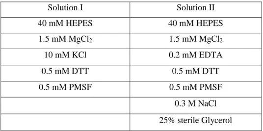
Table 2.3: Solutions for nuclear extract from HeLa cells
Solution I Solution II 40 mM HEPES 40 mM HEPES 1.5 mM MgCl2 1.5 mM MgCl2 10 mM KCl 0.2 mM EDTA 0.5 mM DTT 0.5 mM DTT 0.5 mM PMSF 0.5 mM PMSF 0.3 M NaCl 25% sterile Glycerol
Table 2.4: Materials for preparation of SDS-PAGE resolution gels
Resolution Gel Percentage 7% 10% 12% 15% ddH2O 7.4ml 6.25 ml 5.20 ml 3.65 ml Tris (1M pH:8.8) 3.45 ml 3.45 ml 3.45 ml 3.45 ml 5% SDS 300 ul 300 ul 300 ul 300 ul 30% Acylamide 3.75 ml 5 ml 6 ml 7.5 ml 10% APS 100 ul 100 ul 100 ul 100 ul TEMED 10 ul 10 ul 10 ul 10 ul
Table 2.5: Materials for preparation of SDS-PAGE stacking gel
ddH2O 3 ml Tris (0.5 M pH:6.8) 1.25 ml 5% SDS 100 ul 30% Acylamide 670 ul 10% APS 50 ul TEMED 5 ul
20
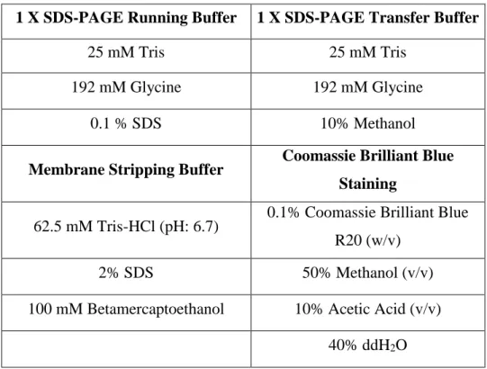
Table 2.6: Buffers required for western blotting
1 X SDS-PAGE Running Buffer 1 X SDS-PAGE Transfer Buffer
25 mM Tris 25 mM Tris
192 mM Glycine 192 mM Glycine
0.1 % SDS 10% Methanol
Membrane Stripping Buffer Coomassie Brilliant Blue Staining
62.5 mM Tris-HCl (pH: 6.7) 0.1% Coomassie Brilliant Blue R20 (w/v)
2% SDS 50% Methanol (v/v)
100 mM Betamercaptoethanol 10% Acetic Acid (v/v)
40% ddH2O
Table 2.7: Other materials and solutions for western blotting
1X PBS-Tween
8 mM Na2HPO4, 150 mM NaCl, 2 mM KH2PO4, 3 mM KCl, 0.1%
Tween20
Destaining Solution 50% H2O, 40% Methanol, 10%
Acetic Acid 30% Acrylamide (v/v) 292,2 gr/L Acrylamide, 7.8 gr/L Bisacrylamide 10% Ammonium Persuşphate (APS) 10 ml ddH2O, 1 gr Ammonium Persulphate 4X SDS Loading Buffer 240 mM Tris-HCl (pH:6.8), 8% SDS (w/v), 40% Glycerol (v/v), 0.04% Bromophenolblue, 5% Betamercaptoethanol
21
Table 2.8: Used Antibodies in Western Blots
Med 12 Home made * Med27 Santa Cruz Cat. No:
Sc-390296
Med13 Home made * Med29 Santa Cruz Cat. No:
SC 393800
Med14 Abcam Cat No:
ab170605 Med30 Home made *
Med15 Proteintech Cat. No:
115661AP RPB1 Home made *
Med16 Santa Cruz RPP62 Home made *
Med17 Home made * CDK7 Cell Signaling
Med19 Home made * CDK8 Home made *
Med23 Home made * CCNC Home made *
Med24 Home made * XPB Cell Signaling
Med25 Santa Cruz Cat. No:
SC393759
Anti-Histidine Cell Signaling
Med26 Home made * Anti-Flag Sigma Cat. No:
F7425
GDOWN1 Home made * TF-Two-E-α Home made *
22
Table 2.9: Mediums required for GTF IIB, IIE and IIF production in BL21 Bacteria
Cells
LB-Agar Plates LB Medium SOC Medium
10 gr Tryptone 10 gr Tryptone 2% Tryptone
5 gr Yeast Extract 5 gr Yeast Extract 0.5% Yeast Extract
10 gr NaCl 10 gr NaCl 0.4% Glucose
15 gr Agar 950 ml ddH2O 10 mM NaCl
951 ml ddH2O 2.5 mM KCl
5mM MgSO4
5mM MgCl2
Table 2.10: Other used materials and Kits
Anti-flag M2 Agarose Affinity
Agarose Beads Sigma Cat. No: A4596
Flag Peptide Sigma Cat. No: F3290
Streptavidin Dynebeads M-280 Invitrogen Thermo Fisher Cat. No: 11205D
BCA Protein Kit Assay Kit Thermo Fisher Pierce BCA Kit
Cat. No: 23227
Restriction Value Pack Thermo Fisher Fastdigest
Restriction Set Cat. No. SM1553
Gel Extraction Kit Thermo Fisher Gel Extraction Kit
Cat. No: K0691
Plasmid Isolation Kit Thermo Fisher Gel Extraction Kit
Cat. No: K0502
Lyophilized Lysozyme enzyme Sigma Aldrich Cat. No:
cDNA Synthesis RevertAid RT Reverse Transcription Kit
Thermo Scientific Cat. No: K1691
23
2.2. METHODS
2.2.1. Production and Purification of Mediator Complex Modules
The Head (H), Middle (M), Head+Middle (H+M), Head+Middle+14 (H+M+14) and Head+Middle+14+26 (H+M+14+26) complexes were readily purified as it is stated in Cevher et al. 2014 [49]. The Mediator complex subunits were cloned to the pFBDM and pUCDM Baculovirus expression system vectors with different tags like Flag, 6xHis and HA. Mediator Head module (Med6-8-11-18-19-20-22-30), Middle module (Med4-7-9-10-21-26-31) Med 17 and Med 14 were inserted into the vectors with tags of HA:Med7, His:Med10 and Flag:Med14. Then the Plasmids were integrated into the Bacmid DNA and amplified. After the purification of Bacmid DNA and transfection of Bacmid into the SF9 cells, first generation P0 viruses were created. Then the amplification of the P0 viruses were also made to the second and third generations P1 and P2 with a higher titer of viruses. So as to obtain the proteins, the infections of Head module virus and Middle module virus and co-infections of H+M, H+M+14, H+M+14+26 were done by infecting nearly 100ml 100 million Hi5 cells for 5 to 6 days. The titrations were determined according to the stoichiometric production of the complexes. To collect the cells, the suspension grown Hi5 cells were centrifuged at 1500 rpm for 10 minutes and supernatant was discarded. The pellet was re-suspended in 4 ml BC500 (5 ml BC1000 + 5ml BC0, 5mmDTT, 5mmPMSF) and cells were homogenized by douncing 3times. After centrifugation of the lysate at 14000 rpm for 15 minutes, pellet was discarded and supernatant was diluted to 300mM salt concentration by addition of BC0 in a drop by drop manner. The extract then incubated with the M2 and followed by HA agarose beads at 4oC overnight to pull down the modules from the tagged subunits. After washing the beads with BC300+0.1%NP-40 five times, complexes were eluted with of 0.5mg/ml Flag or HA peptides to the corresponding beads. Elution step was repeated 3 times at 4oC for 45 minutes in rotation and checked with Coomassie brilliant blue staining and Western blots.
24
2.2.1. Depletion of Mediator Complex from HeLa Nuclear Extracts
Depletion of Mediator complex from HeLa cell nuclear extract was done according to the procedure stated in Cevher et al., 2014 [49]. The depletion was checked again with Western blot.
2.2.2. Immobilized Template Recruitment Assay Using Streptavidin Dynabeads
2.2.2.1. Production of Template
In order to produce the TATA containing template DNA fragments, conventional PCR was used. Biotinylated primers were used to allow the binding of templates to the streptavidin beads. The PCR was conducted with the program shown below and the products were extracted from agarose gel with Gel Extraction Kit (Table 2.10).
Table 2.11: PCR program for TATA template multiplication
Reagents 10x Taq Buffer (5U/ul) dNTP (10mM) Forward Primer (10mM) Reverse Primer (10mM) TATA Template (10 ng/ul) MgCl2 (25mM) ddH2O Added Volume (ul) 5 1 1,5 1,5 1 4 36 PCR cycle temperatures (oC) 95 95 68 72 72 4
Time (minutes) 4 0,5 1 1 7 Till taken
34 cycles
2.2.2.2. Fixing the Template to the Streptavidin Dynabeads
Dynabeads (10 ul/rxn) were shaken to homogenize the slurry and 10 ul for each reaction was taken into eppendorf tubes. Beads were washed with 1XB&W buffer (Table 2.1) for 5 times by putting the tubes on the magnetic rack each time for 90 seconds. Then beads were resuspended in 150 ul 2XB&W buffer. Nearly 8 ug template was added on to the beads to immobilize and volume was completed to 300 ul with H2O addition. The beads were incubated at room temperature for 15 minutes by inverting the eppendorf tubes
25
in every 5 minutes to prevent aggregation and heterogenous distribution of the template. Then the tubes were put onto the magnetic rack again and the liquid buffer was taken out but not discarded since the amount of bound template can be checked by finding out the final concentration of the DNA in the buffer. The Dynebeads were then washed with 300ul of 1XB&W buffer with 0.5mg/ml BSA and 0.003% NP-40 for two times. After that the beads were blocked by addition of 100 ul of Blocking buffer and incubation at room temperature for 15 minutes. Finally, the beads were washed with 500 ul washing buffer two times and stored at +4 oC in 150 ul Wash buffer.
2.2.2.3. Immobilized Template Recruitment Assay
6 reactions were planned with HeLa nuclear extract (NE) and Mediator depleted nuclear extract (∆Mediator NE) by the addition Head (H), Head+Middle (H+M), Head+Middle+14 (H+M+14), Head+Middle+14+26 (H+M+14+26) recombinantly and one negative group with no recombinant Mediator modules added. Then the prepared beads were washed 4 times with 1X assay mix including 0.025% NP-40 and 0.25 mg/ml BSA. Then 300 ul 2X assay mix including 60 ug/ml E. coli genomic DNA was put onto the beads and beads were divided into 6 tubes since 6 reactions were planned. After that, stated amount of recombinant proteins were added onto the beads as it is shown in Table 13 below. The reaction was incubated at 30 oC for 50 minutes by mixing in every 10 minutes. Following the incubation, beads were washed with 50mM NaCl and 16 ul 1X SDS loading buffer was added into each reaction tube to further check by western blot.
Table 2.12: Immobilized template recruitment assay plan with added NE and protein
amounts HeLa NE (Full NE) ∆Mediator NE Recombinantly added Mediator Modules ∆Mediator NE + recombinant proteins +
H M 14 26 N. Cont. (∆Mediator NE only)
+ + + + H+M+14+26
+ + + H+M+14
+ + H+M
26
For the Full NE, 180ug is added while for the H, H+M, H+M+14 and H+M+14+26 4500ug, 5940ug, 5400ug and 6300ug were used respectively
2.2.3. Protein Extractions from SF9 Insect Cells
Table 2.13: Produced subunits, their modules and usage amount of viruses
Modules Produced Mediator Subunits Amount of infected Sf9 cells Amount of used virus Kinase Module Med12 25ml (106 cells/ml) 1ml Med13 25ml (106 cells/ml) 1ml CDK8 25ml (106 cells/ml) 1ml CCNC 25ml (106 cells/ml) 1ml Tail Module Med15 25ml (106 cells/ml) 1ml MEd16 25ml (106 cells/ml) 1ml Med23 25ml (106 cells/ml) 1ml Med24 25ml (106 cells/ml) 1ml Med25 25ml (106 cells/ml) 1ml Med27 25ml (106 cells/ml) 1ml Med29 25ml (106 cells/ml) 1ml Head Module Med19 50ml (10 6 cells/ml) 1.5ml
The readily prepared P1 and P2 generation viruses were produced for the Tail and Kinase subunits as it is described in the production of Mediator modules section. After the infections, the cells were centrifuged at 1500 rpm for 10 minutes and supernatant was discarded. The pellet was re-suspended in 4 ml BC500 (5 ml BC1000 + 5ml BC0, 5mmDTT, 5mmPMSF) and cells were lysed by douncing 3times with glass douncers. After centrifugation of the lysate at 14000 rpm for 15 minutes, pellet was discarded and supernatant was diluted to 300mM salt concentration by addition of BC0 in a drop by drop manner. The extract then incubated with the M2 and HA agarose beads at 4oC overnight to pull down the modules from the tagged subunits. After washing the beads with
27
BC300+0.1%NP-40 five times, complexes were eluted by addition of 0.5mg/ml Flag of HA peptides to the corresponding beads. Elution step was repeated 3 times at 4oC for 45 minutes in rotation and checked with Coomassie brilliant blue staining and Western blots.
2.2.4. Production of GTF IIB, IIE and IIF in BL21 Bacteria Cells
TFIIB, TF-Two-E (IIE-αand IIE-β) and TFIIF (Rap30 and Rap74) were readily cloned into Flag and His tagged pET11d and pET15b bacterial expression vectors, respectively. The cloned expression vectors then transformed into the BL21 bacterial cells for protein production. Firstly, a 4ml starter culture was made for each TF with 100ug/ml ampicillin addition. Overnight incubation of the cells allows the production of a very concentrated bacterial culture. Then this starter culture is used before any precipitation happened and 300ml of SOC media was inoculated with that starter culture for each TFs again with the addition of 100ug/ml ampicillin addition. When the OD600 reaches to 0.4, IPTG was added to induce the production of the proteins with a final concentration of 0.8 mM. In order to allow the protein expression, the culture was incubated at 36 oC for 3 hours and then cells were collected by centrifugation at 4500 rpm for 10 minutes. After this point, pellet can be frozen with liquid nitrogen for further use or the lysis procedure can be followed to extract the produced proteins.
The lysis step was conducted with Lysozyme enzyme. A lysozyme stock was prepared with 100 mg/ml concentration. Then the pellet was resuspended in Lysis buffer which is described below. 100 ul of lysozyme stock was added into 10 ml resuspended cells (final concentration of Lysozyme: 1mg/ml). After adding 5 mM of PMSF to the resuspended cells, the cells were rocked for 1 hour at +4 oC.
Table 2.14: Contents of bacterial cell lysis buffer for protein extraction
Lysis buffer (500mM salt) Lysis buffer (0mM salt)
Material Final Concentration Material Final Concentration HEPES (pH:7.9) 40 mM HEPES (pH:7.9) 40 mM KCl 0.5 M KCl - NP-40 0.10% NP-40 0.10%
28
Glycerol 10% Glycerol 10%
DTT 2 mM DTT 2 mM
After 1hour rotation, the mixture was centrifuged at 1800 rpm at +4 oC and the salt concentration was diluted into 300mM salt concentration by addition of lysis buffer prepared without KCl (ddH2O was added instead). Finally, the extracted TF proteins were stored at -80 oC by freezing the diluted, 300 mM salt containing supernatant with liquid nitrogen.
2.2.5. Immunoprecipitation Using M2 Agarose and Sepharose Beads
For each reaction 10 ul of M2 agarose beads were used. The extracts of TF-Two-E -α and –β subunits were mixed to allow dimerization of the protein in vitro. Firstly, the beads were washed with BC300 containing 5mM PMSF, 5mM DTT and 0.1%NP-40 5 times (with 1 ml each time). Then, the flag tagged TF-Two-E α and β extract mixture was put onto the beads and incubated on rotator for 3 hours at +4 oC. Secondly, the TF-Two-E extract was taken out and the beads were washed 2 times with BC150 containing same amount of PMSF, DTT and NP-40. Then 500 ul of Mediator Complex subunit extracts were put onto the beads and incubated again on rotator for 3 hours at +4 oC. After the incubation, the extract was taken out and the beads were washed 4 times with BC150 containing 5mM PMSF, 5mM DTT and 0.05% NP-40 (with 200 ul of BC150 and by inverting the tube 4 times in each wash) Lastly, 16 ul of 2X SDS loading buffer was added onto the beads and protein content was further analyzed by Western blot.
The sepharose beads were used to conduct a reverse IP since the mediator subunits do not contain a Flag or His tag. The procedure has only one additional step of attaching the relevant antibody to the sepharose beads. For that the beads again washed 5 times with the BC300 containing 5mM PMSF, 5mM DTT and 0.1%NP-40. Then 2 ul of antibody was added onto 100 ul of BC300 and the beads. The mixture was incubated on rotator for
29
3 hours at 4+ oC. Then the procedure follows as in M2 beads for attachment of first protein and second protein’s addition.
2.2.6. Cloning of His-tagged RNA Polymerase II Subunits
Table 2.15: Primers used for clonings of RNAP II subunits with 6xHis tags
His-RPB8 EcoRI Forward GCGAATTCATGCATCATCATCATCATCATGC
GGGCATCCTGTTTGAG
RPB8 Hind3 Reverse GCAAGCTTTCAGGCGAGGTTCAGAAGGCT
His-RPB9 EcoRI Forward GCGAATTCATGCATCATCATCATCATCATGA
GCCCGACGGGACTTAC
RPB9 Hind3 Reverse GCAAGCTTTCACTCGGTCCAGCGGTGGCC
His-RPB10 EcoRI Forward GCGAATTCATGCATCATCATCATCATCATAT
CATCCCTGTACGCTGC
RPB10 Hind3 Reverse GCAAGCTTTCACTTCTCCAGGGGTGCATA
His-RPB11 EcoRI Forward GCGAATTCATGCATCATCATCATCATCATAA
CGCCCCTCCAGCCTTC
RPB11 Hind3 Reverse GCAAGCTTCTACTCAATTCCTTCCTGCTT
His-RPB12 EcoRI Forward GCGAATTCATGCATCATCATCATCATCATGA
CACCCAGAAGGACGTT
RPB12 Hind3 Reverse GCAAGCTTTCATCGAGCATCAAAAACGAC
In order to attach the required His tag to the subunits of RNAP II, readily extracted whole mRNAs were used to produce the cDNAs of the related RNAP II subunit. By using the oligodT primes, the cDNAs and following that the dsDNAs of each subunit were produced. The dsDNAs were then digested with the restriction enzymes which are EcoRI and Hind3 for each subunit. by following the instructions of the manufacturer, dsDNAs
30
and pFBDM baculovirus expression vector was digested and ligated. After the ligations, the constructs were transformed to E. coli DH5α cells. Formed colonies were checked again with PCR and positive colonies were grown for miniprep to obtain the insert having plasmids. The PCR programs for each subunit can be found below.
Table 2.16: Program for Template Production with His Tagged Primers
Reagents 10x Taq Buffer (5U/ul) dNTP (10mM) Forward Primer (10mM) Reverse Primer (10mM) Template (10 ng/ul) MgCl2 (25mM) ddH2O Added Volume (ul) 5 1 1,5 1,5 1 4 36 Initial
Denaturation Denaturation Annealing Elongation
Final Extension Storage PCR cycle temperature (oC) 95 95 * 72 72 4 Time (minutes) 4 0,5 1 1 7 34 cycles
*Annealing temperature for the RPB8-9-10-11 and 12 were set as 68, 66, 65, 63 and 58oC respectively.
31
CHAPTER 3
RESULTS
3.1. Purification of Functional Human Core Mediator Complex with Baculovirus Expression System
As mentioned before, Mediator complex has a high heterogeneity and a very dynamic structure which is in a reciprocal relation with the presence of the subcomplexes. Since the presence of endogenous Mediator complex is very low, the studies on these subcomplexes require the recombinant productions of the subcomplexes [48] the Baculovirus expression system provides the needs of a recombinant multisubunit protein production procedure [87]. As explained in Figure-1.7, the Multi-Bac system was used to produce the human Mediator functional core complex with Head, Middle, and Head+Middle combination by Cevher et al. (2014). The Med14 and Med26 were also added to the core due to their roles in the complex to give functionality [49].
(This figure is taken from Cevher et al., 2014 [49].)
Figure-3.1: Coomassie staining result of recombinantly purified Head (H), Middle (M), H+M, H+M+14 and H+M+14+26 core Mediator complex produced by
Multi-32
Bac expression system. H, M, H+M and H+M+14+26 viruses were used to infect Hi5
insect cells and cells were harvested 72 hours after the infection. Then the extracts were incubated with M2 agarose beads and subcomplexes were eluted with 0.5 mg/ml Flag peptide. The Coomassie stain also shows that the subcomplex subunits have an approximate stoichiometry. The corresponding subunits were shown with lines and stars indicate the impurities.
Figure 3.1 shows the produced H, M, H+M, H+M+14 and H+M+14+26 subcomplexes stained with Coomassie brilliant blue. Each protein is shown with the corresponding subunit names and impurities are stated with stars (*). Impurities does not correspond any subunits of those subcomplexes.
1.2. Dependency of PIC Elements to Mediator Complex to be Recruited to the Promoter Site
Since the Mediator complex has the function of helping the formation of PIC at the promoter site [66], immobilized template recruitment assay was performed to see which elements depend on Mediator Complex to be brought to the PIC. Immobilized template recruitment assay is a method in which promoter containing DNA fragments were attached to Dynabeads which are magnetic. Addition of nuclear extracts onto those DNA fragments allows us to detect proteins that are located to promoter region of DNA fragment. Here, TATA promoter was used downstream of p53 enhancer sequence. Two different systems were designed with Mediator containing HeLa nuclear extract (NE) and Mediator depleted HeLa nuclear extract (denoted as ∆Mediator NE) to see the changes in the protein composition in promoter region upon Mediator depletion. The H, H+M, H+M+26 and H+M+14+26 subcomplexes were additionally used to recover the recruitments.
33
Figure-3.2: Immobilized template recruitment assay executed with and without mediator presence. The H, H+M, H+M+26 and H+M+14+26 (functional core) subcomplexes were added separately to see the difference in the recruitment. NE
represents the full HeLa nuclear extract and the ∆Mediator NE is the Mediator depleted HeLa nuclear extract. The system was designed to show the difference in recruitment with one positive control (lane1), one negative control (lane 2) and four different subcomplexes which are H, H+M, H+M+26 and H+M+14+26 in lanes 3, 4, 5 and 6 respectively. RPB1 is a subunit of RNAP II, TAF100 is a subunit of TFIID, CDK7 and p62 are subunits of TFIIH and TF-Two-E -α is a subunit of TF-Two-E. (This figure was done by Ms.
Yasemin Barış)
In Figure 3.2, lane 1 shows the detected proteins which were recruited to the TATA promoter site. The negative control (which contains no additional Mediator subcomplexes), lane 2, showed no presence of RPB1 and TF-Two-E -α when Mediator is fully depleted. Additions of H, H+M, H+M+26 did not recover the recruitment of RPB1
![Table 1.1: The elements of human transcription machinery and their functions in general transcription [13]](https://thumb-eu.123doks.com/thumbv2/9libnet/5788845.117712/17.918.172.827.292.856/table-elements-human-transcription-machinery-functions-general-transcription.webp)
![Figure 1.1: The panel A show the sequential assembly of PIC in the described order and panel B shows the Holoenzyme model in two different subpopulations [13]](https://thumb-eu.123doks.com/thumbv2/9libnet/5788845.117712/19.918.369.702.525.702/figure-panel-sequential-assembly-described-holoenzyme-different-subpopulations.webp)
![Figure 1.2: Representation of modular structure of Mediator Complex and some distinct subunits identified for regulatory pathways [50]](https://thumb-eu.123doks.com/thumbv2/9libnet/5788845.117712/22.918.255.762.499.970/representation-structure-mediator-complex-distinct-subunits-identified-regulatory.webp)
![Figure 1.3: Representation of Mediator-dependent Transcription in PIC formation, initiation and elongation [71]](https://thumb-eu.123doks.com/thumbv2/9libnet/5788845.117712/25.918.166.812.426.626/figure-representation-mediator-dependent-transcription-formation-initiation-elongation.webp)
![Figure 1.4: Represented amino acid sequences of both subunits of Transcription Factor- Factor-Two-E and their predicted interaction sites in corresponding domains [13]](https://thumb-eu.123doks.com/thumbv2/9libnet/5788845.117712/26.918.181.862.719.985/figure-represented-sequences-subunits-transcription-predicted-interaction-corresponding.webp)
![Figure 1.5: The cartoon representation of human Transcription Factor-Two-E α, human TFIIS, archaeal TFIIB and yeast RPB9 [80]](https://thumb-eu.123doks.com/thumbv2/9libnet/5788845.117712/27.918.187.793.707.991/figure-cartoon-representation-transcription-factor-tfiis-archaeal-tfiib.webp)
![Figure 1.6: The representation of indicated functions of Transcription Factor-Two-E subdomains according to the computer based studies and predictions [78]](https://thumb-eu.123doks.com/thumbv2/9libnet/5788845.117712/28.918.255.721.662.969/representation-indicated-functions-transcription-subdomains-according-computer-predictions.webp)