O
riginala
rticle296 Arq Bras Oftalmol. 2017;80(5):296-9 http://dx.doi.org/10.5935/0004-2749.20170072
ABSTRACT
Purpose: To estimate the prevalence of external punctal stenosis (EPS) in the elderly population and investigate associated factors.
Methods: A total of 278 patients ≥65 years of age were evaluated for evidence of EPS from January to July 2016. Associated systemic, ocular, demographic, and lifestyle factors were investigated. Multiple logistic regression analyses were applied to evaluate the factors related to having EPS. P values <0.05 were considered statistically significant.
Results: The prevalence of EPS in this study population was 63.3%, with a mean age of 70.67 ± 7.85 (65-92 years). The ocular factor that was most commonly related to EPS was chronic blepharitis (48.9%). EPS was also associated with taking glaucoma medications (95% CI, 0.08-0.96, p=0.043), smoking history (95% CI, 0.13-0.84, p=0.021), ectropion (95% CI, 0.004-0.26, p=0.001), complaints of tearing (95% CI, 1.11-3.52, p=0.02), and outdoor occupational activity (95% CI, 3.42-9.97, p<0.05).
Conclusions: EPS is more common in elderly patients than in the general popu-lation. Outdoor occupational activity, taking antiglaucomatous medications, ectropion, and smoking are significantly associated with EPS. In addition, surgical treatment decisions should be made after complete evaluation and interviewing the patients.
Keywords: Lacrimal apparatus; Lacrimal duct obstruction; Blepharitis; Dry eye syndromes; Aged; Turkey/epidemiology
RESUMO
Objetivo: Estimar a prevalência de estenose externa do ponto lacrimal (EEPL) na
po-pulação idosa e investigar os fatores associados.
Métodos: Foram avaliados 278 pacientes ≥65 anos com estenose externa do ponto
lacrimal de janeiro a julho de 2016. Todos os fatores sistêmicos, oculares, demográ-ficos e de estilo de vida associados foram investigados. Foram utilizadas análises de regressão logística múltipla para avaliar os fatores relacionados a estenose externa do ponto lacrimal, valores de p<0,05 foram considerados estatisticamente significativos.
Resultados: A prevalência de estenose externa do ponto lacrimal foi de 63,3%, com
idade média de 70,67 ± 7,85 (65-92 anos). O fator ocular mais relacionado com estenose externa do ponto lacrimal foi blefarite crônica. (IC de 95%, 0,08-0,96, p=0,043), história de tabagismo (IC 95%, 0,13-0,84, p=0,021), ectrópio (IC 95%, 0,004-0,26, p=0,001), queixa de lacrimejamento (95% IC, 1,11-3,52, p=0,02) e atividade ocupacional ao ar livre (IC 95%, 3,42-9,97, p<0,05).
Conclusão: A estenose externa do ponto lacrimal é um distúrbio mais comum em
pacientes idosos do que na população em geral. Atividade ocupacional ao ar livre, medicação antiglaucomatosa, ectrópio e tabagismo foram significativamente associados com estenose externa do ponto lacrimal. A decisão sobre tratamento cirúrgico deve ser dada após a avaliação completas dos fatores associados em cada paciente.
Descritores: Aparelho lacrimal; Obstrução dos ductos lacrimais; Blefarite; Síndromes
do olho seco; Idoso; Turquia/epidemiologia
Prevalence and associated factors of external punctal stenosis among
elderly patients in Turkey
Prevalência e fatores associados à estenose externa do ponto lacrimal em pacientes idosos na Turquia
MahMut Oğuz ulusOy1, MehMet AtAkAn2, sertaç argun Kıvanç3
Submitted for publication: February 14, 2017 Accepted for publication: April 6, 2017
1 Department of Ophthalmology, School of Medicine, Konya Research Hospital, Başkent University, Konya, Turkey.
2 Department of Ophthalmology, Aksaray State Hospital, Aksaray, Turkey.
3 Department of Ophthalmology, School of Medicine, Uludağ University, Bursa, Turkey.
Funding: No specific financial support was available for this study.
Disclosure of potential conflicts of interest: None of the authors have any potential conflict of interest to disclose.
Corresponding author: Mahmut Oğuz Ulusoy. Başkent Üniversitesi Tıp Fakültesi. Konya Uygulama ve Araştırma Hastanesi - E-mail: [email protected]
Approved by the following research ethics committee: Başkent University Institutional Review Board and Ethics Committee (#94603339-604.01.02/2131).
INTRODUCTION
External punctal stenosis (EPS) is a common disorder of the punctum. Regardless of the cause of EPS, this condition, which can be congenital or acquired, can cause epiphora as a result of blockage of the lacrimal passage. The acquired form can originate from topical or systemic medication use, various infections, lid malposition, or different forms of trauma and tumors(1-3). Changes
caused by aging are also a risk factor for EPS(4). The prevalence of
EPS increases with age, and has been reported to be between 17.3% and 54.3%(5,6).
Because of the lack of research in this field, we aimed to evaluate the prevalence of EPS and associated factors in elderly patients in Turkey.
METHODS
This cross-sectional study was performed in Konya, Turkey. Patients who were older than 65 years were recruited from the
patient population that visited our general ophthalmology clinic from January to July 2016. The participants were questioned about basic demographic data, complaints of epiphora, systemic disease, glaucoma therapy, smoking history (patients were categorized accor-ding to their smoking habits into three groups as follows: (1) current smokers (people who smoke any tobacco product occasionally), (2) nonsmokers, and (3) ex-smokers (people who had smoked regularly in the past and had quit smoking at least 1 year before the study), and occupational history (grouped as outdoor or indoor activity).
A slit-lamp biomicroscopy was used to evaluate the external punctum. EPS was visually graded according to the criteria used by Kashkouli et al.(7). All patients with EPS had a grade 1 (severe punctal
stenosis) or grade 2 (less severe punctal stenosis) punctal opening (Table 1). In addition, the presence of pterygium, eyelid malpositions (ectropion, entropium, trichiasis, and distichiasis), chronic da -cryocystitis, cataract, and age-related macular degeneration were also documented. Chronic dacryocystitis was revealed by performing nasolacrimal duct irrigation. A dilated biomicroscopic examination
UlU s oy Mo, e t a l.
297 Arq Bras Oftalmol. 2017;80(5):296-9 Table 1. Grading of different external punctal opening sizes
Grade Clinical findings Methods of inserting A #00 bowman probe 0 No papilla and punctum
(punctal atresia) Need to create a papilla by surgery 1 Papilla is covered by a
membrane (exudative or true membrane) or fibrosis, and is
difficult to recognize
Need a no. 25 needle, followed by a punctal finder and then standard
punctum dilator 2 Smaller than normal size, but
recognizable Need a punctal finder and then a standard punctum dilator
3 Normal Need a regular punctum dilator
4 Small slit (<2 mm) No need for intervention
5 Large slit (≥2 mm) No need for intervention
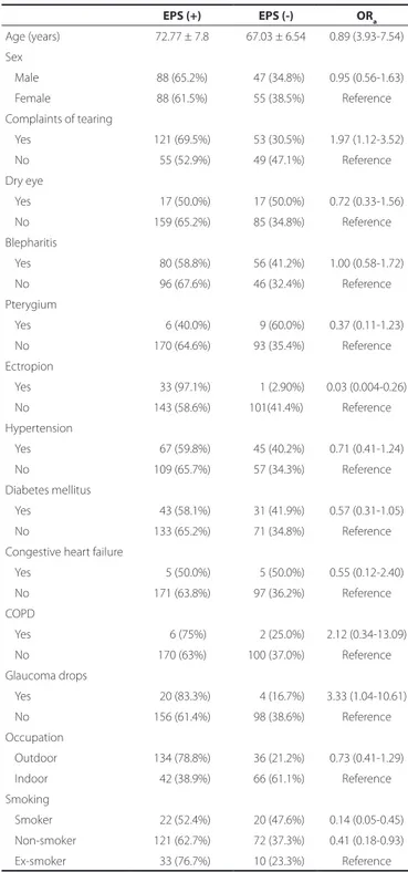
Table 2. Demographic, lifestyle, ocular, and systemic factors associa-ted with external punctal stenosis
EPS (+) EPS (-) ORa Age (years) 72.77 ± 7.8 67.03 ± 6.54 0.89 (3.93-7.54) Sex Male 88 (65.2%) 047 (34.8%) 0.95 (0.56-1.63) Female 88 (61.5%) 055 (38.5%) Reference Complaints of tearing Yes 121 (69.5%) 053 (30.5%) 1.97 (1.12-3.52) No 055 (52.9%) 049 (47.1%) Reference Dry eye Yes 017 (50.0%) 017 (50.0%) 0.72 (0.33-1.56) No 159 (65.2%) 085 (34.8%) Reference Blepharitis Yes 080 (58.8%) 056 (41.2%) 1.00 (0.58-1.72) No 096 (67.6%) 046 (32.4%) Reference Pterygium Yes 006 (40.0%) 009 (60.0%) 0.37 (0.11-1.23) No 170 (64.6%) 093 (35.4%) Reference Ectropion Yes 033 (97.1%) 001 (2.90%) 0.03 (0.004-0.26) No 143 (58.6%) 101(41.4%) Reference Hypertension Yes 067 (59.8%) 045 (40.2%) 0.71 (0.41-1.24) No 109 (65.7%) 057 (34.3%) Reference Diabetes mellitus Yes 043 (58.1%) 031 (41.9%) 0.57 (0.31-1.05) No 133 (65.2%) 071 (34.8%) Reference
Congestive heart failure
Yes 005 (50.0%) 005 (50.0%) 0.55 (0.12-2.40) No 171 (63.8%) 097 (36.2%) Reference COPD Yes 006 (75%) 002 (25.0%) 2.12 (0.34-13.09) No 170 (63%) 100 (37.0%) Reference Glaucoma drops Yes 020 (83.3%) 004 (16.7%) 3.33 (1.04-10.61) No 156 (61.4%) 098 (38.6%) Reference Occupation Outdoor 134 (78.8%) 036 (21.2%) 0.73 (0.41-1.29) Indoor 042 (38.9%) 066 (61.1%) Reference Smoking Smoker 022 (52.4%) 020 (47.6%) 0.14 (0.05-0.45) Non-smoker 121 (62.7%) 072 (37.3%) 0.41 (0.18-0.93) Ex-smoker 033 (76.7%) 010 (23.3%) Reference
EPS= external punctal stenosis; COPD= chronic obstructive pulmonary disease.
of the fundus with a +78 diopters lens was performed to detect age-related macular degeneration.
Suspected dry eye was also evaluated with tear film breakup time (BUT), fluorescein and rose bengal staining, and Schirmer’s test. The BUT was also measured by touching the inferotemporal bulbar conjunctiva with a fluorescein sodium strip wetted with a preservative-free isotonic saline. Patients were asked to blink, and the precorneal tear film was examined under blue light illumination using a slit-lamp biomicroscope. The mean value of three measu-rements was recorded. The BUT was considered low when it was less than 10 seconds. The corneal surface was then examined for fluorescein staining, and its presence or absence was recorded. The cases were labeled as asymptomatic if they did not complain of epiphora and their tear meniscus height was less than 2 mm. The patients were labeled as having dry eye if they had a BUT less than 10 s and positive corneal staining with fluorescein. Schirmer’s test was performed in both eyes. Five minutes after the instillation of one drop of oxybuprocaine hydrochloride 0.4%, the inferior fornix was gently blotted, and a precalibrated standard filter strip was placed in the lower temporal fornix for 5 min. During this time, the participants were instructed to look slightly upward and blink normally. After removing the strip, the length of the strip that was wet was measured. The test result was considered positive if this measurement was ≤5 mm.
This study was approved by Baskent University Institutional Review Board and Ethics Committee (Project no: 94603339-604.01.02/2131) and supported by the Baskent University Research Fund. Informed consent was obtained from all participants. The study adhered to the tenets of the Declaration of Helsinki.
S
tatiSticalanalySiSStatistical data were analyzed using SPSS version 21.0 (IBM Corp., Armonk, NY, USA). Values were expressed as the mean ± standard deviation. The chi-square test with 95% confidence intervals (CIs) and Fisher’s exact test were used to assess the effects of sex, systemic disease, topical or systemic medication, smoking, and occupational activity. Multiple logistic regression analyses were used to evaluate the independent factors related to having EPS. P values <0.05 were considered statistically significant.
RESULTS
A total of 278 patients were recruited, with a mean age of 70.67 ± 7.85 (65-92 years). The prevalence of EPS was 63.3% (176/278) among this elderly population visiting our general eye clinic for routine ophthalmologic examination. One hundred thirty-five of the patients were men (48.6%) and 143 were women (51.4%). One
hundred twelve (40.3%) patients had bilateral EPS. The presence of epiphora was consistent with the EPS ratio (p=0.004). No sex domi-nance was observed for EPS (p=0.45).
Chronic blepharitis was found in 45.5% (80/176) of the patients with EPS (95% CI, 0.58-1.72, p=0.13), and this was the most common ocular factor associated with EPS. The associations between the presence of EPS and other demographic, occupational, ocular, and systemic factors are presented in table 2.
Pr e va l e n c e a n d a s s o c i at e d fac t o r s o f e x t e r n a l P u n c ta l s t e n o s i s a m o n g e l d e r ly Pat i e n t s i n tu r k e y
298 Arq Bras Oftalmol. 2017;80(5):296-9
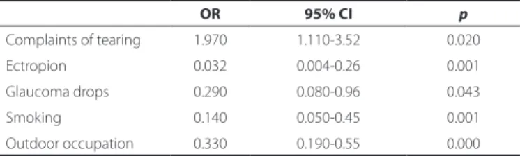
Univariate and multivariate logistic regression analyses were used to identify the independent predictors of EPS (Table 3). Com-plaints of tearing (95% CI, 1.11-3.52, p=0.02), ectropion (95% CI, 0.004-0.26, p=0.001), glaucoma drop usage (95% CI, 0.08-0.96, p=0.043), smoking (95% CI, 0.13-0.84, p=0.021), and outdoor occu-pational activity (95% CI, 3.42-9.97, p<0.05) were significantly asso-ciated with EPS according to the results of the multivariate logistic regression analysis. In contrast, we did not find significant associa-tions between EPS and smoking or outdoor occupational activity. DISCUSSION
To date, this is the first study to evaluate EPS in elderly patients only. The prevalence of EPS was found to be 63.3% in this patient population. EPS is a common cause of epiphora, and recent studies have reported a wide range in the prevalence of the latter, from 8% to 54.3%(5,6,8,9). Similarly, we had found a prevalence of EPS of 25.8%
in our previous study(10). All of these previous studies reported a
positive correlation between the prevalence of EPS and increasing age. This association is thought to be the result of changes caused by aging and fibrosing of the tissue surrounding the punctum(4).
Studies that evaluated the histopathologic changes in EPS speci-mens reported that fibrosis is the most prevalent finding, followed by chronic inflammation(11,12). The higher prevalence of EPS in our
study can be explained according to the findings reported in these previous studies.
Previous studies reported female dominancy in EPS(7,13), and
suggested that postmenopausal hormonal changes may be respon-sible for this sex difference; however, other reports did not show sex differences(5,6). These authors suggested that this lack of sex
diffe-rence could be related to differing sampling methods used in these studies. Similarly, we did not find any sex difference in our study. Our sample group was recruited from patients attending a general ophthalmology clinic, and we believe that this is an ideal method of recruitment to minimize bias.
Chronic blepharitis was diagnosed in 45.5% of our patients, and it was the most commonly related factor to EPS. However, we did not find any significant association between blepharitis and EPS. Conversely, the authors of previous studies showed that the most common underlying factor for EPS was blepharitis(5-7); similarly, no
significant association was found between EPS and blepharitis in these studies. However, the authors suggested that chronic blepha-ritis predisposes individuals to EPS on the basis of the inflammatory and cicatricial changes. Consistent with these reports, recent studies have showed that chronic inflammation is the most common finding in EPS specimens(11,12). It is clear that the chronic inflammatory process
resulting from chronic blepharitis plays an important role, even if not completely, in the development of EPS.
We also evaluated the association between EPS and dry eye, pterygium, and other levels of lacrimal pathway stenosis; however, we did not find any significant relationships. On the other hand, previous studies have reported an association between EPS and nasolacrimal ductal stenosis; topical medication use, systemic
che-motherapy, trachoma, and cicatrizing diseases of the conjunctiva are thought to be responsible for this relationship(13-16). However,
Kashkouli et al. did not find any association between EPS and lacri-mal pathway stenosis(7). Although an association between EPS and
pinguecula was found in a recent study, the authors did not find any relationship between EPS and pterygium, as we did in our study(5).
They suggested that aging and sun exposure could be the common etiologic factors for these diseases.
Eyelid malposition, as seen in ectropion, may cause punctal ste-nosis, possibly due to underuse of an external punctum unopposed to the tear meniscus, or perhaps secondary to local inflammation(17).
Damasceno et al. revealed a decrease in elastic fibers in the pretarsal orbicularis oculi muscle, in the tarsal stroma, and in the eyelid skin in patients suffering from age-related involutional ectropion and entropion; these changes might have caused the fibrotic changes in the EPS specimens observed in previous research(18). In our study,
18.8% (33/143) of the patients with EPS had ectropion (p=0.002). Previous studies that studied EPS did not show any association between these two situations (i.e.; EPS and ectropion). Presumably, the authors felt that this relationship did not need to be evaluated because of the already well-known and strong association between these pathologies. However, the difference in treatment between EPS and ectropion means that it is important to show the relationship between these diseases.
Long-term treatment with several topical antiglaucoma agents has also been associated with punctal stenosis(15). In addition, the
conjunctiva and Tenon’s capsule of the patients receiving long-term latanoprost therapy exhibit inflammatory changes(19). However, this
association has not been shown in population-based EPS studies until now. In our study group, EPS was significantly more prevalent in patients taking glaucoma medication (p=0.033). Nonetheless, it is important to take into consideration the lack of discrimination of the type of glaucoma drops and the medication period.
There are two main types of risk factors: intrinsic and extrinsic. Extrinsic factors include parameters such as alcohol use, chronic exposure to sunlight, smoking, and nutrition deficit. The most impor-tant parameter, contributing to 80% of skin aging, appears to be sun exposure. Ultraviolet A rays induce the formation of reactive oxygen species that readily react with membrane lipids and amino acids, and are suspected of playing a substantial role in skin photoaging(21). Skin
photoaging can lead to fibrotic changes in punctal tissue and EPS. Therefore, we evaluated and found a significant association between outdoor occupational activity and EPS (95% CI, 3.42-9.97, p<0.05). Agricultural workers constituted the majority of our study group. Similarly, Viso et al. found a significant relationship between these parameters and EPS(5).
Previous studies that included histopathologic analysis have revealed that 10-15% of EPS specimens show squamous metapla-sia(11,12). Squamous metaplasia is known to be an epithelial response
to chronic irritant exposure, and is typically seen in the airways of smokers. Therefore, we evaluated the effect of smoking on EPS, and found that current smokers have a significantly greater EPS ratio than non-smokers and ex-smokers (95% CI, 0.13-0.84, p=0.021). If we had had a chance to assess the histopathology of these patients, we suspect that we might have found a high rate of squamous metaplasia. Conversely, a recent study did not find this difference in smokers compared to non-smokers(5).
Dry eye is a common ocular surface disease in elderly pa-tients(22). The effectiveness of punctal occlusion in dry eye patients
is reportedly comparable to that of other treatment modalities(23).
In our study, complaints of epiphora were significantly associated with the presence of EPS. However, 31.3% (55/176) of the patients with EPS had no complaints of epiphora, although increased tear meniscus was found after punctal occlusion in previous research(24).
In addition, 92.7% (51/55) of these patients had no dry eye findings. These results indicate that EPS may help protect against dry eye in
Table 3. Multivariate logistic regression analysis of potential associa-tions with external punctal stenosis
OR 95% CI p Complaints of tearing 1.970 1.110-3.52 0.020 Ectropion 0.032 0.004-0.26 0.001 Glaucoma drops 0.290 0.080-0.96 0.043 Smoking 0.140 0.050-0.45 0.001 Outdoor occupation 0.330 0.190-0.55 0.000
UlU s oy Mo, e t a l.
299 Arq Bras Oftalmol. 2017;80(5):296-9
elderly patients. According to the data, we suggest that surgical in-tervention for EPS in elderly patients should not be overly promoted to avoid impairment of this suspected protective mechanism.
In conclusion, to the best of our knowledge, this was the first study to evaluate EPS in elderly patients. We showed that its pre-valence was higher in this group than in the general population. Chronic inflammation, antiglaucomatous medication use, ectropion, smoking, and sun exposure as a result of outdoor occupation activity are significant etiologic factors in the development of EPS. However, further studies with a large sample size supported by histopathologic analysis are needed in future to verify these associations.
REFERENCES
1. Seiff SR, Shorr N, Adams T. Surgical treatment of punctalcanalicular fibrosis from 5-fluorouracil therapy. Cancer. 1985;56(8):2148-9.
2. Tabbara KF, Bobb AA. Lacrimal system complications in trachoma. Ophthalmology. 1980;87(4):298-301.
3. Brink HM, Beex LV. Punctal and canalicular stenosis associated with systemic fluo-rouracil therapy. Report of five cases and review of the literature. Doc Ophthalmol. 1995;90(1):1-6.
4. Kristan RW. Treatment of lacrimal punctal stenosis with a one-snip canaliculotomy and temporary punctal plugs. Arch Ophthalmol. 1988;106(7):878-9.
5. Viso E, Rodríguez-Ares MT, Gude F. Prevalence and associations of external punctal stenosis in a general population in Spain. Cornea. 2012;31(11):1240-5.
6. Bukhari A. Prevalence of punctal stenosis among ophthalmology patients. Middle East Afr J Ophthalmol. 2009;16(2):85-7.
7. Kashkouli M, Beigi B, Murthy R, Astbury N. Acquired external punctalstenosis: etiology and associated findings. Am J Ophthalmol. 2003;136(6):1079-84.
8. Nemet AY. The etiology of epiphora: a multifactorial issue. Semin Ophthalmol. 2016; 31(3):275-9.
9. Mainville N, Jordan DR. Etiology of tearing: a retrospective analysis of referrals to a tertiary care oculoplastics practice. Ophthal Plast Reconstr Surg. 2011;27(3):155-7.
10. Ulusoy MO, Kıvanç SA, Atakan M, Akova-Budak B. How important is the etiology in the treatment of epiphora? J Ophthalmol. 2016;2016:1438376.
11. Port AD, Chen YT, Lelli GJ Jr. Histopathologic changes in punctal stenosis. Ophthal Plast Reconstr Surg. 2013;29(3):201-4.
12. Ali MJ, Mishra DK, Baig F, Lakshman M, Naik MN. Punctal stenosis: histopathology, immunology, and electron microscopic features-a step toward unraveling the mys-terious etiopathogenesis. Ophthal Plast Reconstr Surg. 2015;31(2):98-102. 13. Esmaeli B, Valero V, Ahmadi MA, Booser D. Canalicular stenosis secondary to docetaxel
(Taxotere), a newly recognized side effect. Ophthalmology 2001;108(5):994-5. 14. Tabbara KF, Bobb AA. Lacrimal system complications in trachoma. Ophthalmology.
1980;87(4):298-301.
15. McNab AA. Lacrimal canalicular obstruction associated with topical ocular medica-tion. Aust NZ J Ophthalmol. 1998;26(3):219-23.
16. Weston BC, Loveless JW. Canalicular stenosis due to topical use of fortified antibiotics. Can J Ophthalmol. 2000;35(6):334-5.
17. Soiberman U, Kakizaki H, Selva D, Leibovitch I. Punctal stenosis: definition, diagnosis, and treatment. Clin Ophthalmol. 2012;6:1011-8.
18. Damasceno RW, Heindl LM, Hofmann-Rummelt C, Belfort R, Schlötzer-Schrehardt U, Kruse FE, et al. Pathogenesis of involutional ectropion and entropion: the involvement of matrix metalloproteinases in elastic fiber degradation. Orbit. 2011;30(3):132-9. 19. Sherwood MB, Grierson I, Millar L, Hitchings RA. Long-term morphologic effects of
antiglaucoma drugs on the conjunctiva and Tenon’s capsule in glaucomatous pa-tients. Ophthalmology. 1989;96(3):327-35.
20. Damasceno RW, Avgitidou G, Belfort R Jr, Dantas PE, Holbach LM, Heindl LM. Eyelid aging: pathophysiology and clinical management. Arq Bras Oftalmol. 2015;78(5):328-31. 21. Matsumura Y, Ananthaswamy HN. Toxic effects of ultraviolet radiation on the skin.
Toxicol Appl Pharmacol. 2004;195(3):298-308.
22. Viso E, Rodriguez-Ares MT, Gude F. Prevalence of and associated factors for dry eye in a Spanish adult population (the Salnes Eye Study). Ophthalmic Epidemiol. 2009; 16(1):15-21.
23. Roberts CW, Carniglia PE, Brazzo BG. Comparison of topical cyclosporine, punctal occlusion, and a combination for the treatment of dry eye. Cornea. 2007;26(7):805-9. 24. Ibrahim OM, Dogru M, Kojima T, Matsumoto Y, Wakamatsu TH, Tsubota K, et al. OCT
assessment of tear meniscus after punctal occlusion in dry eye disease. Optom Vis Sci. 2012;89(5):E770-6.