Ankara Üniv Vet Fak Derg, 61, 153-154, 2014
Kısa Bilimsel Çalışma / Short Communication
Calcinosis circumscripta in a dog
Nihat YUMUŞAK1, Mehmet Eray ALÇIĞIR2, Murat ÇALIŞKAN3, Osman KUTSAL2
1Harran University,Faculty of Veterinary Medicine, Department of Pathology, Sanliurfa; Ankara University, Faculty of Veterinary
Medicine, 2Department of Pathology; 3Department of Surgery, Ankara, Turkey.
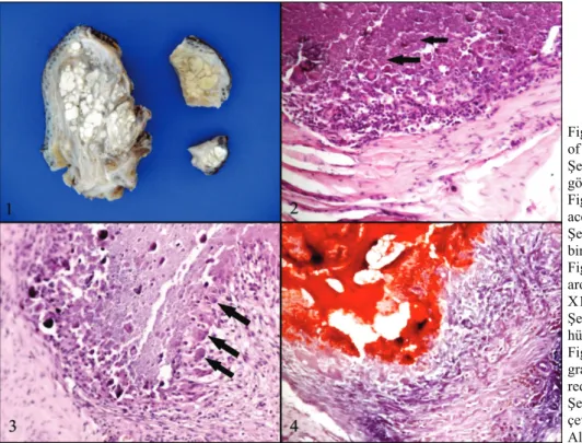
Summary: In the case, calcinosis circumscripta are described with macroscopical and microscopical findings on proximal of right limb in a 2.5 years old, male Rotweiller dog. Macroscopically, all masses were covered with skin and had firm. The cut sections were seen lobullary and composed of well defined areas and vicious and white fluid. Microscopically, it was detected that numbered of restricted areas, where the center had calcification in dark blue stained as haematoxylin eosin (HE) and red stained with Alizerine red S., connective tissue and mononuclear cell infiltration consisting of macrophages, lymphocytes and plasma cells and giant cells.
Key words: Calcinosis circumscripta, dog, histopathology.
Bir köpekte kalsinozis sirkumskripta
Özet: Bu olguda Rottweiler ırkı, 2,5 yaşlı, erkek bir köpeğin sağ ön bacağın proksimalinde kalsinozis sirkumskripta olgusu tanımlandı. Makroskobik olarak; kitler aynı özellikte ve üzerleri deriyle kaplı, oldukça sert kıvamlı, kesit yüzleri ise çok sayıda, lobuler tarzda, sınırları belirgin ve içlerinde koyu kıvamlı beyaz renkte içerik bulunan yapılardan oluşmaktaydı. Mikroskobik incelemede; dermiste, ortalarında hematoksilen eosin (HE) ile koyu mor renkte Alizerine red S. boyamasında ise kırmızı renkte boyanmış kalsiyum birikimlerinin çevrelerinden makrofajlar ve lenfositlerle birlikte dev hücrelerininde olduğu bağ doku hücreleri ile sınırlandırılmış yapılar görüldü.
Anahtar sözcükler: Histopatoloji, kalsinozis sirkumskripta, köpek.
Calcinosis circumscripta is an uncommon tumor like lesions characterizated solid or multiple masses by ectopic, dystrophic, metastatic and iatrogenic deposition of calcium salts in soft tissues (2, 8-11). The pathogenesis of this lesions are unknown, but dog may develop suffering from serious illness such as chronic renal disease (11). It is occurred mostly in horse (between 1 and 13 years old), large dog breeds (<2 years old) which localized in pad nodullary (3, 8, 10). Pathogenesis is still unclear, but it has been thought due to trauma or excessive secrection of apocrin glands in dog and horse (5, 6, 9, 10). Especially, it is frequently seen in Magyar and German shepherd dog and made an assumption about susceptible to these kind of lesion for the breeds (4, 7, 9, 10). In this case, provides the macroscopically and histopathologically description of calcinosis circumscripta in a dog.
The biopsy materials taken from proximal of right limb of a 2.5 year old, male Rotweiller dog with suffering from a swelling and difficulty in walking. Macroscopically, masses from stated regions were weighed of 97 g, 11 g and 2 g and diameter in 10x6x4, 5x3x2 ve 2x2x1 cm. masses were covered with skin and had firm consistency. The cut sections were seen
lobullary and composed of well defined areas and white colored vicious fluid (Figure 1). Tissues were fixed in formalin %10, processed routinly and embedded in parafin. The sections were cut in 5 µm thickness to Alzerine Red S. staining for calcium salts identification and also to haematoxylin eosin (HE) staining. Microscopically, it was detected that numbered of restricted areas, where the center had calcification in dark blue (Figure 2) stained as haematoxylin eosin (HE) and red stained with Alizerine red S. (Figure 4), with connective tissue and inflammatory cell infiltration consisting of macrophages, lymphocytes and plasma cells and giant cells (Figure 3).
Calcinosis circumscripta is mainly evaluated in terms of causes as metastatic, dystrophic, idiopathic and iatrogenic. Metastatic calcinosis is mostly seen in human beings while the other types is usually encountered in dogs and cats (1, 3, 9, 10). Metastatic calcinosis is frequently occured in calcium-phosphate metabolic disorders such as hypercalcemia, hyperphospahatemia and chronic renal disorders (2, 4, 5, 8, 10). However, distrophic calcinosis is followed to necrosis, inflammation and neoplasia inspite of beings normally calcium-phosphat levels in blood. In idiopathic type of calcinosis,
Nihat Yumuşak - Mehmet Eray Alçığır - Murat Çalışkan - Osman Kutsal 154
causes has not entirely been known but, breed predisposition has been thought since it is frequently seen in German and Magyar shepherd dogs (4, 7, 9). However, it is described in Rotweiller dog in the. calcinosis circumscripta is usually occured focally and localizated in pad of large breed dog (<2 year-old dogs) (2, 6, 10). But, it has often been confused with synovial osteochondromatositosis and chondral sarkoma (5, 6, 9-11). In present case, it was detected that typical lesions of calcinosis, which had same characteristic, was circumscripted in multiple masses in proximal of right limb of 2.5 years old Rotweiller dog. On the other hand, it is mentioned about neither neoplasia nor metastasize. However, in the case presented herein, despite the malignancy of the masses, its recurrence and the rather bad prognosis, no such metastasis was detected. In conclusion, calcinosis circumscripta can be diagnosis by radiographic and histopatlogic examinations. And also surgical resection is a helpful for cure. This study will provide guidance for veterinary clinic practices.
References
1. Antonella MA, Benjamin WM, Premal AP, Joanne F, Nicholas MPC (2012): Digital calcinosis circumscripta:
case series and review of the literature. Journal of
Pediatric Orthopaedics B, 21, 443-447.
2. Burns RE, Bicknese EJ, Westropp JL, Shiraki R, Stalis IH (2013): Tumoral calcinosis form of hydroxyapatite
deposition disease in related red-bellied short-necked turtles, emydura subglobosa. Veterinary Pathology, 50,
443-450.
3. Collados J, Bertos A, Peña L, Rodríguez-Quirós J, San Roman F (2002): Lingual calcinosis
circumscripta in a dog. J Vet Dent, 19, 19-21.
4. Davidson EB, Schulz KS, Wisner ER, Schwartz JA (1998): Calcinosis circumscripta of the thoracic wall in a
German shepherd dog. J Am Anim Hosp Assoc, 34,
153-156.
5. Engel S, Randall EK, Cuddon PA, Webb BT, Aboellail TA (2013): Imaging diagnosis: multiple cartilaginous
exostoses and calcinosis circumscripta occurring simultaneously in the cervical spine of a dog. Vet Radiol
Ultrasound, Doi: 10.1111/vru.12066.
6. Komori S, Washizu M (2001): Metastatic calcinosis
circumscripta treated with an oral charcoal absorbent in a dog. J Vet Med Sci, 63, 913-916.
7. Lin JYC, Tsai HJ, Hsu KS, Wang FI (2009): Calcinosis
cutis, calcinosis circumscripta, and “Mille Feuille” lesions. JVCS, 2, 79-87.
8. Pool RR, Thompson KG (2002): Tumors of joints. 199-243. In: DJ Meuton, (ed), Tumors in Domestic Animals. 4 th ed. Iowa State Press, Iowa.
9. Stampley A, Bellah JR (1990): Calcinosis circumscripta
of the metacarpal pad in a dog. J Am Vet Med Assoc, 196,
113-114.
10. Szczepaniak AL, Orzelski M, Smiech A (2008): Canine
calcinosis circumscripta- retrospective studies. Medycyna
Wet, 64, 1397-1400.
11. Tafti AK, Hanna P, Bourque AC (2005): Calcinosis
circumscripta in the dog: A Retrospective Pathological Study. J Vet Med, 52, 13-17.
Geliş tarihi: 21.08.2013 / Kabul tarihi: 12.12.2013
Address for correspondence:
Nihat Yumuşak Harran University,
Faculty of Veterinary Medicine Department of Pathology, 63000, Sanliurfa-TURKEY e-mail: [email protected]
Figure 1. Macroscopical appearance of the masses.
Şekil 1. Kitlelerin makroskobik görünümü.
Figure 2. Granular calcium accumulation (arrows), HE, X100. Şekil 2. Granuler tarzda kalsiyum birikimi (oklar), HE, X100. Figure 3. Giant cells (arrows) around of calcium region, HE, X100.
Şekil 3. Kalsiyum çevresinde dev hücreler (oklar), HE, X100. Figure 4. Calcium accumulation and granulomatous reaction, Alizerine red S., X100.
Şekil 4. Kalsiyum birikimi ve çevresinde granulomatöz reaksiyon, Alizerine red S., X100.