Address for Correspondence: Dr. Serpil Eroğlu, Başkent Üniversitesi Tıp Fakültesi, Kardiyoloji Anabilim Dalı, Fevzi Çakmak Cad. 10. Sok. No:45, Bahçelievler, Ankara-Türkiye
Phone: +90 312 212 68 68 E-mail: serpileroglu@gmail.com Accepted Date: 19.12.2014
©Copyright 2015 by Turkish Society of Cardiology - Available online at www.anatoljcardiol.com DOI:10.5152/akd.2015.5991
Serpil Eroğlu
Department of Cardiology, Faculty of Medicine, Başkent University; Ankara-Turkey
How do we measure epicardial adipose tissue thickness by
transthoracic echocardiography?
A
BSTRACTEpicardial adipose tissue (EAT) is a component of the visceral adiposity located between the heart and pericardium. It is associated with cer-tain diseases, such as metabolic syndrome, coronary artery disease, and hypertension. Therefore, measurement of EAT thickness has recently gained importance. Examination by transthoracic echocardiography for measuring EAT thickness is preferable because of easy availability and low cost. The present review focuses on the method of measuring EAT thickness by transthoracic echocardiography as well as the issues of concern. (Anatol J Cardiol 2015; 15: 416-9)
Keywords: epicardial adipose tissue, epicardial fat, echocardiography
Introduction
Epicardial adipose tissue (EAT) is part of visceral adipose tissue localized between the heart and pericardium, particularly in the atrioventricular and interventricular sulcus, lateral wall of the right ventricle, and around the coronary arteries (1-3). EAT has endocrine, paracrine, vasocrine, and inflammatory charac-teristics (4-6) and is associated with metabolic syndrome (7), insulin resistance (8), coronary artery disease (9, 10), and hyper-tension (11, 12). Therefore, measurement of EAT thickness has gained importance. EAT thickness can be measured by transtho-racic echocardiography, cardiac computed tomography (CT), and cardiac magnetic resonance imaging (MRI) methods. Evaluation by transthoracic echocardiography has come to the forefront because of many advantages, such as easy availability, low cost, no radiation exposure, fastness, and reproducibility. Measurement of EAT thickness by transthoracic echocardiogra-phy is discussed in this article.
EAT measurement by echocardiography was first defined in 2003 by Iacobellis et al. (13). They expressed EAT as an echo-free space above the right ventricular echo-free wall by transthoracic echocardiography and measured the thickness from the anterior aspect of the right ventricular free wall through parasternal long and short axis windows (13). They stated that the reason for
them to prefer this point was the highest EAT thickness in that area and optimal cursor beam orientation in each view (13). By this method, they determined that EAT measurements are cor-related with MRI measurements and confirmed the accuracy of measurements by echocardiography (13). Further studies began to measure EAT thickness considering this method, which was recommended by Iacobellis et al. (13), as the reference.
How is the epicardial adipose tissue measured and from where?
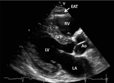
EAT demonstrated by transthoracic echocardiography is the echo-lucent area between the epicardium of the right ventricle and parietal pericardium, which is seen as a thick line above the right ventricular free wall on echo (Fig. 1, 2) (7, 9).
For EAT measurement, the individual is placed in the left lateral decubitus position, and an optimal parasternal long-axis view is tried to be obtained through the left sternal 2-3 intercos-tal space. Interventricular septum and particularly the aortic root are considered as the reference points for the measure-ment from the parasternal long-axis view. Taking the aortic root as the reference, measurement is made by putting the right ventricular free wall and the aortic annulus in the midline of ultrasound waves (Fig. 3) (14-18). The hypoechoic area extend-ing from epicardial surface to the parietal pericardium in front of
Education
416
the right ventricular free wall is vertically measured at the thick-est level (Fig. 3). More sensitive measurements can be made by enhancing depth setting and magnifying the view to assess EAT thickness more clearly (Fig. 4) (9). Some references recommend the measurements to be made in three (3, 15, 17, 18), some refer-ences recommend to be made in six (9), and some referrefer-ences recommend to be made in 10 (3) cardiac cycles. Making the measurements in at least three cardiac cycles, calculating the mean value, and not being satisfied with a single measurement would be convenient for accurate measurement.
After measuring from the parasternal long-axis, the probe is switched to 90° clockwise and parasternal short-axis view is obtained. In the parasternal short-axis view, mid-chordal region (19), the tip of the papillary muscles (18, 19), and interventricular
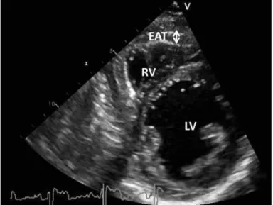
septum (19) can be regarded as reference points (19). In general, measurement from 2 cm away from the interventricular septum (14, 16) and from the parasternal short-axis mid-ventricular level is recommended (20). EAT is measured from the echo-lucent area between the right ventricle and parietal pericardium on the parasternal short axis section as shown in Figure 5 (3). Parasternal long- and short-axis measurements must be aver-aged to obtain the mean thickness.
In the initial publications, although EAT was most frequently measured during end-systole due to deformation and pressure on EAT in the distal aspect (13, 14), it is measured, in some pub-lications during end-diastole in order to be consistent with car-diac CT and MRI (9, 15, 19). In our studies, we prefer measuring EAT thickness during diastole (10, 11, 16). Performing EAT
mea-Eroğlu S. Measure epicardial adipose tissue
Anatol J Cardiol 2015; 15: 416-9
417
Figure 1. Transthoracic echocardiographic view of epicardial adipose tissue. Epicardial adipose tissue is an echo-lucent area between epicardial surface and parietal pericardium in front of the right ventricular free wall and is pointed by a white arrow
Ao- aorta; EAT- epicardial adipose tissue; IVS- interventricular septum; LA- left atrium; LV- left ventricle; RV- right ventricle
Figure 3. Making measurement by taking the aortic root as the reference and placing in the midline between the right ventricular free wall and aortic annulus. Vertical length between the right ventricular free wall and parietal pericardium is measured. The area between the white arrows indicates epicardial adipose tissue thickness
Ao- aorta; EAT- epicardial adipose tissue; RV- right ventricle
Figure 2. The view of epicardial adipose tissue(zoomed in). Epicardial adipose tissue is an echo-lucent area between the epicardium of the right ventricle and parietal pericardium in front of the right ventricular free wall
EAT- epicardial adipose tissue; RV- right ventricle
Figure 4. The borders of epicardial adipose tissue in magnified size and measurement of epicardial adipose tissue. White arrow points out the measurement of epicardial adipose tissue thickness
surement during end-diastole just before the R-wave on the ECG would be convenient for the standardization of publications and measurements (3). It should be kept in mind that end-systolic measurements will reveal higher values as compared with end-diastolic measurements. Therefore, it is necessary to pay atten-tion whether measurements have been made during end-systole or -diastole while interpreting EAT thickness in the publications. How can we differentiate epicardial adipose tissue from pericardial adipose tissue?
One of the important issues to pay attention while measuring EAT thickness is to make a clear differentiation between peri-cardial adipose tissue and EAT. Periperi-cardial adipose tissue and EAT arise from different embryonic origins; their local circulation
and biomolecular features are different (21). EAT should be dif-ferentiated from pericardial adipose tissue because it is a real visceral adipose tissue (21).
Pericardial adipose tissue is the hypoechoic area in front of EAT and parietal pericardium (Fig. 6) (19). Although different from EAT, it does not alter with cardiac cycle (19, 21). It can be easily differentiated from EAT with these features.
Differentiation between epicardial adipose tissue and peri-cardial effusion
Differentiating EAT from pericardial effusion is of great importance. Even though the EAT is viewed as a hypoechoic area, it has different features from pericardial effusion. EAT has specific echo-density that comprises echo-lucent areas and whitish-speckled appearance (16). However, pericardial effusion has more hypoechoic appearance. In addition, although EAT is limited to the front of the right ventricle, pericardial effusion reveals wider spread, and usually it is more prominent in the posterolateral aspect of the left ventricle while the patient is left lateral decubitis position.
What is the normal value of epicardial adipose tissue thickness?
There is yet no definite value considered normal for EAT thickness. There are inconsistencies in the literature regarding EAT thickness. Iacobellis et al. (13) found that EAT thickness measured during end-systole to be minimum 1 mm and maximum 22.6 mm with a mean value of 7 mm in males and 6.5 mm in females among individuals evaluated by echocardiography for standard clinical indications (19). When measured in end-dias-tole, Jeong et al. (15) found a mean value of 6.38 mm (1.1-16.6 mm) in 203 individuals referred to coronary angiography, and Nelson et al. (22) found a mean of 4.7±1.5 mm in 356 asymptom-atic patients. Mookadam et al. (23) stated that an EAT thickness >5 mm during end-diastole is associated with cardiac abnor-malities (left atrial dilatation, lower ejection fraction, increased left ventricular mass, and diastolic dysfunction) that have been detected by echocardiography. In another study, 7.6 mm and higher values measured during end-diastole (15) and in our pre-vious study, 5.2 mm and higher values measured in end-diastole (10) were found as threshold limit values associated with the presence of coronary artery disease. Likewise, threshold limit values for subclinical atherosclerosis (24), metabolic syndrome (19), low coronary flow reserve (16), and hypertension (11) have also been defined. Bertaso et al. (3) suggested that in system-atic review measurements >5 mm should represent a relevant cutoff to define increased EAT thickness, particularly in low-risk populations. Possibly measurements >5 mm during end-diastole could be a cut-off value increased epicardial fat, but that value should be supported by a large studies.
While interpreting these threshold limit values, it should be kept in mind that EAT thickness could be influenced by age, gen-der, and race and whether the measurement was done during end-systole or -diastole.
Eroğlu S.
Measure epicardial adipose tissue Anatol J Cardiol 2015; 15: 416-9
418
Figure 5. Measurement of epicardial adipose tissue thickness from parasternal short axis view. Epicardial adipose tissue thickness is marked by white two-sided arrow
EAT - epicardial adipose tissue; LV - left ventricle; RV - right ventricle
Figure 6. Differentiation between epicardial adipose tissue and pericardial adipose tissue. Epicardial adipose tissue is pointed out by white arrow, and pericardial adipose tissue is pointed out by dashed-line arrow
Limitations to measuring epicardial adipose tissue thickness by transthoracic echocardiography
There are several limitations to the measurement of EAT thickness by transthoracic echocardiography. First, we only par-tially measure EAT by transthoracic echocardiography. In con-trast, both EAT thickness and volume can be measured by car-diac CT and MRI precisely and more accurately than echocar-diography. Echocardiographic measurements are not as repro-ducible as cardiac CT and MRI. Another limitation is the rela-tively poor inter-observer and intra-observer variability as com-pared with cardiac MRI and CT. The foremost limitation is the lack of certain threshold values to predict in pathologies. EAT thickness appears to increase with age, and it could be influ-enced with gender and ethnicity. Although EAT measurement by echocardiography has some limitations, it has the advantage of being an easy, readily available, repeatable, and low cost modal-ity without radiation exposure.
Conclusion
EAT, which still remains a mystery despite new information determined with each passing day, can be easily, cost affec-tively, and reproducibly evaluated by transthoracic echocardiog-raphy. EAT assessment is still a subject of research; however, it appears to be an additional promising marker in assessing car-diovascular and metabolic risks in daily clinical practice.
Conflict of interest: None declared. Peer-review: Externally peer-reviewed.
Acknowledgement: The author thanks to Prof. Dr. Leyla Elif Sade for her scientific support and experiences in preparing the present manuscript.
References
1. Singh N, Singh H, Khanijoun H K, Iacobellis G. Echocardiographic Assessment of Epicardial Adipose Tissue - A Marker of Visceral Adiposity. MJM 2007; 101: 26-30.
2. Wu FZ, Chou KJ, Huang YL, Wu MT. The relation of location-specif-ic eplocation-specif-icardial adipose tissue thlocation-specif-ickness and obstructive coronary artery disease: systemic review and meta-analysis of observa-tional studies. BMC Cardiovasc Disord 2014; 14: 62. [CrossRef] 3. Bertaso AG, Bertol D, Duncan BB, Foppa M. Epicardial fat:
defini-tion, measurements and systematic review of main outcomes. Arq Bras Cardiol 2013; 101: e18-28. [CrossRef]
4. Iacobellis G, Corradi D, Sharma AM. Epicardial adipose tissue: anatomic, biomolecular and clinical relationships with the heart. Nat Clin Pract Cardiovasc Med 2005; 10: 536-43. [CrossRef] 5. Sacks HS, Fain JN. Human epicardial adipose tissue: a review. Am
Heart J 2007; 153: 907-17. [CrossRef]
6. Iacobellis G, Bianco AC. Epicardial adipose tissue: emerging physiological, pathophysiological and clinical features. Trends Endocrinol Metab 2011; 22: 450-7. [CrossRef]
7. Iacobellis G, Ribaudo MC, Assael F, Vecci E, Tiberti C, Zappaterreno A, et al. Echocardiographic epicardial adipose tissue is related to
anthropometric and clinical parameters of metabolic syndrome: a new indicator of cardiovascular risk. J Clin Endocrinol Metab 2003; 88: 5163e8.
8. Iacobellis G, Leonetti F. Epicardial adipose tissue and insulin resistance in obese subjects. J Clin Endocrinol Metab 2005; 90: 6300-2. [CrossRef] 9. Ahn SG, Lim HS, Joe DY, Kang SJ, Choi BJ, Choi SY, et al.
Relationship of epicardial adipose tissue by echocardiography to coronary artery disease. Heart 2008; 94: e7. [CrossRef]
10. Eroğlu S, Sade LE, Yıldırır A, Bal U, Özbiçer S, Özgül AS, et al. Epicardial adipose tissue thickness by echocardiography is a marker for the presence and severity of coronary artery disease. Nutr Metab Cardiovasc Dis 2009; 19: 211-7. [CrossRef]
11. Eroğlu S, Sade LE, Yıldırır A, Demir O, Müderrisoğlu H. Association of epicardial adipose tissue thickness by echocardiography and hypertension. Turk Kardiyol Dern Ars 2013; 41: 115-22. [CrossRef] 12. Dicker D, Atar E, Kornowski R, Bachar GN. Increased epicardial adipose
tissue thickness as a predictor for hypertension: a cross-sectional obser-vational study. J Clin Hypertens (Greenwich) 2013; 15: 893-8. [CrossRef] 13. Iacobellis G, Assael F, Ribaudo MC, Zappaterreno A, Alessi G, Di
Mario U, et al. Epicardial fat from echocardiography: a new method for visceral adipose tissue prediction. Obes Res 2003; 11: 304e310. 14. Chaowalit N, Somers VK, Pellikka PA, Rihal CS, Lopez-Jimenez F.
Subepicardial adipose tissue and the presence and severity of coro-nary artery disease. Atherosclerosis 2006; 186: 354-9. [CrossRef] 15. Jeong JW, Jeong MH, Yun KH, Oh SK, Park EM, Kim YK, et al.
Echocardiographic epicardial fat thickness and coronary artery disease. Circ J 2007; 71: 536-9. [CrossRef]
16. Sade LE, Eroğlu S, Bozbaş H, Özbiçer S, Hayran M, Haberal A, et al. Relation between epicardial fat thickness and coronary flow reserve in women with chest pain and angiographically normal coronary arteries. Atherosclerosis 2009; 204: 580-5. [CrossRef] 17. Momesso DP, Bussade I, Epifanio MA, Schettino CD, Russo LA,
Kupfer R. Increased epicardial adipose tissue in type 1 diabetes is associated with central obesity and metabolic syndrome. Diabetes Res Clin Pract 2011; 91: 47-53. [CrossRef]
18. Mustelier JV, Rego JO, González AG, Sarmiento JC, Riverón BV. Echocardiographic parameters of epicardial fat deposition and its relation to coronary artery disease. Arq Bras Cardiol 2011; 97: 122-9. [CrossRef] 19. Iacobellis G, Willens HJ, Barbaro G, Sharma AM. Threshold values
of high-risk echocardiographic epicardial fat thickness. Obesity (Silver Spring) 2008; 16: 887-92. [CrossRef]
20. Malavazos AE, Di Leo G, Secchi F, Lupo EN, Dogliotti G, Coman C, et al. Relation of echocardiographic epicardial fat thickness and myo-cardial fat. Am J Cardiol 2010; 105: 1831-5. [CrossRef]
21. Iacobellis G, Willens HJ. Echocardiographic epicardial fat: a review of research and clinical applications. J Am Soc Echocardiogr 2009; 22: 1311-9. [CrossRef]
22. Nelson MR, Mookadam F, Thota V, Emani U, Al Harthi M, Lester SJ, et al. Epicardial fat: an additional measurement for subclinical atherosclerosis and cardiovascular risk stratification? J Am Soc Echocardiogr 2011; 24: 339-45. [CrossRef]
23. Mookadam F, Goel R, Alharthi MS, Jiamsripong P, Cha S. Epicardial fat and its association with cardiovascular risk: a cross-sectional observational study. Heart Views 2010; 11: 103-8. [CrossRef] 24. Natale F, Tedesco MA, Mocerino R, de Simone V, Di Marco GM,
Aronne L, et al. Visceral adiposity and arterial stiffness: echocar-diographic epicardial fat thickness reflects, better than waist cir-cumference, carotid arterial stiffness in a large population of hypertensives. Eur J Echocardiogr 2009; 10: 549-55. [CrossRef]
Eroğlu S. Measure epicardial adipose tissue

