Yazışma Adresi/Address for Correspondence: Dr. Derviş Mansuri Yılmaz, Cukurova University School of Medicine,
OLGU SUNUMU / CASE REPORT
Lumbar intervertebral disc herniation in childhood: a report of 7 cases
Çocukluk çağında lomber intervertebral disk herniasyonu: 7 olgunun sunumu
Derviş Mansuri Yılmaz
1, Ersin Hacıyakupoğlu
2, Ömer Neşet Kişi
3, Erol Akgül
4,
Sebahattin Hacıyakupoğlu
51Cukurova University School of Medicine, Department of Neurosurgery, Adana, Turkey 2Klinik für Wirbelsaulen Chirurgie und Neurotraumatologie, Zwickau, Germany 3Mersin State Hospital, Deparment of Neurosurgery, Mersin, Turkey
4Medipol University, School of Medicine, Department of Radiology, Medipol Hospital, İstanbul, Turkey
5Acıbadem University School of Medicine, Department of Neurosurgery, Acibadem Adana Hospital, Adana, Turkey
Cukurova Medical Journal 2018;43(1):197-201.
Abstract Öz
Lumbar disc herniation is a rare pathology in adolescent age and these patients only constitute 1-5% of all patients who underwent surgery for disc herniation. Trauma and family history are reported as important initiating factors for disc herniations but degenerative changes are less important than adults. The objective of this study was to treat the lumbar intervertebral disc herniation in childhood.Between 2005 and 2016, we performed interlaminarmicrodiscectomy to 7 patients younger than 15 years of age for lumbar disc herniation. These patients were evaluated for clinical features, radiological features, operative findings and outcome of surgery. All patients were complaining of low back pain, six of them had radicular pain. Although most of our patients have significant trauma histories there are no evidences of severe trauma on imaging studies. We had excellent results in all patients for the improvement of low back pain and leg pain but neurological deficits were more resistant to improvement. We had excellent results with limited laminectomy and microdiscectomy without fusion. In all cases pains improved postoperatively and they got back to their normal lifestyle in one month
Lomber disk hernisi adolesan yaşta görülen nadir bir patolojidir ve bu hastalar disk hernisi için ameliyat edilen tüm hastaların sadece % 1-5'ini oluşturur. Travma ve aile öyküsü disk hernileri için önemli faktörlerdir. Fakat dejeneratif değişiklikler yetişkinlerden daha az önemlidir. Bu çalışmanın amacı çocukluk çağında lumbar intervertebral disk herniasyonunu tedavi etmekti. 2005-2016 yılları arasında lumbar disk hernisi için 15 yaşından küçük 7 hastaya interlaminar mikrodiskektomi uyguladık. Bu hastalar klinik özellikler, radyolojik özellikler, ameliyat bulguları ve ameliyatın sonucu açısından değerlendirildi. Tüm hastalarda bel ağrısı şikayetleri vardı. Hastaların altısında radiküler ağrı şikayeti mevcuttu. Hastalarımızın çoğunda belirgin travma öyküsü olsa da, görüntüleme çalışmalarında ciddi travma olduğuna dair herhangi bir kanıt bulunmamaktadır. Tüm hastalarda bel ağrısı ve bacak ağrısı şikayetlerinin düzelmesi açısından mükemmel sonuçlar aldık, ancak nörolojik defisitin düzelmesi daha dirençli idi. Sınırlı laminektomi ve mikrodiskektomi ile füzyonsuz mükemmel sonuçlarımız vardı. Tüm olguların şikayetleri ameliyat sonrası düzeldi ve bir ay içinde normal yaşam tarzlarına döndüler.
Key words: Lumbar intervertebral, disc herniation,
childhood, low back pain, microdiscectomy Anahtar kelimeler: herniasyonu, çocukluk çağı, bel ağrısıLomber intervertebral, disk
INTRODUCTION
Lumbar disc herniation is a common vertebral column disease in adults whereas this disease is rare in childhood and adolescent age. Since it was first described by Wahren as a case report1 in 1945 there
was only few patients described in the literature. In only 13% of these cases magnetic resonance imaging studies showed degenerative changes, end plaque defects and asymmetric joint facets as to be adults. It was affected at single level in 88% and at two or three levels in 12% of these patients. While disc
were remained subligamentous in 80%, extruded in 4%, intact annulus in 16%, it was a rarely announced calcification of disc2-5. In 30-60% of patients, it was
reported existence of a significant trauma in historical examination, in addition familial predisposition and congenital anomalies were determined as important factors in young patients3,4,6-9.
The most common complaint of these patients was low back pain. Radiculopathy, neurological compromise due to lumbar disc herniation compressing, sensory loss, cauda equina syndrome, sphincteric disturbances and scoliosis were major findings in physical examination2,3,5. In previously
series, macroscopic examination of herniated discs represent different to those observed in adults.
Discs were soft, rubbery and hydrated in 92% while hard or partially calcified in 8%3,10. The objective of
this study was to show method of the lumbar disc herniation treatment with limited laminectomy and microdiscectomy without fusion in childhood.
CASES
Between 2005 and 2016, seven patients younger than 15 years of age were surgically treated for lumbar disc herniation in our clinic (Table 1). All patients presented low back pain due to lumbar disc herniation. Other causes of low back pain (spondylolisthesis, congenital malformations, infections of spine, inflammation and neoplastic pathologies) were excluded.
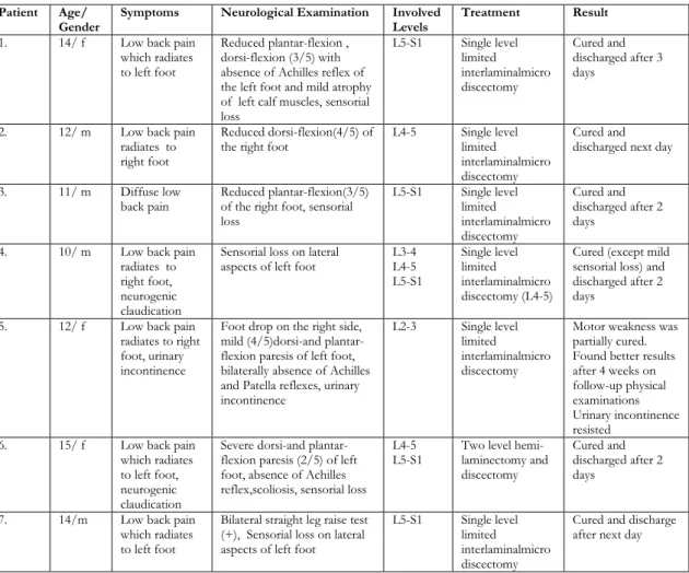
Table 1. Age, gender, clinical features, radiological involved levels and treatment results of patients.
Patient Age/
Gender Symptoms Neurological Examination Involved Levels Treatment Result 1. 14/ f Low back pain
which radiates to left foot
Reduced plantar-flexion , dorsi-flexion (3/5) with absence of Achilles reflex of the left foot and mild atrophy of left calf muscles, sensorial loss L5-S1 Single level limited interlaminalmicro discectomy Cured and discharged after 3 days
2. 12/ m Low back pain radiates to right foot
Reduced dorsi-flexion(4/5) of
the right foot L4-5 Single level limited interlaminalmicro discectomy
Cured and discharged next day 3. 11/ m Diffuse low
back pain Reduced plantar-flexion(3/5) of the right foot, sensorial loss L5-S1 Single level limited interlaminalmicro discectomy Cured and discharged after 2 days
4. 10/ m Low back pain radiates to right foot, neurogenic claudication
Sensorial loss on lateral
aspects of left foot L3-4 L4-5 L5-S1
Single level limited interlaminalmicro discectomy (L4-5)
Cured (except mild sensorial loss) and discharged after 2 days
5. 12/ f Low back pain radiates to right foot, urinary incontinence
Foot drop on the right side, mild (4/5)dorsi-and plantar-flexion paresis of left foot, bilaterally absence of Achilles and Patella reflexes, urinary incontinence
L2-3 Single level limited interlaminalmicro discectomy
Motor weakness was partially cured. Found better results after 4 weeks on follow-up physical examinations Urinary incontinence resisted
6. 15/ f Low back pain which radiates to left foot, neurogenic claudication
Severe dorsi-and plantar-flexion paresis (2/5) of left foot, absence of Achilles reflex,scoliosis, sensorial loss
L4-5
L5-S1 Two level hemi-laminectomy and discectomy
Cured and discharged after 2 days
7. 14/m Low back pain which radiates to left foot
Bilateral straight leg raise test (+), Sensorial loss on lateral aspects of left foot
L5-S1 Single level limited interlaminalmicro discectomy
Cured and discharge after next day
These patients were evaluated for clinical features, radiological features, operative findings and outcome of surgery. Although all of these seven patients were applied rest and conservative treatment, they continued to complain of radicular pain, persistent back pain and restriction of activity. Four of patients were boy and three were girls. The mean ages of the patients were 12.5 years. Six of our patients had radicular pain. Six patients had a history of significant trauma, three of them had nonspecific sport injuries, and other three patients have fallen down from tree and stairs.
The last patients historical and physical examination also exposed being heavy lifting. There wasn’t any family history of lumbar disc herniation. We performed single level interlaminar microdiscectomy to six patients and two level microdiscectomy to one patient.The patient with three levels of herniated disc underwent surgery for exceedingly protruded L4-5 level; L3-4, L5-S1 levels were not operated.
Figure 1A. Sagittal T2-MRI lumbar disc herniation showing a marked extrusion at L2–3.
Figure 1B. Axial T2-M lumbar disc herniation showing a marked extrusion at L2–3.
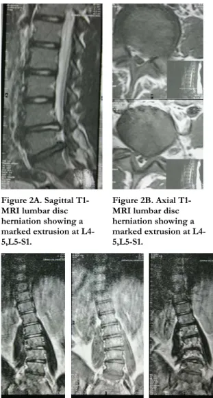
Five patients had lower limb weakness, in two patient there was no motor weakness and three patients had no sensorial loss. Scoliosis was present in one case.The most commonly involved level was L5-S1 which occurs in five patients. Two cases were affected multiple levels, one of them was L4-5, L5-S1(Figure 2A-B-C) and the other one was L3-4, L4-5, L5-S1(Figure 3A-B).
In all cases of magnetic resonance imaging (MRG) disclosed centrolateral placed discs. Single level discectomy was performed in 5 cases and in one case we performed two level discectomy. At operation, all disc substances were soft, rubbery and dehydrated. Before discharge, their low back pain were improved. Except the patient which L2-3 disc was affected, their motor deficits improved.
herniation was better after operation and dorsiflexion loss was minimally improved. In all patients claudication distance and scoliosis improvements were observed. Out of fourpatients’ sensorial loss improved only in one patient.
Figure 2A. Sagittal T1-MRI lumbar disc herniation showing a marked extrusion at L4-5,L5-S1.
Figure 2B. Axial T1-MRI lumbar disc herniation showing a marked extrusion at L4-5,L5-S1.
Figure 2C. Coronal T1-MRI lumbar scoliosis
In eleven years, we operated seven patients for incapacitating, low back pain, radicular pain or neurological deficit due to lumbar disc herniation. The mean duration of symptoms was 5.6 months. All patients had low back pain, which was diffuse low back pain in one and radicular in six patients. Neurogenic claudication was present in two patients. Incomplete cauda equine syndromes and sphinteric disturbances were present in one patient at L2-3 level (Figure1A-B).
DISCUSSION
Lumbar disc herniation rarely occurs in childhood and adolescence, it represents only 0.5-3.8%of all cases. This rate is raised among Japanese and it constitutes around 15.4% of disc disease5,11. There
are reported only four cases in age 9 and less5. In
adults, lumber disc herniation results from lifting and twisting, whereas major causes are repetitive trauma by sport, being heavy lifting and having tall stature in childhood, also in previously reports most cases are seemed to be related to fractures and dislocations which associated with major traumas11,12,13. It is exceptional to happen disc
herniation without vertebral osseos injury in adolescence period. In 13% of cases there exists degenerative changes and only 2% of these cases are aggravated by trauma and it has suggested that decreased blood flow in end plaque results degeneration of nucleus pulposus. 60-68%of cases have a family history of lumbar disc herniation. It might be a result of weak connective tissue and early predisposition of degenerative changes in the spine in these families, and leading to disc herniation in relatively young age13,14.
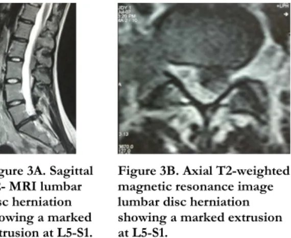
Figure 3A. Sagittal T2- MRI lumbar disc herniation showing a marked extrusion at L5-S1.
Figure 3B. Axial T2-weighted magnetic resonance image lumbar disc herniation showing a marked extrusion at L5-S1.
Six of our cases have a story of significant trauma: four of them were sport related incidents, one of them was falling down from a tree and the last one was falling down from stairs. The sixth patient has a complaint of heavy lifting. None of the patients had any family story, nor repetitive traumas. It was previously reported that severe trauma is associated with structural changes such as fractures and dislocations, furthermore occurrence of disc herniation without any osseous injury is unusua5,15.
Immature ligamentous flaccidity, immature muscular structures, incomplete ossification and increased
mobility due to immature facet joints might prevent fractures and dislocations in childhood. Although most of our cases have a story of trauma, imaging studies and operational findings showed not any vertebral injury. It is supporting previously reports that suggests gross changes are not necessarily contributory factors for lumbar disc herniation in adolescent age group3,4,16, also microtrauma which
may not be seen radiologically but aggravates disc herniation is a significant factor in these cases. In contrast to previously reports which suggests congenital anomalies and discogenic diseases play an important role, in our series there was not any4,8.
It was suggested that the most vulnerable level is L4-5 in childhood3,5.However, in our series the most
commonly involved level was L5-S1. The L2-3 level was involved in one case. Multiple level involvement was seen in two cases (Table1). Their major complaint was low back pain. Pain was diffuse in one patient and radicular in six patients. We have found motor weakness in five cases, sensorial loss in five cases and severe sphincteric disturbances in one case. As previously reported, after surgical treatment, we demonstrated excellent results especially for the improvement of the pain. Only one patient with L2-3 level involvement was partially cured, motor weakness was partially improved but urinary incontinence resisted to treatment. These results were similar to previously reports which demonstrated that neurologic deficits are more resistant for improvement3,17,18.
We performed interlaminar microdiscectomy to our patients. Discs were soft, rubbery, hydrated therefore hard to excise. All of these operations were performed unilaterally where major neurologic deficit locates.The cases with multiple levels of herniated lumbar discs were treated at the level of where MRI studies demonstrated significant protruded disc. We only performed multilevel discectomy (L4-5,L5-S1)to the case with foot drop and diminished achilles reflex together.It was endoscopic discectomy, chemonucleolysis and other noninvasive techniques and anterolateral approaches were previously reported as useful methods.Some senior authors suggest to use fusion techniques. We had excellent results with limited laminectomy and discectomy without fusion. The pain of the all cases were improved postoperatively, the patients returned to normal lifestyle in one month after surgery. We performed interlaminar microdiscectomy for lumbar disc herniation under
15 years of age. We had excellent results with limited laminectomy and microdiscectomy without fusion. In all cases, pain improved postoperatively and they got back to their normal lifestyle in one month.
REFERENCES
1. Wahren H. Herniated nucleus pulposus in a child of twelve years. ActaOrthopScand. 1945;16:40-42. 2. Aulisa L, Pitta L, Aulisa AG, Mastantuoni G, Pola E,
Leone A. Lumbar nerve root “walled” by a calcified herniated mass in a young patient.Child NervSyst. 2003;19:384-86.
3. Kumar R, Kumar V, Das NK, Behari S, Mahapatra AK. Adolescent lumbar disc disease: findings and outcome. Child NervSyst. 2007;23:1295–9.
4. Lee JY, Ernestus RI, Schröder R, Klug N. Histological study of lumbar intervertebral disc herniation in adolescents.ActaNeurochir. 2000;142:1107-10.
5. Martinez-Lage JF, Cornejo VF, Lopez F, Poza M. Lumbar disc herniation in early childhood:case report and literature review. Child NervSyst. 2003;19:258–60.
6. Bradford DS, Garcia A. Herniations of the lumbar intervertebral disc in children and adolescents. JAMA. 1969;210:2045-51.
7. Callahan DJ, Pack LL, Bream RC, Hensinger RN. Intervertebraldisc impingement syndrome in a child. Spine. 1986;11:402-4.
8. Grobler LJ, Simmons EH, Barrington TW. Intervertebral disc herniation in the adolescent. Spine. 1979;4:267-78.
9. Heitho KB, Gundry CR, Burton CV, Winter RB. Juvenile discogenic disease. Spine. 1994;19:335-40. 10. Kumar R, Kalra SK, Vaid VK. Multiple factor
analysis ofprognosis in lumbar disc herniation and assessment of long-term outcome. Pan Arab J of Neurosurg. 2007;11:56-64.
11. Martinez-Lage JF, Martinez Robledo A, Lopez F, Poza M. Disc protrusion in the child. Particular features and comparison with neoplasms. Child NervSyst. 1997;13:201-7.
12. Kurihara A, Kataoka O. Lumbar disc herniation in children and adolescents. A review of 70 operated cases and their minimum 5-year follow-up studies. Spine. 1980;5:443-51.
13. Shillito J Jr. Pediatric lumbar disc surgery: 20 patients under 15 years of age. Surg Neurol. 1996;46:14-8. 14. Pietila TA, Stendel R, Kombos T, Ramsbacher J,
Schulte T, Brock M. Lumbar disc herniation in patients up to 25 years of age. Neurol Med-Chir. 2001;41:340-4.
15. King AB. Surgical removal of a ruptured intervertebral disc in early childhood. J Pediatr. 1959;55:57-62.
16. Boos N, Weissbach S, Rohrbach H, Weiler C, Spratt KF, Nerlich AG. Classification of age-related changes in lumbar intervertebral discs: 2002 Volvo Award in basic science. Spine. 2002;27:2631-44. 17. Borgesen SE, Vang PS. Herniation of the lumbar
intervertebral disc in children and adolescents. ActaOrthopScand. 1974;45:540–9.
18. DeLuca PF, Mason DE, Weiand R, Howard R, Bassett GS. Excision of herniated nucleus pulposus in children and adolescents. J PediatrOrthoped. 1994;14:318-22.