INVESTIGATION OF NOVEL TUMOR MARKERS
BASED ON HYBRIDOMA TECHNOLOGY
A THESIS SUBMITTED TO
THE DEPARTMENT OF MOLECULAR BIOLOGY AND GENETICS
AND THE INSTITUTE OF ENGINEERING AND SCIENCE OF
BILKENT UNIVERSITY
IN PARTIAL FULFILLMENT OF THE REQUIREMENTS
FOR THE DEGREE OF MASTER OF SCIENCE
BY
HİLAL ÇELİKKAYA
AUGUST, 2004
I certify that I have read this thesis and in my opinion it is fully adequate, in scope and in
quality, as a thesis for the degree of Master of Science.
Assoc. Prof. Ayhan Kubar
I certify that I have read this thesis and in my opinion it is fully adequate, in scope and in
quality, as a thesis for the degree of Master of Science.
Assist. Prof. Kamil Can Akçalı
I certify that I have read this thesis and in my opinion it is fully adequate, in scope and in
quality, as a thesis for the degree of Master of Science.
Assist. Prof. Tamer Yağcı
Approved for the Institute of Engineering and Science.
Prof. Dr. Mehmet Baray
ABSTRACT
Investigation of Novel Tumor Markers
Based On Hybridoma Technology
Hilal Çelikkaya
M.Sc. in Molecular Biology and Genetics
Supervisor: Assist. Prof. Tamer Yağcı
2004, 75 Pages
Hybridoma technology is a highly specific technique utilized for the production of
monoclonal antibodies. In cancer research, monoclonal antibodies are used as tumor
markers for diagnosis of malignant tissue versus benign or normal, for differential
diagnosis of tumor type, for pathological grading of tumor biopsy specimens, for
detection of the antigens that are up- or down-regulated in tumor cells or in sera of
cancer patients, and for detection of primary or metastatic lesions. Additionally,
monoclonal antibodies are employed in predicting the course of the disease, in
diagnostic approaches for carrying imaging reagents to tumors, and in therapy for
targeting cytotoxic reagents to and triggering or blocking cell surface molecules. This
study involved generation of 6D5 and 9C11 monoclonal antibodies against apoptosis
induced hepatocellular carcinoma cell line HuH-7, followed by characterization
experiments. 6D5 antibody recognized 5 different epitopes in a panel of 28 cell lines in
Western blotting experiments. In immunohistochemistry studies, 6D5 demonstrated
positive staining in cirrhotic and cancerous cells of liver cancer tissue samples. On the
other hand, 9C11 antibody recognized a single band in the same panel of 28 cell lines
but it was not immunoreactive in immunoperixodase studies of liver cancer tissue
samples, under our experimental conditions.
ÖZET
Yeni Tümör Belirleyicilerin
Hibridoma Teknolojisine Dayalı Olarak Araştırılması
Hilal Çelikkaya
Yüksek Lisans Tezi, Moleküler Biyoloji ve Genetik Bölümü
Tez Yöneticisi: Yard. Doç. Tamer Yağcı
2004, 75 Sayfa
Hibridoma teknolojisi, monoklonal antikor üretimi için yararlanılan oldukça özgün bir
tekniktir. Monoklonal antikorlar kanser araştırmalarında; kanserli dokunun benign veya
normal dokuya karşı ayırıcı tanısında, tumor tipinin ayırıcı tanısında, tumor biyopsisi
örneklerinin evrelerinin belirlenmesinde, tumor hücreleri veya kanserli hasta
serumlarında artan veya azalan biçimde düzenlenen antijenlerin saptanmasında ve
birincil veya metastazik lezyonların saptanmasında tumor belirleyici olarak
kullanılmaktadır. Bunun yanı sıra monoclonal antikorlar; hastalık seyrinin öngörüsünde,
görüntüleme ayıraçlarının tumor hücrelerine hedeflenmesi gibi tanısal yaklaşımlarda ve
terapide, sitotoksik ayıraçların hücre yüzey moleküllerine yönlendirilmesi ve hücre
yüzey moleküllerinin tetiklenmesi veya bloke edilmesi icin kullanılmaktadır. Bu
çalışma, apoptoz tetiklenmiş hepatoselüler karsinom hücre hattı HuH-7’a karşı 6D5 ve
9C11 monoklonal antikorlarının üretimini takiben, karakterizasyon deneylerini
içermektedir. 6D5 antikoru, Western blotlaması deneylerinde kullanılan 28 hücre
hattından oluşan bir panelde 5 farklı epitopu tanımaktadır. Immünohistokimya
çalışmalarında 6D5, karaciğer kanseri doku örneklerindeki sirozlu ve kanserli hücreleri
pozitif boyamaktadır. Diğer yandan, 9C11 antikoru, 28 hücre hattından oluşan aynı
panelde tek bir bant tanımakta ancak kullandığımız deneysel koşullar altındaki
immunohistokimya çalışmalarında, karaciğer kanseri doku örneklerinde
immunoreaktivite göstermemektedir.
ACKNOWLEDGEMENT
First of all, I would like to express my sincere gratitude to my advisor Assist. Prof.
Tamer Yağcı for being supportive of my studies, for his precious guidance, and for
always being patient with me through my graduate study as an apprentice scientist.
I am grateful to Prof. Dr. Mehmet Öztürk for keeping an eye on my studies and helping
me with his guidance whenever I needed.
I also would like to thank to my colleague Ceren Çıracı for her most valuable support in
my experiments and pleasant friendship.
I am thankful to Assoc. Prof. Emin Öztaş from Gülhane Military Medical Academy,
Department of Medical Histology and Embryology for helping me with optimization of
immunoperoxidase assays. I am also thankful to Assist. Prof. Ayhan Özcan from
Gülhane Military Medical Academy, Department of Pathology for providing us liver
tissue samples with Assoc. Prof. Emin Öztaş, as well as his pathological evaluation of
those tissues and taking picture of the stained sections.
I appreciate guidance of Assist. Prof. Can Akcali in immunoperoxidase studies and I am
thankful to him for answering my questions about immunohistochemistry protocol.
I would like to thank to Molecular Oncology Group members, particularly to Nuri
Öztürk, and to all members of MBG Department for sharing their scientific experience
with me and for their friendship.
My special thanks and deepest gratitude are for my family and for my dearest friends,
who beautify my life with their unconditional love.
TABLE OF CONTENTS
SIGNATURE PAGE……….I
ABSTRACT……….II
ÖZET………...III
ACKNOWLEDGEMENT………...IV
TABLE OF CONTENTS………..V
LIST OF TABLES……….VIII
LIST OF FIGURES………..IX
ABBREVIATIONS………..XI
1. INTRODUCTION……….1
1.1. Understanding Cancer………...1
1.2. Cancer Incidence and Mortality Rates………....1
1.3. Cancer Development………...2
1.4. Cancer Genetics………...5
1.4.1. Tumor Suppressor Genes……….5
1.4.2. Oncogenes………....6
1.5. Selected Cancer Types……….11
1.5.1. Bone Cancer (Sarcomas of the Bone)………11
1.5.2. Breast Cancer……….11
1.5.3. Cervical Cancer………..13
1.5.4. Colon and Rectum Cancer………..13
1.5.5. Kidney Cancer………....14
1.5.6. Liver Cancer………...15
1.5.7. Myeloma……….16
1.5.8. Malignant Melanoma………..17
1.5.9. Prostate Cancer………...17
1.6. Hybridoma Technology and Monoclonal Antibody Production…………...18
1.7. Tumor Markers………..20
1.8.1. CA 125………...22
1.8.2. Carcinoembryonic Antigen (CEA)……….22
1.8.3. Alpha-Fetoprotein (AFP)………...23
1.8.4. CD20………...23
1.8.5. Cytokeratins………....24
1.8.6. HER2………..25
1.8.7. Prostate-Specific Antigen (PSA)………26
1.8.8. Prostate-Specific Membrane Antigen (PSMA)………..26
2. AIM OF THE STUDY……….27
3. MATERIALS AND METHODS……….28
3.1. Production of 6D5 and 9C11 Monoclonal Antibodies………..28
3.1.1. Production of 6D5 and 9C11 Monoclonal Antibody
Producing Hybridomas………...28
3.1.2. Culturing 6D5 and 9C11 Hybridoma Cells for
Antibody Production………..29
3.2. Western Blotting With 6D5 and 9C11 Monoclonal Antibodies…………...30
3.2.1. Tissue Culture Studies………...30
3.2.1.1 Thawing of Cells………..32
3.2.1.2. Subculturing of Cells………...32
3.2.1.3. Freezing of Cells………..33
3.2.2. Protein Extraction from Cells……….33
3.2.3. Bradford Assay for Protein Quantification………34
3.2.4. SDS-Polyacrylamide Gel Electrophoresis
(SDS-PAGE) of Proteins…...36
3.2.5. Transfer of Proteins from SDS-Polyacrylamide
Gel to PVDF Membrane……….40
3.2.6. Immunological Detection of Immobilized Proteins
(Western Blotting)………..41
3.3. Immunoperoxidase Staining of Paraffin-Embedded
Liver Cancer Tissue Samples………42
4. RESULTS……….44
4.1. Production of 6D5 and 9C11 Monoclonal Antibodies………..44
4.2. Biochemical Characterization of 6D5 and 9C11
Monoclonal Antibodies……….44
4.2.1 Western Blotting With 6D5 Antibody……….45
4.2.2 Western Blotting with 9C11 Antibody………52
4.2.3. Banding Pattern of Cell Lines in Western Blotting
With 6D5 and 9C11 Antibodies……….55
4.3. Immunohistochemistry with 6D5 and 9C11 Antibodies………...58
5. DISCUSSION AND FUTURE PERSPECTIVES………...61
LIST OF TABLES
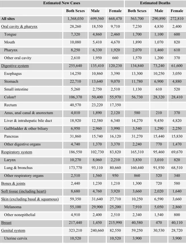
Table 1.1: Estimated new cancer cases and deaths by sex
for all body sites, U.S., 2004………....3
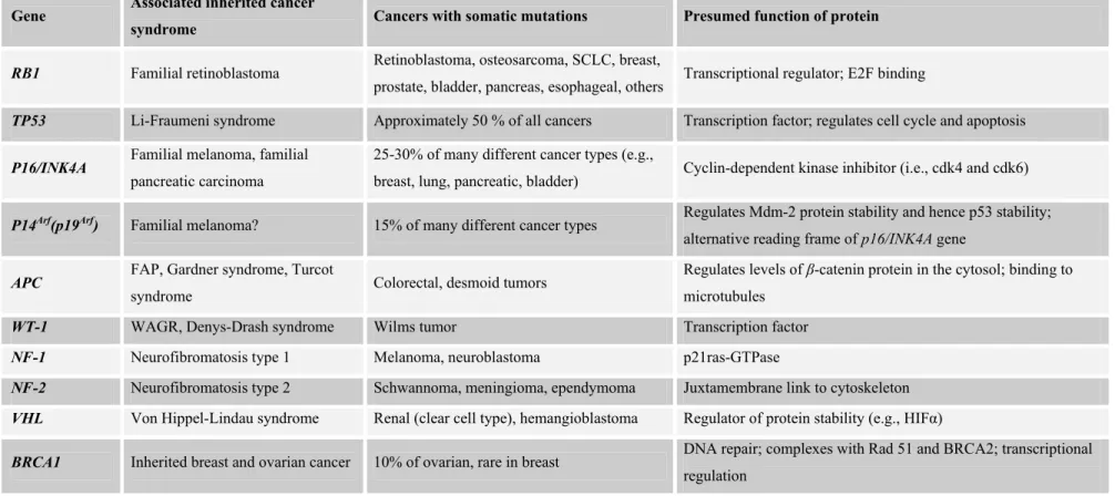
Table 1.2: Some tumor suppressor genes and tumor suppressor proteins………....7
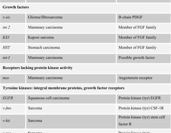
Table 1.3: Oncogenes………....9
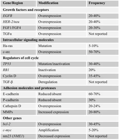
Table 1.4: Somatic alterations in breast cancer………...12
Table 1.5: Clinical applications of monoclonal antibodies (mAbs) in cancer………....19
Table 1.6: Some serum and immunohistochemical markers………...21
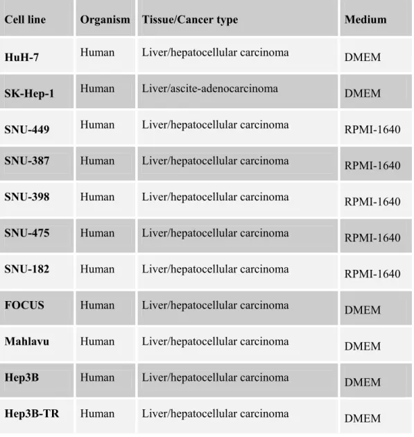
Table 3.1: The cell lines used in Western blotting with 6D5 and 9C11 antibodies…....30
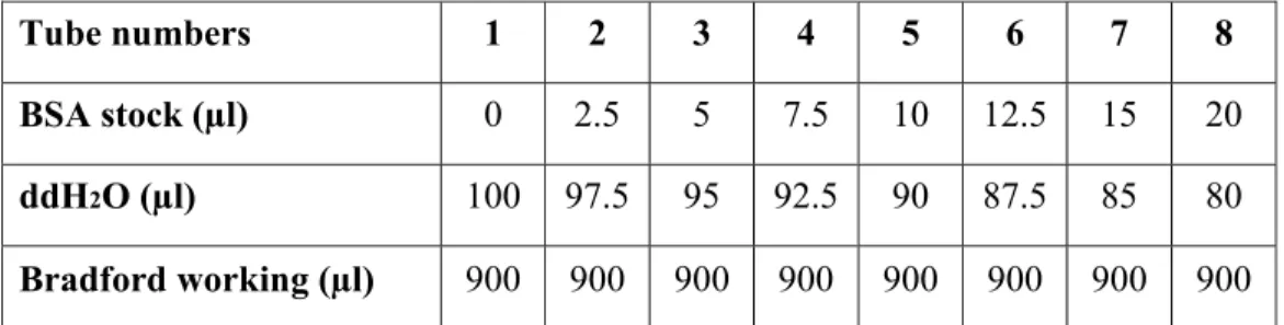
Table 3.2: Preparation of BSA samples………..35
Table 3.3: Preparation of protein samples………..35
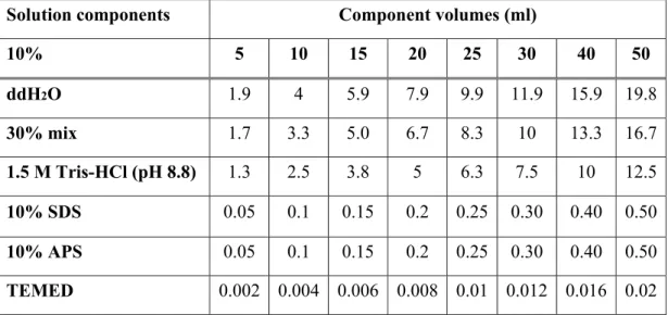
Table 3.4: Components of 10% resolving gel for tris-glycine SDS-PAGE………36
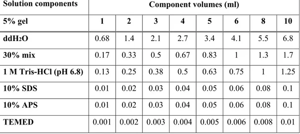
Table 3.5: Components of 5% stacking gel for tris-glycine SDS-PAGE………37
LIST OF FIGURES
Figure 4.1: 6D5 Western blotting with 9 different HCC cell lines……….46
Figure 4.2: 6D5 expression pattern of 4 different HCC cell lines………...47
Figure 4.3: 6D5 Western blotting with cell lines from 7 different
tissues-20 µg ………....48
Figure 4.4: 6D5 Western blotting with cell lines from 7 different
tissues-50 µg ………...….………49
Figure 4.5: 6D5 expression analysis of human and non-human
origin cell lines ………...50
Figure 4.6: Differential expression of Hep3B and Hep3B-TR
cell lines with 6D5………..………..51
Figure 4.7: 9C11 Western blotting of 5 different HCC cell lines…………..………….52
Figure 4.8: 9C11 expression pattern of 8 different HCC cell lines ………...…….53
Figure 4.9: 9C11 Western blotting with cell lines from 7 different tissues ……….…..54
Figure 4.10: 9C11 expression analysis of human and non-human
origin cell lines……….………..…55
Figure 4.11: Immunoperoxidase stainings of paraffin-embedded
liver carcinoma tissue sample S1………...59
Figure 4.12: Immunoperoxidase stainings of paraffin-embedded
liver carcinoma tissue sample S2………..60
Figure 4.13: Immunoperoxidase stainings of paraffin-embedded
ABBREVIATIONS
ACF
Aberrant Crypt Foci
AFP Alpha-Fetoprotein
APS Ammonium
Persulphate
BSA
Bovine Serum Albumin
C Degree
Celsius
CD
Cluster of differentiation
CK Cytokeratin
cm² square
centimeter
CYFRA
Cytokeratin 19 Fragments in Serum
dH
2O Distilled
Water
ddH
2O
Doluble Distilled Water
DAB
Diaminobenzidine Tetrahydrochloride
DMSO Dimethyl
Sulfoxide
DNA Deoxyribonucleic
Acid
DMEM
Dulbecco's Modified Eagle Medium
ECL Enhanced
Chemiluminescence
EDTA
Ethylenediamine Tetraacetic Acid
EGF
Epidermal Growth Factor
EGFR
Epidermal Growth Factor Receptor
ELISA
Enzyme-Linked Immunosorbent Assay
FCS
Fetal Calf Serum
FDA
Food and Drug Administration
FLC Fibrolamellar
Carcinoma
HAT
Hypoxanthine Aminopterin Thymidine
HCC
Hepatocellular Carcinoma
HGPRT
Hypoxanthine Guanine Phosphoribosyl Transferase
HPV
Human Papilloma Virus
HRP
Horse Radish Peroxidase
IgG Immunoglobulin
G
kDa kilodalton
L Liter
LOH
Loss of heterozygosity
mA Miliampere
mAb Monoclonal
antibody
ml Mmililiter
mm Milimeter
mM
Milimolar
M-protein Monoclonal
(or Myeloma) Protein
µg Microgram
µl Microliter
nm Nanometer
NSCLC
Non-Small Cell Lung Cancer
PAGE
Polyacrylamide Gel Electrophoresis
Pap Papanicolaou
PBS
Phosphate Buffered Saline
PSA Prostate-Specific
Antigen
PSMA
Prostate-Specific Membrane Antigen
PVDF Polyvinyl
Difluoride
rpm Revolutions
per
Minute
RTK
Receptor Tyrosine Kinase
SDS Sodium
Dodecyl
Sulfate
TAA Tumor-Associated
Antigen
TBS
Tris-Buffered Saline
TBS-T Tris-Buffered
Saline-Tween-20
TEMED N,N,N',N'-Tetramethylethylenediamine
TGF-β
1Transforming Growth Factor β1
U Unit(s)
UV Ultraviolet
v Volume
w Weight
W Watt
1. INTRODUCTION
1.1. Understanding Cancer
Among the 75 to 100 trillion of an estimated number of cells in the human body, it is not surprising that one or some of our cells may gain an aberrant proliferative activity in the course of a lifetime. Beyond this, any individual may contain hereditary defects leading to abnormal proliferative activity of her/his cells. However, not every aberrant proliferation of cells is referred to as cancer. Excessive division of the cells gives rise to an abnormal mass of tissue, called tumor or neoplasm. If the cells of the tumor stay clustered together in this tumor mass without spreading to surrounding tissue or farther, the neoplasm is said to be benign. On the contrary, malignant (cancerous) tumors invade the nearby tissue and metastasize to distant parts of the body (American Cancer Society, 2004; Alberts et al., 2002; DeVita et al., 2001; http://www.nci.nih.gov).
1.2. Cancer Incidence and Mortality Rates
In American population, the lifetime risk of a male individual of developing cancer is nearly 1 in 2 and it is nearly 1 in 3 for females. American Cancer Society states that about 1,368,030 new cancer cases are expected to be diagnosed in 2004 in the United States (U.S.) excluding carcinoma in situ of any site except urinary bladder and cancers of basal and squamous cells of skin (Table 1.1). The estimated number of basal and squamous cell skin cancers in 2004 is more than one million (American Cancer Society, 2004).
According to National Vital Statistics Reports of leading causes of deaths for 2001 (Arias et al., 2001), malignant neoplasms rank in the second order coming after the diseases of heart as being the first cause of deaths during 2000-2001 in the U.S. Malignant neoplasms are still ranking in the second place in the published preliminary data of National Vital Statistics Reports for the year 2002. The
estimation of American Cancer Society for 2004 reaches a consensus on ranking of cancer deaths with those previous reports. As a result, every year, 1 of every 4 people in the U.S. dies of cancer. 563,700 cancer deaths are expected to occur in U.S. in 2004, which means more than 1,500 people dying of cancer per day (American Cancer Society, 2004).
1.3. Cancer Development
It should be noted that anyone can develop cancer. There is a diverse set of genomic abnormalities in cancer cells and those abnormalities affect the phenotype of those cells as well. Loss of differentiation, increased motility leading to invasion and metastasis, deregulation of cell cycle control, self-sufficiency in growth signals, escape from apoptosis, sustained angiogenesis and decreased drug sensitivity are among the significant characteristics of cancer cells. As DeVita et al. (2001) underlines, there is a common misconception such as “cancer cells replicate faster than their normal counterparts”. However, the growth abnormality of cancer cells is not a result of faster replication but lack of cell cycle control and insensitivity to anti-growth and apoptotic signals. Understanding the interconnections between anti-growth inhibitory and growth stimulatory pathways would reveal the complexity of cell growth control networks.
The progression of cancer from a normal tissue is a multistep process and it takes place between 5 to 20 years. During the years of cancer progression, accumulation of mutations occurs resulting in malignant phenotype and this suggests that genetic instability is an early occurring event during tumor progression. Despite this fact, the exact number of mutations leading to malignancy is not known. Cancer progression is influenced by both inherited factors and somatic genetic changes, which is affected by both intrinsic (hormones, immune conditions, mutations occurring as a result of metabolic activity) and extrinsic factors (tobacco, chemicals, radiation, infectious organisms). About 5% to 10% of cancers are caused by hereditary defects.
Table 1.1: Estimated new cancer cases and deaths by sex for all body sites, U.S., 2004* (taken from American Cancer Society, 2004)
Estimated New Cases Estimated Deaths
Both Sexes Male Female Both Sexes Male Female
All sites 1,368,030 699,560 668,470 563,700 290,890 272,810
Oral cavity & pharynx 28,260 18,550 9,710 7,230 4,830 2,400
Tongue 7,320 4,860 2,460 1,700 1,100 600
Mouth 10,080 5,410 4,670 1,890 1,070 820
Pharynx 8,250 6,330 1,920 2,070 1,460 610
Other oral cavity 2,610 1,950 660 1,570 1,200 370
Digestive system 255,640 135,410 120,230 134,840 73,240 61,600 Esophagus 14,250 10,860 3,390 13,300 10,250 3,050 Stomach 22,710 13,640 9,070 11,780 6,900 4,880 Small intestine 5,260 2,750 2,510 1,130 610 520 Colon† 106,370 50,400 55,970 56,730 28,320 28,410 Rectum 40,570 23,220 17,350
Anus, anal canal & anorectum 4,010 1,890 2,120 580 210 370 Liver & intrahepatic bile duct 18,920 12,580 6,340 14,270 9,450 4,820 Gallbladder & other biliary 6,950 2,960 3,990 3,540 1,290 2,250 Pancreas 31,860 15,740 16,120 31,270 15,440 15,830 Other digestive organs 4,740 1,370 3,370 2,240 770 1,470 Respiratory system 186,550 102,730 83,820 165,310 95,460 69,670
Larynx 10,270 8,060 2,210 3,830 3,010 820
Lung & bronchus 173,770 93,110 80,660 160,440 91,930 68,510 Other respiratory organs 2,510 1,560 950 860 520 340
Bones & joints 2,440 1,230 1,210 1,300 720 580
Soft tissue (including heart) 8,680 4,760 3,920 3,660 2,020 1,640 Skin (excluding basal & squamous) 59,350 31,640 27,710 10,250 6,590 3,660
Melanoma 55,100 29,900 25,200 7,910 5,050 2,860
Other nonepithelial 4,910 2,400 2,510 2,340 1,540 800
Breast 217,440 1,450 215,990 40,580 470 40,110
Genital system 323,210 240,660 82,550 59,250 30,530 28,720
Uterine corpus 40,320 40,320 7,090 7,090
Ovary 25,580 25,580 16,090 16,090
Vulva 3,970 3,970 850 850
Vagina & other genital, female 2,160 2,160 790 790
Prostate 230,110 230,110 29,900 29,900
Testis 8,980 8,980 360 360
Penis & other genital, male 1,570 1,570 270 270
Urinary system 98,400 68,290 30,110 25,880 17,060 8,820 Urinary bladder 60,240 44,640 15,600 12,710 8,780 3,930 Kidney & renal pelvis 35,710 22,080 13,630 12,480 7,870 4,610 Ureter & other urinary organs 2,450 1,570 880 690 410 280
Eye & orbit 2,090 1,130 960 180 110 70
Brain & other nervous system 18,400 10,540 7,860 12,690 7,200 5,490 Endocrine system 25,520 6,950 18,570 2,440 1,440 1,300 Thyroid 23,600 5,960 17,640 1,460 620 840 Other endocrine 1,920 990 930 980 520 460 Lymphoma 62,250 33,180 29,070 20,730 11,090 9,640 Hodgkin disease 7,880 4,330 3,550 1,320 700 620 Non-Hodgkin lymphoma 54,370 28,850 25,520 19,410 10,390 9,020 Multiple myeloma 15,270 8,090 7,180 11,070 5,430 5,640 Leukemia 33,440 19,020 14,420 23,300 12,990 10,310
Acute lymphocytic leukemia 3,830 2,110 1,720 1,450 820 630 Chronic lymphocytic leukemia 8,190 5,050 3,140 4,800 2,730 2,070 Acute myeloid leukemia 11,920 6,280 5,640 8,870 4,810 4,060 Chronic myeloid leukemia 4,600 2,700 1,900 1,570 940 630 Other leukemia 4,900 2,880 2,020 6,610 3,690 2,920 Other & unspecified primary sites‡ 31,090 15,930 15,160 45,170 22,10 23,160
* Rounded to the nearest 10; excludes basal and squamous cell skin cancers and in situ carcinomas except urinary bladder. Carcinoma in situ of breast accounts for about 59,390 new cases annually, and in situ melanoma accounts for about 40,780 new cases annually.
† Estimated deaths for colon and rectum cancers are combined.
‡ More deaths than cases suggests lack of specificity in recording underlying causes of death on death certificates.
As a result of the fact that cancer progresses in a time-dependent manner including several rate-limiting steps, many types of cancers show an increase of incidence in an age-dependent manner. Most cancer cases affect adults beginning in middle age and about 76% of all cancers are diagnosed at age 55 and older (American Cancer Society, 2004; Qin and Tang, 2002; Vogelstein and Kinzler, 2002; Weber, 2002; DeVita et al., 2001; Röcken and Carl-McGrath, 2001; Hanahan and Weinberg 2000; Hunter, 1997; Kinzler and Vogelstein, 1996).
1.4. Cancer Genetics
1.4.1. Tumor Suppressor Genes
There is a big group of genes whose inactivation directly contributes to neoplasmic growth including TP53, RB1, VHL, WT, PTEN, NF-1, NF-2 and APC (Table 1.2). They function in growth control of cells and are said to be “gatekeepers” to prevent uncontrolled cell growth and named as tumor suppressors. Tumor suppressor genes are targets of loss-of-function mutations in cancer cells. They function in a diverse set of signaling and growth regulatory networks including cell cycle control, differentiation, cell-cell adhesion, apoptosis and maintenance of genomic integrity. As an example, in patients suffering from familial adenomatous polyposis (FAP), an inherited defect in the APC tumor suppressor gene results in development of hundreds of adenomatous polyps. The great number of those polyps guarantees the progression of some of them to malignant neoplasm.
There is also an ever-increasing number of susceptibility genes including XPB, ATM,
MSH2, and MLH1 that indirectly suppress neoplasia. Those genes are said to be
“caretakers” of the genome and admitted to be a subset of tumor suppressor genes. They locate in DNA repair pathways, not in cell growth regulation or differentiation. While inactivation of a gatekeeper directly contributes to oncogenesis, inactivation of a caretaker contributes to increased mutation rate in the genome. For example, hereditary nonpolyposis colorectal cancer (HNPCC) leads to formation of adenomatous polyps in an individual at about the same rate as the general population
but since those patients are more open to have mutations because of defective mismatch repair, those polyps progress to cancer much more often when compared to general population (Vogelstein and Kinzler, 2002; Kinzler and Vogelstein, 1998; Kinzler and Vogelstein, 1996).
The retinoblastoma gene (RB1) is the first tumor suppressor to be cloned and it is found to be mutated in retinoblastoma, small cell lung carcinoma (SCLC) and osteosarcoma. pRb has a role in cell cycle control, DNA replication, differentiation and apoptosis, interacting with more than 100 different cellular proteins. In pRb deficient cells, proliferation rate increases whereas differentiation decreases contributing to malignant phenotype (Classon and Harlow, 2002).
TP53 is the most commonly mutated gene in human cancers with at least 50% of the
tumors having abnormal TP53 gene. Moreover, there are some inactivating mutations of p53 function in some cancers. One of the examples is inactivation of p53 by HPV infection in cervical carcinoma. The reason of importance of TP53 lies in its multiple involvements in cellular processes. p53 functions in cell cycle control, apoptosis and maintenance of genetic stability. Therefore, loss of p53 activity is extremely dangerous in relation to cancer (Alberts et al., 2002; DeVita et al., 2001).
1.4.2. Oncogenes
RAS, MYC, EGFR, HER2, BCL-2, CCND-1, β-catenin, RET, SMO, MDM2 are some
of the oncogenes that are frequently mutated in human cancers (Table 1.3). Oncogenes are altered forms of proto-oncogenes that are normal cellular genes whose proteins are highly conserved in evolution. Proto-oncogenic proteins function in networks that regulate cell cycle, apoptosis, cell division and differentiation. As a result, their expression is carefully regulated in different cell types during different stages of cell cycle and embryonic development. In oncogenesis, some of the oncogenes rescue cells from apoptosis and senescence by blocking cell differentiation whereas some of them liberate cells from their growth factor requirements. The activation of proto-oncogenes most frequently occur by
Table 1.2: Some tumor suppressor genes and tumor suppressor proteins (taken from Vogelstein and Kinzler, 2002)
Gene Associated inherited cancer
syndrome Cancers with somatic mutations Presumed function of protein
RB1 Familial retinoblastoma Retinoblastoma, osteosarcoma, SCLC, breast,
prostate, bladder, pancreas, esophageal, others Transcriptional regulator; E2F binding
TP53 Li-Fraumeni syndrome Approximately 50 % of all cancers Transcription factor; regulates cell cycle and apoptosis
P16/INK4A Familial melanoma, familial
pancreatic carcinoma
25-30% of many different cancer types (e.g.,
breast, lung, pancreatic, bladder) Cyclin-dependent kinase inhibitor (i.e., cdk4 and cdk6)
P14Arf(p19Arf) Familial melanoma? 15% of many different cancer types Regulates Mdm-2 protein stability and hence p53 stability;
alternative reading frame of p16/INK4A gene
APC FAP, Gardner syndrome, Turcot
syndrome Colorectal, desmoid tumors
Regulates levels of β-catenin protein in the cytosol; binding to microtubules
WT-1 WAGR, Denys-Drash syndrome Wilms tumor Transcription factor
NF-1 Neurofibromatosis type 1 Melanoma, neuroblastoma p21ras-GTPase
NF-2 Neurofibromatosis type 2 Schwannoma, meningioma, ependymoma Juxtamembrane link to cytoskeleton
VHL Von Hippel-Lindau syndrome Renal (clear cell type), hemangioblastoma Regulator of protein stability (e.g., HIFα)
BRCA1 Inherited breast and ovarian cancer 10% of ovarian, rare in breast DNA repair; complexes with Rad 51 and BRCA2; transcriptional regulation
BRCA2 Inherited breast (both female and
male), pancreatic cancer, others? Rare mutations in pancreatic, others? DNA repair; complexes with Rad 51 and BRCA1
MEN-1 Multiple endocrine neoplasia type 1 Parathyroid adenoma, pituitary adenoma,
endocrine tumors of the pancreas Not known
PTCH Gorlin syndrome, hereditary basal
cell carcinoma syndrome Basal cell skin, medulloblastoma
Transmembrane receptor for sonic hedgehog factor; negative regulator of smoothened protein
PTEN/MMAC1 Cowden syndrome, sporadic cases
of juvenile polyposis syndrome
Glioma, breast, prostate, follicular thyroid,
head and neck squamous Phosphoinositide 3-phosphatase; protein tyrosine phosphatase
DPC4 Familial juvenile polyposis
syndrome
50% of pancreatic, 10-15% of colorectal, rare
in others Transcriptional factor in TGF-β signaling pathway
E-CAD Familial diffuse-type gastric cancer Gastric (diffuse type), lobular breast, rare in
other types (e.g., ovarian) Cell-cell adhesion molecule
LKB1/STK1 Peutz-Jeghers syndrome Rare in colorectal, not known in others Serine/threonine protein kinase
EXT1 Hereditary multiple exostoses Not known Glycosyltransferase; heparan sulfate chain elongation
EXT2 Hereditary multiple exostoses Not known Glycosyltransferase; heparan sulfate chain elongation
TSC1 Tuberous sclerosis Not known Not known; cytoplasmic vesicle localization
TSC2 Tuberous sclerosis Not known Putative GTPase-activating protein for Rap1 and rab5; golgi localization
MSH2, MLH1, PMS1, PMS2, MSH6
Hereditary nonpolyposis colorectal
Table 1.3: Oncogenes (taken from Vogelstein and Kinzler, 2002)
Oncogene Neoplasm Proto-oncogene
Growth factors
v-sis Glioma/fibrosarcoma B-chain PDGF
int 2 Mammary carcinoma Member of FGF family
KS3 Kaposi sarcoma Member of FGF family
HST Stomach carcinoma Member of FGF family
int-I Mammary carcinoma Possible growth factor
Receptors lacking protein kinase activity
mas Mammary carcinoma Angiotensin receptor
Tyrosine kinases: integral membrane proteins, growth factor receptors
EGFR Squamous cell carcinoma Protein kinase (tyr) EGFR
v-fms Sarcoma Protein kinase (tyr) CSF-1R
v-kit Sarcoma Protein kinase (tyr) stem cell factor R
v-ros Sarcoma Protein kinase (tyr)
MET MNNG-treated human osteocarcinoma cell line Protein kinase (tyr) HGF/SFR
TRK Colon carcinoma Protein kinase (tyr) NGFR
NEU(HER2) Breast carcinoma , neuroblastoma Protein kinase (tyr)
RET Thyroid carcinoma Protein kinase (tyr) GDNFR
Tyrosine kinases: membrane associated
SRC Colon carcinoma Protein kinase (tyr)
v-yes Sarcoma Protein kinase (tyr)
v-fgr Sarcoma Protein kinase (tyr)
v-fps Sarcoma Protein kinase (tyr)
v-fes Sarcoma Protein kinase (tyr)
Membrane-associated G proteins
H-RAS Colon, lung, pancreas carcinoma GTPase
K-RAS Acute myelogenous leukemia, thyroid carcinoma,
melanoma GTPase
N-RAS Neuroblastoma, acute myeloid leukemia, multiple
myeloma, melanoma GTPase
gsp Thyroid carcinoma G6α
gip Ovary, adrenal carcinoma G1α
GEF family of proteins
Dbl Diffuse B-cell lymphoma GEF for Rho and Cdc42Hs
Ost Osteosarcomas GEF for RhoA and Cdc42Hs
Tiam-1 T lymphoma GEF for Rac and Cdc42Hs
Vay Hematopoietic cells GEF for Ras?
Lbc Myeloid leukemias GEF for Rho
Serine/threonine kinases: cytoplasmic
v-mos Sarcoma Protein kinase (ser/thr)
v-raf Sarcoma Protein kinase (ser/thr)
pim-1 T-cell lymphoma Protein kinase (ser/thr)
Nuclear protein family
N-MYC Neuroblastoma, lung carcinoma Transcription factor
L-MYC Lung carcinoma Transcription factor
v-myb Myeloblastosis Transcription factor
v-fos Osteosarcoma Transcription factor API
v-jun Sarcoma Transcription factor API
v-ski Carcinoma Transcription factor
v-rel Lymphatic leukemia Mutant NFκB
v-ets Myeloblastosis Transcription factor
amplification of those genes, by point mutations, by proviral insertion and gene rearrangements.
The complex network of growth inhibitory and growth stimulatory pathways necessitates the interconnection of tumor suppressors and oncogenes on the same pathway for a regular control of cell growth. This fact underlines the importance of such interconnected pathways. Therefore, the acquisition of sequential alterations involving both oncogenes and tumor suppressor genes is required for the progression of the malignant phenotype (Vogelstein and Kinzler, 2002; Hunter, 1997).
1.5. Selected Cancer Types
1.5.1. Bone Cancer (Sarcomas of the Bone)
The most common bone tumor is osteosarcoma and it generally occurs in childhood and adolescence. Osteosarcoma is a high-grade, malignant spindle cell tumor with poor prognosis accounting for 5% of the tumors in childhood. Bones of the knee joint are the most common sites for this cancer. Generally, 80-90% of osteosarcomas arise in the long tubular bones. The most important prognostic feature of the tumor is its resectability because this tumor is highly resistant to radiotherapy. Osteosarcoma is found to be not associated with a specifically recurrent translocation or any other specific chromosomal rearrangements. However, inactivation of RB1, TP53, INK2A,
INK4A genes, deregulation of CDK4, and over-expression of MDM2, MYC, HER2
and CCND1 genes are reported (Sandberg and Bridge, 2003; DeVita et al., 2001; http://www.cancer.gov/cancertopics/pdq/treatment/osteosarcoma/healthprofessional).
1.5.2. Breast Cancer:
Breast cancer is the most frequent non-skin cancer type among women. It ranks second among cancer deaths in females. Increased risk-associated factors for the disease are a personal or family history of breast cancer (e.g., carrying mutated
BRCA1 and BRCA2 genes, which are high-penetrance breast cancer susceptibility
genes), atypical hyperplasia in the breast, a long menstrual history, obesity, postmenopausal hormone therapy of combination of estrogen and progestin, never having children, and excessive consumption of alcoholic beverages.
Most of the breast cancers are ductal type. Hereditary breast cancer accounts for 5 to 15% of all breast cancer cases. Breast cancer patients who carry an altered gene related to the disease have an increased risk of developing ovarian cancer. Somatic alterations in breast cancer include the genes that are displayed in Table 1.4 (American Cancer Society, 2004; Vogelstein and Kinzler, 2002, DeVita et al., 2001; http://www.cancer.gov/cancertopics/pdq/treatment/breast/patient).
Table 1.4: Somatic alterations in breast cancer (taken from Vogelstein and Kinzler, 2002)
Gene/Region Modification Frequency
Growth factors and receptors
EGFR Overexpression 20-40%
HER-2/neu Overexpression 20-40%
FGF1/FGF4 Overexpression 20-30%
TGFα Overexpression Not reported
Intracellular signaling molecules
Ha-ras Mutation 5-10%
c-src Overexpression 50-70%
Regulators of cell cycle
TP53 Mutation/inactivation 30-40%
RB1 Inactivation 20%
Cyclin D Overexpression 35-45%
TGF-β Deregulation Not reported
Adhesion molecules and proteases
E-cadherin Reduced/absent 60-70% P-cadherin Reduced/absent 30% Cathepsin D Overexpression 20-24% MMPs Increased expression 20-80% Other genes bcl-2 Overexpression 30-45% c-myc Amplification 5-20% nm23 (NME1) Decreased expression Not reported
1.5.3. Cervical Cancer
Cancer of uterine cervix risk is closely related to the sexual behavior of the individual. Sexually transmitted infections caused by several strains of human papilloma virus (HPV), sexual activity starting at an early age, having many sexual partners or having sexual partners who have had many sexual partners increase the risk of cervical cancer. Besides, cigarette smokers and overweight patients are at disadvantage in the disease progression. For women aged between 20 and 39 years, cervical cancer is the second leading cause of cancer-related deaths ranking after breast cancer in U.S. However, Pap test is a routine diagnostic tool that can be simply used as a part of pelvic examination for detection of abnormal cells and fortunately, most cervical precancers grow slowly.
In the disease progression with HPV infection, HPV E6 and E7 oncoproteins interact directly with p53 and pRB tumor suppressor proteins, respectively. This leads to degradation of p53 protein by ubiquitin pathway and inactivation pRB protein. c-myc and HER2 gene amplifications are also reported in cervical cancer cases (American Cancer society, 2004; Vogelstein and Kinzler, 2002; DeVita et al., 2001).
1.5.4. Colon and Rectum Cancers
In U.S., colon and rectum cancers account for the third most commonly diagnosed cancer both in males and females. In addition, it ranks third among mortality rates for both sexes. This ranking accounts for 10% of deaths caused by cancer. The disease is highly age-dependent with more than 90% of cases diagnosed in individuals older than age of 50. Half of the Western population develops benign colorectal tumors (adenomatous polyps) by age 70, and a fraction of those sporadic tumors progress into cancer (American Cancer society, 2004; Vogelstein and Kinzler, 2002; Kinzler and Vogelstein, 1998).
As mentioned before, tumorigenesis is a process involving activation of oncogenes and inactivation of tumor suppressor genes. In colorectal cancer, the presence of
aberrant crypt foci (ACF) is the earliest premalignant lesion. Dysplastic form of ACF is referred to as adenomatous crypts (microadenoma) and the most frequent reason of this form is loss of heterozygosity on 5q of the adenomatous polyposis coli (APC) gene. Dysplastic ACF are precursors of the adenomatous polyps, which further become colon carcinoma lesions. TP53 gene mutations appear to be of significant importance during transition from adenoma to high-grade dysplasia. When compared, TP53 mutation is a late event in contrast to APC gene mutation that is the earliest genetic event in colorectal cancer progression (Luebeck and Moolgavkar, 2002). Among the several inherited predispositions to colorectal cancer, the two best characterized are HNPCC and FAP. Notwithstanding, those two account for a small fraction of all cases and most cases still retain a sporadic character. K-RAS, H-RAS,
N-RAS, CTNNB1 oncogene mutations are frequently observed in colorectal tumors.
The most frequently mutated tumor suppressor genes other than APC and TP53 are
DCC, SMAD4/DPC4, SMAD, TGFBRII, hMSH2, hMSH3, hMSH6, hMLH1 hPMS1, and hMLH1 (Vogelstein and Kinzler, 2002; Grady and Markowitz, 2002; Buda and
Pignatelli, 2002; Röcken and Carl-McGrath, 2001; Kinzler and Vogelstein, 1996; http://www.cancer.gov/cancertopics/pdq/treatment/colon/).
1.5.5. Kidney Cancer
Renal cell cancer, also called renal adenocarcinoma or hypernephroma, is a malignant neoplasm found in the lining epithelium of tubules in the kidney. Similar to many other cancer types, renal cell cancer can often be cured if it is diagnosed and treated at a localized stage to the kidney and to the very nearby surrounding tissue. 96% of the renal carcinoma cases are sporadic and there is a strong correlation between cigarette smoking, exposure to asbestos and development of renal carcinoma. VHL tumor suppressor gene mutations are of significant importance in renal cell tumorigenesis (DeVita et al., 2001; http://www.cancer.gov/cancertopics/ pdq/treatment/renalcell/healthprofessional; http://www.cancer.gov/cancertopics/pdq/ treatment/renalcell/patient).
1.5.6. Liver Cancer
Among all primary liver malignancies, hepatocellular carcinoma (HCC), which is carcinoma of hepatocytes, accounts for 90% of all cases and most of these cases arise from chronic liver infection by Hepatitis B and Hepatitis C viruses, accompanied by underlying cirrhosis. Hepatocyte necrosis, inflammation, regeneration, and fibrosis are associated with hepatitis and it may proceed to cirrhosis. Quiescent cells start to proliferate following liver necrosis, and chronic hepatitis consists of repetitive cycles of necrosis and regeneration that facilitate cancer development. The pathways that are suggested to be important in HCC development are p53, pRB, TGF-β and APC/β-catenin pathways. In addition, c-myc, c-fos, H-ras, N-ras and insulin-like growth factor gene are suggested to involve in oncogenic activation. Furthermore, there are environmental, metabolic (e.g., hemochromatosis, glycogen storage disease
type 1), nutritional (e.g., aflatoxin B1 uptake, excessive alcohol consumption) and
endocrine factors that may contribute to hepatocarcinogenesis.
HCC is a highly malignant disease with poor prognosis and most cases are lately diagnosed. It ranks fifth in frequency in the world and has a male preponderance (2:1; male: female). HCCs are almost always soft tumors with the exception of the fibrolamellar variant (FLC), which is rare. All the same, FLC differs from other HCCs in female preponderance, young mean age of the patients (23 years) and its usual occurrence in the absence of cirrhosis. Serum alpha-fetoprotein is usually normal in FLC contrary to patients with underlying cirrhotic disease who display a progressive increase in alpha-fetoprotein (AFP) and/or in alkaline phosphatase. FLC’s importance results from the fact that an increased proportion of fibrolamellar carcinoma patients may be cured if the tumor can be resected. It also generally exhibits a slower clinical course than the more common hepatocellular carcinoma.
Hepatoblastoma is another malignant tumor of liver and it has embryonic origin with differing patterns of differentiation. Hepatoblastoma usually does not spread outside the liver (Nita et al., 2002; Röcken and Carl-McGrath, 2001; Dürr and Caselmann, 2000; Habib, 2000; Robinson, 1994; http://www.cancer.gov/cancerinfo/pdq/ treatment/adultprimaryliver/HealthProfessional; http://www.cancer.gov/cancertopics/
pdq/treatment/adult-primary-liver/patient; http://www.vh.org/adult/provider/ pathology/LiverPathology/Text/11Epithelial.html).
1.5.7. Myeloma
Myeloma is the systemic neoplasm of B cells, and the most common type is multiple myeloma. In multiple myeloma, cancerous plasma cells are found in the bone marrow. The small tumors made by plasma cells that collect in the bone are called plasmacytomas.
Macroglobulinemia is a type of plasma cell neoplasm in which lymphocytes that make an M-protein build up in the blood. Lymph nodes, liver and spleen may be swollen.
Despite the fact that it is rarely curable, myeloma is a highly treatable disease. Since the cancer arises from a single cell by clonal expansion, too many amount of monoclonal antibody and monoclonal (or myeloma) protein (M-protein) is found in the serum and/or urine of the myeloma patients at the time of diagnosis. Exposure to ionizing radiation is the major environmental cause of myeloma. Other potential environmental risk factors include exposure to nickel, agricultural chemicals, benzene, petroleum products and silicon. More than 40% of all myeloma cases have deletion in RB1 gene. Additionally, scientists report constitutive phosphorylation of pRB. The high frequency of abnormalities in p16 (75%) and p15 (67%) suggests the importance of pRB regulatory pathway in myeloma development. Furthermore, the most common translocation involving 14q32 results in overexpression of cyclin D (DeVita et al., 2001; http://www.cancer.gov/cancertopics/pdq/treatment/myeloma/ healthprofessional; http://www.cancer.gov/cancertopics/pdq/treatment/myeloma/ patient).
1.5.8. Malignant Melanoma
Melanocytes are the pigment producing cells of the skin from which malignant melanoma arises. Melanoma is the most serious type of all skin cancers. Since it is 10 times more common in whites than in African Americans, it is admitted to be a disease of whites. Melanoma has a quickly spreading character but is highly curable when detected in its early, localized stages. Exposure to UV irradiation is the most influencing environmental factor of the disease. There are some loss of heterozygosity (LOH) regions that are frequently coupled to melanoma formation. Three of those regions contain metastasis suppressor gene NM23 and tumor
suppressor genes NF1 and PTEN. MLM, p16, p15, p19ARF and CDK4 are the genes
whose mutations influence melanoma (American Cancer Society, 2004; Vogelstein and Kinzler, 2002; DeVita et al., 2001).
1.5.9. Prostate Cancer
Prostate cancer has the highest incidence among other cancers in men and unfortunately, it is the second leading cause of cancer deaths following lung cancer. Similar to colon and rectum cancers, more than 70% of the individuals diagnosed with this disease are over the age of 65. Histologically, more than 95% of the prostate tumors are adenocarcinomas that arise from acinar cells of the prostate. The genes that are found to be mutated in prostate cancer include p53, PTEN, RB1, RAS,
p16, Bcl-2, E-cadherin and androgen receptor (American Cancer Society, 2004;
1.6. Hybridoma Technology and Monoclonal Antibody Production
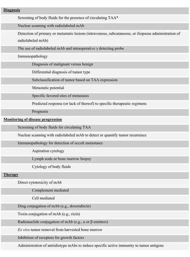
B lymphocytes generate humoral immune response that is polyclonal and therefore, the antibodies present in an individual are heterogeneous. However, one can obtain large amounts of homogeneous antibody reacting with a single epitope (monoclonal) by cloning B lymphocytes. From the time that Koehler and Milstein introduced hybridoma technology based on this fact, scientists have identified a broad range of biological molecules with the help of monoclonal antibodies. Immune stimulatory and regulatory molecules, cellular markers for distinguishing the tissue origin of one cell from another, cellular and tumor markers for characterization of the nature of a tumor biopsy specimen, the antigens that are up- or down-regulated in tumor cells are some of the biomolecules in this range. Additionally, monoclonal antibodies are used in therapeutic approaches to target cytotoxic reagents to and trigger or block cell surface molecules, and in diagnostic approaches to carry imaging reagents to tumors (Table 1.5) (White et al., 2001; DeVita et al., 2001; Goding 1983).
Antibody-secreting hybridoma is the fusion of an indefinitely growing myeloma cell and an immune B lymphoblast expressing a specific antibody gene. Myeloma cells are established to be deficient for hypoxanthine guanine phosphoribosyl transferase (HGPRT) enzyme. Polyethylene glycol, which is a polywax, is used to facilitate cell fusion because it promotes cell adherence and the exchange of nuclei. HAT medium that contains hypoxanthine, aminopterin and thymidine is used to select successfully fused cells because only hybrid cells can grow in this medium. Homogeneous cell clones that secrete only one specific antibody are obtained by limiting dilution (Abbas and Lichtman, 2003; Little et al., 2000).
The culture of a hybridoma cell line can yield 1-10 µg of Ig per ml supernatant. Ascites fluids can produce between 1 and 10 mg of Ig per 1 ml (DeVita et al., 2001).
Table 1.5: Clinical applications of monoclonal antibodies (mAbs) in cancer (taken from DeVita et al., 2001)
Diagnosis
Screening of body fluids for the presence of circulating TAA* Nuclear scanning with radiolabeled mAb
Detection of primary or metastatic lesions (intravenous, subcutaneous, or iliopsoas administration of radiolabeled mAb)
The use of radiolabeled mAb and intraoperative γ detecting probe Immunopathology
Diagnosis of malignant versus benign Differential diagnosis of tumor type
Subclassification of tumor based on TAA expression Metastatic potential
Specific favored sites of metastasis
Predicted response (or lack of thereof) to specific therapeutic regimens Prognosis
Monitoring of disease progression
Screening of body fluids for circulating TAA
Nuclear scanning with radiolabeled mAb to detect or quantify tumor recurrence Immunopathology for detection of occult metastases
Aspiration cytology
Lymph node or bone marrow biopsy Cytology of body fluids
Therapy
Direct cytotoxicity of mAb Complement mediated Cell mediated
Drug conjugation of mAb (e.g., doxorubicin) Toxin conjugation of mAb (e.g., ricin)
Radionuclide conjugation of mAb (e.g., α or β emitters)
Ex vivo tumor removal from harvested bone marrow
Inhibition of receptors for growth factors
Administration of antiidiotype mAbs to induce specific active immunity to tumor antigens
1.7. Tumor Markers
Tumor markers are products that are often detected in higher-than-normal amounts in the blood or other body fluids or tissues of some individuals with certain types of cancer. Tumor markers are produced either by the tumor itself (e.g., tumor antigens) or by the body in response to the presence of cancer. Tumor markers can be classified into three categories in terms of the aim of use:
1. Diagnostic Markers: They give clues for identifying a disease by its signs, symptoms and laboratory findings.
2. Prognostic Markers: They help to predict the course of the disease.
3. Therapeutic Markers: They pertain with the treatment of the disease. They are typically of IgG class molecules.
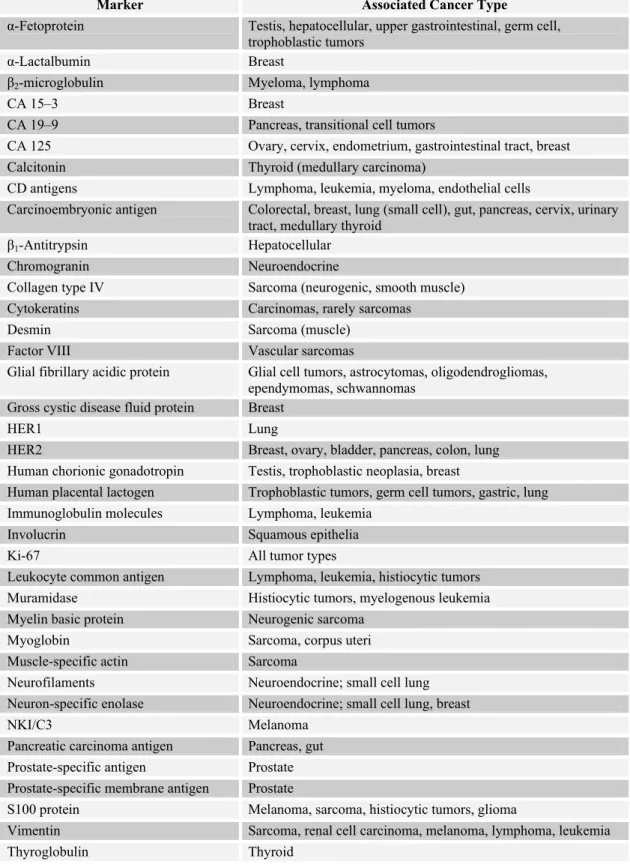
There are cell surface markers (e.g., phenotypic CD antigens of hematopoietic cells), genetic markers (e.g., Philadelphia chromosome), secreted proteins (e.g., paraproteins in the serum), oncofetal proteins (e.g., carcinoembryonic antigen, α-fetoprotein), hormones (e.g. human chorionic gonadotropin), enzymes (e.g., prostate-specific antigen, lactic dehydrogenase, neuron-prostate-specific enolase) and cancer antigens (e.g., CA 125, CA 15-3, CA 19-9) whose elevated levels or abnormal expression pattern is used for diagnostic, prognostic or therapeutic purposes (Table 1.6). Anyone may notice that a tumor marker can have more than one of the features mentioned in the sentence before (e.g., carcinoembryonic antigen is a cell surface protein and an oncofetal protein) (http://lwwoncology.com; http://cis.nci.nih.gov/ fact/5_18.htm). This thesis will focus on protein markers because of the design of the thesis project.
Table 1.6: Some serum and immunohistochemical markers (taken and adapted from http://lwwoncology.com)
Marker Associated Cancer Type
α-Fetoprotein Testis, hepatocellular, upper gastrointestinal, germ cell, trophoblastic tumors
α-Lactalbumin Breast
β2-microglobulin Myeloma, lymphoma
CA 15–3 Breast
CA 19–9 Pancreas, transitional cell tumors
CA 125 Ovary, cervix, endometrium, gastrointestinal tract, breast Calcitonin Thyroid (medullary carcinoma)
CD antigens Lymphoma, leukemia, myeloma, endothelial cells
Carcinoembryonic antigen Colorectal, breast, lung (small cell), gut, pancreas, cervix, urinary tract, medullary thyroid
β1-Antitrypsin Hepatocellular
Chromogranin Neuroendocrine
Collagen type IV Sarcoma (neurogenic, smooth muscle) Cytokeratins Carcinomas, rarely sarcomas
Desmin Sarcoma (muscle)
Factor VIII Vascular sarcomas
Glial fibrillary acidic protein Glial cell tumors, astrocytomas, oligodendrogliomas, ependymomas, schwannomas
Gross cystic disease fluid protein Breast
HER1 Lung
HER2 Breast, ovary, bladder, pancreas, colon, lung Human chorionic gonadotropin Testis, trophoblastic neoplasia, breast
Human placental lactogen Trophoblastic tumors, germ cell tumors, gastric, lung Immunoglobulin molecules Lymphoma, leukemia
Involucrin Squamous epithelia
Ki-67 All tumor types
Leukocyte common antigen Lymphoma, leukemia, histiocytic tumors Muramidase Histiocytic tumors, myelogenous leukemia Myelin basic protein Neurogenic sarcoma
Myoglobin Sarcoma, corpus uteri
Muscle-specific actin Sarcoma
Neurofilaments Neuroendocrine; small cell lung Neuron-specific enolase Neuroendocrine; small cell lung, breast
NKI/C3 Melanoma
Pancreatic carcinoma antigen Pancreas, gut Prostate-specific antigen Prostate Prostate-specific membrane antigen Prostate
S100 protein Melanoma, sarcoma, histiocytic tumors, glioma
Vimentin Sarcoma, renal cell carcinoma, melanoma, lymphoma, leukemia
1.8. Selected Tumor Markers
1.8.1. CA 125
CA 125 is an antigenic determinant on a high molecular weight glycoprotein and one of its two antigenic domains is recognized by the murine monoclonal antibody OC-125 and the other is recognized by the monoclonal antibody M11. Inclusion cysts, metaplasic parts and papillary excrescences found in ovary express CA 125 determinant but it is not expressed in the normal surface epithelium of fetal or adult ovaries. CA 125 is elevated in 85% of epithelial ovarian cancers and 13-21% of squamous carcinoma. In ovarian cancer, preoperative serum level of CA 125 is found to be correlated with tumor stage and histological grade but it is not suggested to be an independent prognostic or diagnostic factor on its own. Elevation of serum CA 125 can also be associated with pancreas, breast, colon, lung malignant neoplasms as well as with some benign neoplasms and physiological states such as pregnancy, and menstruation (Robertson et al., 2002; Meyer and Rustin, 2000; http://lwwoncology.com). The major contribution of CA 125 as a tumor marker is said to be its utility in the monitoring of tumor response to chemotherapy (Verheijen
et al., 1999).
1.8.2. Carcinoembryonic Antigen (CEA)
CEA is an oncofetal serum antigen and it is present in small amounts in adult colon. If an increased serum level of CEA is detected in a pretreatment stage, it implies a negative prognostic significance for the patient. Serum level elevation of CEA is related to many cancer types including colon, rectum, gallbladder, pancreas and stomach. It is frequently used in the management of patients with rectal, colon, gallbladder and stomach cancers but it is suggested that CEA is not useful by itself for screening those cancers and use of CEA alone for monitoring response to treatment is not recommended. This is as a result of lack of tumor type specificity of CEA. Additionally, benign diseases of liver, gastrointestinal tract, lung and cigarette smoking may cause elevation of CEA levels decreasing its tumor specificity
(http://lwwoncology.com; http://www.cancer.gov/cancerinfo/pdq/treatment/rectal/ HealthProfessional).
1.8.3. Alpha-Fetoprotein (AFP)
AFP is an oncoprotein, too. This glycoprotein is produced by fetal yolk sac, liver, and upper gastrointestinal tract. Pregnancy and benign liver disease also cause the elevation of AFP. Serum levels of this protein are increased in most of liver tumors and in some of the gastric, pancreatic, colon, and bronchogenic cancers. AFP is accepted to be the best diagnostic serum tumor marker for primary liver cancer. It is suggested that AFP is a reliable factor for monitoring therapeutic response and detecting recurrences in women with endodermal sinus tumors and embryonic carcinomas (Uenishi et al., 2003; http://lwwoncology.com).
1.8.4. CD20
CD20 is a transmembrane protein present on almost all of the B cells from the time of their commitment to B cell development until the time that the protein is down-regulated in the differentiated, antibody-secreting plasma cells. Therefore, CD20 is accepted as a pan-B cell antigenic marker. The expression of CD20 is quite heterogeneous both in the tumor sample of an individual and among different lymphoma types.
The first monoclonal antibody to be approved by U.S. Food and Drug Administration (FDA) for the therapy of cancer is rituximab (Rituxan®, 1997). Rituximab is used to target CD20 that is present in many B cells of non-Hodgkin’s lymphoma subtypes. It induces apoptosis, antibody-dependent cell cytotoxicity and complement mediated cytotoxicity in these tumors. When used in combination with other chemotherapeutics, it significantly improves disease-free survival rates (Smith, 2003; Ross et al., 2003; White et al., 2001; Glennie et al., 2000; Maloney et al., 1994).
1.8.5. Cytokeratins
Cytokeratins are intermediate filaments that build cytoskeleton, being a member of it in all epithelial cells. They are specific markers of epithelial differentiation and although not the same, they are continued to be expressed after malignant transformation. More than 20 cytokeratins have been described in humans. One of the applications of those proteins is their use in differential diagnosis of epithelial tumors and detecting their differentiation status. Cytokeratin fragments can be detected in the serum by monoclonal antibodies because in malignant epithelial cells, activated proteases degrade cytokeratins into fragments that are soluble in the serum. CYFRA 21-1 is an assay developed to measure a soluble fragment of CK 19 in the serum. In non-small cell lung cancer (NSCLC), CYFRA 21-1 is more sensitive than any other established markers. Moreover, it is found to be a useful marker for cervical, esophageal, breast, gastric, and bladder cancers. Uenishi et al. (2003) suggest CYFRA 21-1 as a useful diagnostic test for intrahepatic cholangiocarcinoma patients because CK 19 is differentially expressed in bile duct cells of normal liver whereas normal hepatocytes do not express it (Upasani et al., 2004; Uenishi et al., 2003; Young et al., 2002; Kamoi et al., 2002; http://lwwoncology.com).
In a study of differential expression of cytokeratin 18 (CK 18) in NSCLC subtypes of adenocarcinoma, squamous cell carcinoma and adenosquamous carcinoma, it was found that CK 18 expression was strongest in adenocarcinoma, weak in adenosquamous carcinoma and undetectable in squamous cell carcinoma concluding that CK18 might be of diagnostic value for those subtypes (Young et al., 2002). In another study involving anal carcinoma, the authors concluded that loss of expression of CK 18 and CK 19 is a marker of dedifferentiation in anal carcinoma (Behrendt and Hansman, 2001). Ordonez (2002) underlines that CK 5/6 is among the most useful positive immunohistochemical markers for epitheloid mesothelioma diagnosis.
1.8.6. HER2
HER2 (erbB2) belong to the erbB family of tyrosine kinase receptors (RTKs) -also known as type I receptor tyrosine kinases or EGF receptor family- and it is a relative of HER1. In normal cells, activation of erbB RTKs triggers a broad network of signaling pathways controlling cell growth, differentiation, motility and adhesion. Deregulation of HER2 receptor is observed in many cancers leading to a more aggressive clinical outcome of the disease. In breast cancer, nearly 25% to 30% of patients have overexpression of HER2 caused by amplification of HER2 gene. This amplification is associated with a poor prognosis and chemoresistance of tumor in breast cancer patients. In addition, HER2 has a metastasis-promoting effect promoting the secretion of matrix metalloproteases.
The hypothesis that the blockage of binding of epidermal growth factor (EGF) to its receptor might prevent cell proliferation by inhibiting receptor activation influenced production of anti-EGF receptor monoclonal antibodies. HER2 is the target of the first monoclonal antibody approved by FDA as an erbB receptor inhibitor, trastuzumab (Herceptin®; Genentech, Inc.; South San Francisco, CA). The metastatic breast cancer patients with a profile of HER2 over-expression benefit from therapies involving trastuzumab. HER2-targeted therapies can lead to tumor regression, delay of tumor growth and some symptomatic improvements. There is a consensus among researchers on prognostic value of HER2 amplification/expression in node-positive breast cancer patients. In addition, HER2 over-expression has a negative predictive value for the responsiveness of the cancer patients to chemotherapy and endocrine therapy indicating the probability of resistance to those therapies. Apart from this, the approval of trastuzumab provided the use of an immunohistochemical diagnostic test (HercepTest™). HercepTest™ is designed specifically to detect HER2 protein over-expression in routinely processed breast cancer tissues. HercepTest™ is approved by FDA for the measurement of HER2 status when deciding whether patients are eligible for Herceptin® therapy. In Europe, patients are accepted to be eligible for treatment if they demonstrate IHC 3+ HER2 over-expression and in the U.S. patients are eligible if they demonstrate 2+ or 3+ HER2 over-expression. Therefore, HercepTest™ aims a reliable detection of
HER2 over-expression for the success of Herceptin® therapy.(Menard et al., 2003; Rowinsky 2003; Ross et al., 2003; White et al., 2001; Hanahan and Weinberg, 2000; Mendelsohn and Baselga, 2000; Glennie and Johnson, 2000; http://www.dakocytomation.com/dako_facts_1final.pdf).
1.8.7. Prostate-Specific Antigen (PSA)
The measurement of PSA levels in serum is utilized for screening and early detection of prostate cancer. However, PSA is not a highly specific tumor marker. Serum contains two forms of PSA; complexed PSA and free PSA. There are three forms of free PSA and the one that is a proenzyme is associated with cancer of prostate. The ratio of elevated serum level of truncated proenzyme PSA to total serum PSA correlates with an increased risk of prostate cancer (Mikolajczyk et al., 2002; Peter et
al., 2001; Mikolajczyk et al., 2000).
1.8.8. Prostate-Specific Membrane Antigen (PSMA)
PSMA is a tissue-specific protein that is accepted to be a very good target for imaging and therapy because of its prostate specificity and its extracellular large domain. Although PSMA is expressed in kidney, proximal small intestine, salivary gland and brain, those tissues have a thousand-fold smaller expression levels than that of prostate. PSMA is up-regulated many folds in prostate cancer and interestingly, it is expressed in neovascularizations of most of the solid tumors but not in neovascularizations of normal tissues (Ghosh and Heston, 2004).
2. AIM OF THE STUDY
Utilization of tumor markers in cancer research and in clinical applications provides benefit of differential diagnosis of cancer versus normal or benign tissue, differential diagnosis of tumor degree, tumor types and subtypes, predicting the prognosis of the disease, and targeted treatment of the tumor versus normal tissue, selectively. Although there are many tumor markers investigated on the basis of monoclonal antibody production, very few markers are specific in terms of cancer and tissue type. This feature restricts the use of tumor markers leading to lack of routinely used informative tumor markers in many cancer types. Therefore, investigation of novel, informative tumor markers is in great demand.
This study aims production of monoclonal antibodies that may be candidates for fulfilling the requirements of an ideal tumor marker. To serve this purpose, two monoclonal antibodies were produced against apoptosis induced hepatocellular carcinoma cell line HuH-7. Next, the monoclonal antibodies were subjected to characterization experiments to explore their immunoreactivity in different cell, tissue and tumor types.
3. MATERIALS AND METHODS
3.1. Production of 6D5 and 9C11 Monoclonal Antibodies
3.1.1. Production of 6D5 and 9C11 Monoclonal Antibody Producing
Hybridomas
6D5 and 9C11 producing hybridomas were previously produced by Tamer Yagci. Ten million of cells of apoptosis induced (by UV-C irradiation, 60-120 mJ/cm²) HuH-7 hepatocellular carcinoma cell line was lysed in 2 ml PBS and 0.5 ml of lysate was injected into peritoneal cavity of Balb/c mice. Mice were immunized twice more at two weeks intervals and hybridomas were prepared by spleen cells after the animals were sacrificed. Sp2/0-Ag14 mouse myeloma cell line was used as fusion partner.
Partaking in the project at the time of culturing after the fusion, the antibody producing hybridoma cells were selected by enzyme-linked immunosorbent assay (ELISA). HuH-7 cells were grown in 96-well tissue culture plates in DMEM medium (supplemented with 10% FCS, 100 U/ml penicillin, 100 µg/ml streptomycin, 2 mM L-glutamine and 0.1 mM non-essential amino acids) in tissue culture incubator that
was set to 37 ºC and 5% CO2.When they reached to desired number, they were fixed
with 4% formalin in PBS for 15 minutes in dark at room temperature. Afterwards, they were permeabilized with 0.2% Triton X-100 in 1X PBS for 5 minutes at room temperature. Then, the plates were washed two times with distilled water and dried gently by tapping on a paper towel. 2% BSA in PBS was used for blocking and the cells were incubated with this blocking solution for 1 hour at 37 ºC. Blocking was followed by addition of hybridoma supernatants to the wells that contain HuH-7 cells. The cells were incubated with the supernatants for 1 hour at 37 ºC. Following incubation, wells were washed 2 times with distilled water and secondary antibody (goat anti-mouse IgG-alkaline phosphatase, Sigma) was added as 1:1000 diluted. After an incubation period of 1 hour at 37 ºC, the wells were washed 4 times with distilled water. Next, substrate tablet (pNPP disodium hexahydrate, phosphatase
substrate, Sigma) was dissolved in 20 ml ddH2O in dark at room temperature and added to each well. At the end of 20-45 minutes of incubation, spectrophotometric measurement was performed at A405 at Beckman Biomek ELISA reader. DMEM containing wells were used as negative control. The supernatants were accepted to produce antibody if they had an optic density twice of that of the negative control.
After ELISA assay, the positive hybridoma cells were cloned by limiting dilution. The hybridoma cells were counted and they were plated to 96-well plates with a calculated theoretical amount of 0.5 cell per well. Recloning was done to ensure clonality of the cells.
Clones, producing different antibodies of single cell origin, were screened again by the same ELISA method described before. The antibody producing clones to be selected for further studies were determined from the ones that revealed a measurement result of three times that of the negative control.
Two of the antibody producing clones, namely 6D5 and 9C11, were chosen to be used for further studies.
3.1.2. Culturing 6D5 and 9C11 Hybridoma Cells for Antibody Production
6D5 and 9C11 hybridoma cells were cultured in high glucose containing DMEM medium supplemented with 20% FCS, 100 U/ml penicillin, 100 µg/ml streptomycin, and 0.1 mM non-essential amino acids. The incubator where the cells were grown was set to 37 ºC and 5% CO2. The hybridoma supernatants were collected by centrifugation of the cultures at 1500 rpm for 5 minutes at room temperature. They were buffered with 20 mM Tris (pH 8) and 0.02% sodium azide (w/v) was added as preservative to prevent microorganism contamination. The supernatants were stored at -20 ºC if they were not going to be used immediately. New stocks were prepared by freezing 10 million hybridoma cells in 90% FCS and 10% DMSO containing freezing medium.