Mid-Term Results of Two Different Fixation Methods for Chevron
Osteotomy for Correction of Hallux Valgus
Baran Komur, MD
1, Baris Yilmaz, MD
2, Erdem Kaan, MD
3, Bulent Yucel, MD
1, Tahir Mutlu Duymus, MD
1,
Guzelali Ozdemir, MD
2, Olcay Guler, MD
41Orthopedist, Kanuni Sultan Suleyman Training and Research Hospital, Istanbul, Turkey 2Orthopedist, Fatih Sultan Mehmet Training and Research Hospital, Istanbul, Turkey 3Orthopedist, Dr. Ersin Arslan Training and Research Hospital, Gaziantep, Turkey
4Orthopedist, Department of Orthopaedics and Traumatology, Medipol University, Istanbul, Turkey
A R T I C L E I N F O Level of Clinical Evidence:3 Keywords: bioabsorbable pins cannulated screws chevron osteotomy fixation methods hallux valgus A B S T R A C T
We compared 2 different fixation methods (bioabsorbable pins and cannulated screws) after chevron os-teotomy for the treatment of hallux valgus. We reviewed consecutive proximal chevron osteotomies in 80 patients (100 feet) performed by 2 surgeons. Of the 100 feet (80 patients), 48 feet (40 patients) were stabilized with bioabsorbable pins, and 52 feet (40 patients) were stabilized with cannulated screws. In the pin group, 8 patients were male (20%) and 32 were female (80%). In the screw group, 10 patients were male (25%) and 30 were female (75%). The mean patient age was 43.1 (range 24 to 60) years in the pin group and 43.5 (range 20 to 60) years in the cannulated screw group. The visual analog scale, intermetatarsal angle, and hallux valgus angle decreased significantly and the American Orthopaedic Foot and Ankle Society scores increased significantly in all patients in both groups after surgery (p< .05). No statistically significant differences were found between the 2 groups (p> .05). Both fixation methods were found to be safe and reliable under the appropriate conditions and when performed by an experienced surgeon.
© 2018 by the American College of Foot and Ankle Surgeons. All rights reserved.
Hallux valgus is the most common problem of the forefoot in adults
(1). It is progressive and involves several stages, beginning with lateral deviation of the great toe (hallux) and medial deviation of the first metatarsal (metatarsus primus varus)(2). Management of hallux valgus generally begins with conservative treatment, especially in juvenile hallux valgus. Surgical correction is indicated for cases of failed con-servative management, progressive and painful deformity, or disruption of lifestyle or activity(3).
More than 140 surgical procedures have been described to correct hallux valgus. The chevron osteotomy has become widely accepted for correction of mild to moderate hallux valgus deformities(4). This technique includes removal of the medial eminence and a horizon-tally directed V-shaped osteotomy of the distal first metatarsal(5). The indications for this procedure include the following: failed con-servative treatment, mild to moderate deformity (metatarsophalangeal
angle<35° and an intermetatarsal angle [IMA] <15°), and the absence of arthritis in the first metatarsophalangeal joint.
Currently, nondegradable implants are primarily made of steel or titanium. Although these implants provide maximum stability, the dis-advantages include interference with imaging modalities such as direct radiography and magnetic resonance imaging. In addition, they might require an undesirable second operation for hardware removal. More-over, the mechanical properties of nondegradable implants are quite different from those of cortical bone, potentially resulting in inho-mogeneous stress transfer and limited bone healing. This constellation of effects is referred to as “stress shielding.” Therefore, it might be ben-eficial to use implants with a Young’s modulus close to that of cortical bone. Biodegradable implants are currently in clinical use for fixa-tion in distal chevron osteotomies. These implants are mechanically weaker than their metallic counterparts and have been associated with foreign body reactions and osteolysis(6,7). However, bioabsorbable pins have been shown to provide a similar correction of the IMA and to have comparable rates of complications compared with cannu-lated screws(8).
The aim of the present study was to compare the outcomes using cannulated screws and bioabsorbable pins for fixation after chevron osteotomy in the surgical treatment of hallux valgus.
Financial Disclosure: None reported. Conflict of Interest: None reported.
Address correspondence to: Baran Komur, MD, Kanuni Sultan Suleyman Training and Research Hospital, Turgut Ozal Street, No:1, Halkali, Kucukcekmece, Istanbul, Turkey.
E-mail address:[email protected](B. Komur).
1067-2516/$ - see front matter © 2018 by the American College of Foot and Ankle Surgeons. All rights reserved. https://doi.org/10.1053/j.jfas.2018.03.021
Contents lists available atScienceDirect
The Journal of Foot & Ankle Surgery
Patients and Methods
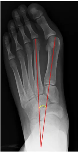
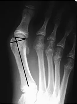
We reviewed 100 distal chevron osteotomies in 80 patients performed by 2 sur-geons (B.K., B.Y.) from March 2014 to February 2016. Our local ethics committee approved the study. The mean follow-up period was 14 (range 2 to 27). All the patients provid-ed written informprovid-ed consent before enrollment in the study. Of the 100 feet, 48 feet (40 patients) were stabilized with bioabsorbable pins by 1 surgeon (B.K.) and 52 feet (40 patients) were stabilized with cannulated screws performed by 1 surgeon (B.Y.). This method was chosen to minimize surgical bias. In the pin group, 8 patients were male (20%) and 32 were female (80%). In the screw group, 10 patients were male (25%) and 30 were female (75%). The mean patient age was 43.1 (range 24 to 60) years in the pin group and 43.5 (range 20 to 60) years in the cannulated screw group. Patients with rheumatoid arthritis, hallux rigidus, or failed previous hallux valgus surgery were excluded from the present study. Clinical results were obtained using the American Orthopaedic Foot and Ankle Society (AOFAS) ankle-hindfoot score and the visual analog scale (VAS) for pain. Radiographically, the hallux valgus angle (HVA) (Fig. 1), IMA (Fig. 2), and distal metatarsal articular angle (DMAA) (Fig. 3) were measured and compared between the 2 groups. The HVA was measured as the angle between the center of the longitudinal axis of the first metatarsal and the axis of the great toe. The IMA was mea-sured as the angle between the line of the first metatarsal and the line bisecting the diaphyseal portions of the second metatarsal. Finally, DMAA was calculated as the angle between the articular surface of the distal first metatarsal and the longitudinal axis of the first metatarsal.
Surgical Technique
The surgical technique used for both study groups was the same except that dif-ferent implants were used, as described. The chevron osteotomy was performed in both groups through a medial longitudinal incision starting from the base of the proximal phalanx and extending 5 to 7 cm proximally to the metatarsal head. The subcutane-ous tissue and bursa were dissected, and the capsule of the joint was identified. The capsule of the joint was incised in a Y-shaped fashion, and a straight longitudinal in-cision was continued out to the bone. The toe was then adducted, and a bunionectomy was performed using an oscillating saw. The medial eminence was removed, and the “V” osteotomy was performed with an angle of 50° to 60° between the cuts. After the osteotomy had been completed, the distal metatarsal head was slid laterally and
displaced 3 to 4 mm. Once this displacement was complete, the osteotomy site was first fixed with Kirschner wires, after which bioabsorbable pins composed of biodegradable copolymers L-lactide, D,L-lactide, and trimethylene carbonate (OTPS Bio-degradable Pins, Inion, Tampere, Finland) or cannulated screws (Herbert headless cannulated titanium screws, TST Tibbi Aletler San., Istanbul, Turkey) were applied to hold the fragments in position (Fig. 4). Postoperatively, all operative feet in both groups were placed in a splint for ~3 weeks, and weightbearing on the first toe was not allowed until the seventh postoperative week (Fig. 5). No differences were found in terms of the surgical approach or postoperative management for the patients in either group.
Statistical Analysis
Descriptive statistics were used to describe the continuous dependent variables (mean, standard deviation, minimum, median, and maximum). The Wilcoxon signed-rank test was used to compare the dependent data that were not normally distributed, and the Mann-Whitney U test was used to compare the independent data that were not normally distributed. Data were deemed statistically significant when p< .05. All analyses were performed using MedCalc Statistical Software, version 12.7.7 (MedCalc, Ostend, Belgium; available at:http://www.medcalc.org; 2013).
Results
We compared the age and sex distribution between the bioabsorbable pin and cannulated screw groups and found no statis-tically significant difference between them (Tables 1 and 2).
Changes in the HVA, IMA, and DMAA with respect to the time after surgery in the 2 groups are presented inTables 3–5. Preoperatively,
Fig. 1. Plain radiograph showing hallux valgus angle of the first metatarsal.
no differences were found in any of these angles between the 2 groups. Postoperatively, a significant decrease in the HVA was observed in both the bioabsorbable pin and the cannulated screw groups, and the re-duction was similar between the 2 groups (p= .858). The IMA in the pin and screw groups decreased significantly after surgery com-pared with the preoperative measurements (p< .001, for both groups), and the reduction was similar for both groups (p= .495). The DMAA in the pin and screw groups also decreased significantly postopera-tively compared with preoperapostopera-tively (p< .001, for both groups). Also, the reduction was similar for both groups (p= .618;Tables 3–5).
The VAS scores in the pin and screw groups had decreased sig-nificantly postoperatively compared with preoperatively (p< .001, for both groups). The decrease was also similar for both groups (p= .629;
Table 6).
The AOFAS ankle-hindfoot scale scores in the pin and screw groups were significantly increased postoperatively compared with before
surgery (p< .001, for both groups). This increase was also similar for both groups (p= .119;Table 7).
Four patients in group 1 and two in group 2 developed superfi-cial wound infections that healed after superfisuperfi-cial debridement. Loss of correction was not observed in any patient.
Discussion
Since Austin and Leventen first described the chevron osteotomy for the treatment of symptomatic hallux valgus, the procedure has been
Fig. 3. A hallux valgus radiograph of a juvenile showing the distal metatarsal articu-lar angle of the hallux.
Fig. 4. Intraoperative photograph showing insertion of the bioabsorbable pin across the osteotomy site after bunionectomy.
Fig. 5. Postoperative radiographs of a chevron osteotomy with bioabsorbable pins used for fixation.
Table 1
Gender distribution stratified by study group
Fixation Method Male Patients Female Patients Total
Pin 8 (20) 32 (80) 40 (100) Screw 10 (25) 30 (75) 40 (100) Total 18 (22.5) 62 (77.5) 80 (100) Data presented as n (%). p= .592, χ2test. Table 2
Demographic information of study groups stratified by age
Variable Age (y)
Pin Group Screw Group Whole Group
Patients (n) 40 40 80 Mean 43.1 43.5 43.3 Median 43.0 45.5 43.0 Standard deviation 11.5 11.8 11.6 Minimum 24.0 20.0 20.0 Maximum 60.0 60.0 60.0 p= .857. Mann-Whitney U test.
modified to improve stability by incorporating various internal fixa-tion methods(7,8). These have included Kirschner wires, metal screws, metal plates, staples, and, more recently, bioabsorbable pins(7,8).
Screw fixation has become widely used owing to its ease of use and the absence of an externally protruding wire, which decreases the risk of skin irritation and pin tract infection(9). This technique has
generally been performed on younger patients who have good bone quality. Herbert screws, cortical screws, and Acutrak compression screws can be used for fixation of chevron osteotomies. The cannu-lated Acutrak screws have the advantage of providing greater compression and more solid bony union. Furthermore, removal of the screw has only been required in rare cases. Toorney and McGarvey
Table 3
Preoperative and postoperative hallux valgus angle stratified by treatment group
Variable Pin Group Screw Group
Pre-HVA Post-HVA Difference Pre-HVA Post-HVA Difference
Patients (n) 40 40 40 40 40 40 Mean 32.850 12.825 −20.0 32.800 12.725 −20.07 Median 33.000 13.000 −20.0 33.000 13.500 −20.00 Standard deviation 1.1886 0.6751 1.1 2.2326 1.9214 3.30 Minimum 30.0 12.0 −21.0 29.0 10.0 −26.00 Maximum 34.0 14.0 −17.0 36.0 15.0 −14.00 p Value* < .001 < .001
Abbreviations: HVA, hallux valgus angle; Pre-HVA, preoperative HVA; Post-HVA, postoperative HVA. * Wilcoxon signed rank test.
Table 4
Preoperative and postoperative intermetatarsal angle stratified by treatment group
Variable Pin Group Screw Group
Pre-IMA Post-IMA Difference Pre-IMA Post-IMA Difference
Patients (n) 40 40 40 40 40 40 Mean 17.72 7.50 −10.22 17.525 7.65 −9.87 Median 18.00 7.50 −11.00 18.00 8.00 −10.0 Standard deviation 0.9334 0.5064 1.0497 1.4848 0.5796 1.712 Minimum 16.0 7.0 −11.00 14.0 7.0 −12.00 Maximum 19.0 8.0 −8.00 19.0 9.0 −6.00 p Value* < .001 < .001
Abbreviations: IMA, intermetatarsal angle; Pre-IMA, preoperative IMA; Post-IMA, postoperative IMA. * Wilcoxon signed rank test.
Table 5
Preoperative and postoperative distal metatarsal articular angle stratified by treatment group
Variable Pin Group Screw Group
Pre-DMAA Post-DMAA Difference Pre-DMAA Post-DMAA Difference
Patients (n) 40 40 40 40 40 40 Mean 15.600 9.000 −6.60 15.57 9.075 −6.500 Median 15.000 9.000 −7.00 15.00 9.000 −7.000 Standard deviation 0.7779 0.9058 0.9554 0.6751 1.1851 1.3587 Minimum 150 8.0 −8.00 15.0 7.0 −9.00 Maximum 17.,0 11.0 −5.00 17.0 11.0 −4.00 p Value* < .001 < .001
Abbreviations: DMAA, distal metatarsal articular angle; Pre-DMMA, preoperative DMMA; Post-DMMA, postoperative DMMA. * Wilcoxon signed rank test.
Table 6
Preoperative and postoperative visual analog scale scores stratified by treatment group
Variable Pin Group Screw Group
Pre-VAS Post-VAS Difference Pre-VAS Post-VAS Difference
Patients (n) 40 40 40 40 40 40 Mean 75.000 18.750 −56.2500 76.375 21.875 −54.5000 Median 75.000 25.000 −55.0000 75.000 25.000 −55.0000 Standard deviation 5.5470 11.8619 12.39055 4.5273 9.1769 10.36513 Minimum 65.0 0.0 −80.00 65.0 0.0 −80.00 Maximum 80.0 30.0 −40.00 90.0 30.0 −40.00 p Value* < .001 < .001
Abbreviations: VAS, visual analog scale; Pre-VAS, preoperative VAS; Post-VAS, postoperative VAS. * Wilcoxon signed rank test.
(10)summarized the advantages of the cannulated screw. These include stable osteotomy compression and fixation, the ability to check the osteotomy alignment and position before final fixation, precision of drilling and screw insertion, and the presence of a low-profile screw head that eliminates potentially prominent hardware(10). We did not observe any implant failure in the screw group.
In contrast, the use of permanent metal implants has been asso-ciated with drawbacks such as the potential need for hardware removal and the recently discovered possibility of metal hypersensitivity. The incidence of metal implant removal has ranged from 2% to 15%, ac-cording to various investigators, and, in some cases, this can be a complex procedure(6). Therefore, these challenges have led to im-provements in the development of bioabsorbable implants. Bioabsorbable pins are advantageous in that they have a lower elas-ticity modulus, and their mechanical properties are more similar to that of cancellous bone(11). During our follow-up period, we removed the bioabsorbable pins from 3 patients because of irritation.
The field of bioabsorbable implants for fixation in orthopedics is new and rapidly growing(12–14). The bioabsorbable implants that are commercially available primarily consist of 1 or 2 of 3 polymers: polyglycolic acid (PGA), polylactic acid, and polydioxanone. These poly-mers are part of a group known as α-polyesters or poly(α-hydroxy acids)(15).
Pelto-Vasenius et al(16)reported 21 episodes (22%) of osteolysis among 94 chevron osteotomies that were fixed with PGA pins. They found no association between osteolysis and the development of foreign body reactions, infection, avascular necrosis, loss of hallux valgus cor-rection, or osteoarthritis. They concluded that the osteolytic changes did not require treatment(16). We did not observe any osteolytic changes in our patients. Pihlajamäki et al(17)reported a series of 27 patients with osteotomies treated by internal fixation with absorb-able self-reinforced poly-l-lactide pins. No displacement occurred in any patient, and no signs of inflammatory foreign body reaction were noted(17).
Although more expensive than cannulated screws, bioabsorbable pins seem to be adequate for the fixation of small fragments such as the metatarsal heads and the radial head. They have the advantage of not resulting in inflammatory sinus tracts, osteolysis, or loss of fix-ation(18). Plaass et al(19)compared bioabsorbable magnesium screws and titanium screws using magnetic resonance imaging and found that magnesium screws are comparable to titanium screws and more suit-able for radiologic analysis. Successful use of bioabsorbsuit-able pins is likely related to the size of the bone and the implant, the inherent stabili-ty of the osteotomy, and the stabili-type of implant used.
One weakness of our study was that we did not have long-term follow-up data for our patients. Thus, we could not observe all pos-sible complications in each of our groups. We have continued to monitor these patients in the long term.
In the present study, we compared the use of absorbable pins and cannulated screws for fixation of distal chevron osteotomies of the first metatarsal. We found no significant difference between these im-plants. Both implants led to a decreased HVA, DMAA, and IMA. We believe these implants (bioabsorbable pins and cannulated screws) result in no significant differences in outcomes for fixation of chevron osteotomy. They each have individual advantages and disadvan-tages. It has been our impression that bioabsorbable pin fixation is a safe and effective method of fixing relatively stable osteotomies in the first metatarsal; however, the costs are greater than those with can-nulated screws.
In conclusion, both fixation methods are safe and reliable fixa-tion methods after chevron osteotomy for correcfixa-tion of hallux valgus under appropriate conditions and when performed by an experi-enced surgeon. The 2 fixation methods each have specific advantages and disadvantages.
References
1. Hecht PJ, Lin TJ. Hallux valgus. Med Clin North Am 98:227–232, 2014.
2. Hardy RH, Clapham JC. Observations on hallux valgus; based on a controlled series. J Bone Joint Surg Br 33:376–391, 1951.
3. Mann RA, Coughlin MJ. Hallux valgus—etiology, anatomy, treatment and surgical considerations. Clin Orthop Relat Res 157:31–41, 1981.
4. Trnka HJ, Hofstaetter S. The chevron osteotomy for correction of hallux valgus. Interact Surg 2:52–61, 2007.
5. Johnson JE. Chevron osteotomy for hallux valgus: surgical indications and technique. Oper Tech Orthop 2:177–183, 1992.
6. Windhagen H, Radtke K, Weizbauer A, Diekmann J, Noll Y, Kreimeyer U, Schavan R, Stukenborg-Colsman C, Waizy H. Biodegradable magnesium-based screw clinically equivalent to titanium screw in hallux valgus surgery: short term results of the first prospective, randomized, controlled clinical pilot study. Biomed Eng Online 12:62, 2013.
7. Morandi A, Ungaro E, Fraccia A, Sansone V. Chevron osteotomy of the first metatarsal stabilized with an absorbable pin: our 5-year experience. Foot Ankle Int 34:380–385, 2013.
8. Alcelik I, Alnaib M, Pollock R, Marsh DJ, Tulloch DJ. Bioabsorbable fixation for Mitchell’s bunionectomy osteotomy. J Foot Ankle Surg 48:9–14, 2009.
9. Andrews BJ, Fallat LM, Kish JP. Screw versus plate fixation for chevron osteotomy: a retrospective study. J Foot Ankle Surg 55:81–84, 2016.
10. Toorney EP, McGarvey SR. Fixation of the proximal first metatarsal crescentic osteotomy with the small cannulated screw system. Foot Ankle 11:397–399, 1991.
11. Côrte-Real N, Vasco Silva J, Esquível Pereira J, Caramelo J. Chevron osteotomy fixed with biodegradable screws in the treatment of Hallux valgus. Foot Ankle Surg 4:87–91, 1998.
12. Pentikainen I, Ojala R, Ohtonen P, Piippo J, Leppilahti J. Preoperative radiological factors correlated to long-term recurrence of hallux valgus following distal Chevron osteotomy. Foot Ankle Int 35:1262–1267, 2014.
13. Caminear DS, Pavlovich R Jr, Pietrzak WS. Fixation of the Chevron osteotomy with an absorbable copolymer pin for treatment of hallux valgus deformity. J Foot Ankle Surg 44:203–210, 2005.
14. Ford TC, Maurer LM, Myrick KW. Cortical bone pin fixation: a preliminary report on fixation of digital arthrodeses and distal Chevron first metatarsal osteotomies. J Foot Ankle Surg 41:23–29, 2002.
Table 7
Preoperative and postoperative American Orthopaedic Foot and Ankle Society scale score stratified by treatment group
Variable Pin Group Screw Group
Pre-AOFAS Post-AOFAS Difference Pre-AOFAS Post-AOFAS Difference
Patients (n) 40 40 40 40 40 40 Mean 30.40 82.125 51.73 31.275 80.00 48.73 Median 30.000 85.000 53.00 30.000 80.000 48.50 Standard deviation 6.7967 3.9039 6.41 6.1602 4.9355 7.372 Minimum 22.0 75.0 41.00 22.0 75.0 33.00 Maximum 41.0 85.0 63.00 42.0 90.0 63.00 p Value* < .001 < .001
Abbreviations: AOFAS, American Orthopaedic Foot and Ankle Society (ankle-hindfoot scale); Pre-AOFAS, preoperative AOFAS; Post-AOFAS, postoperative AOFAS. * Wilcoxon signed rank test.
15. Raikin SM, Ching AC. Bioabsorbable fixation in foot and ankle. Foot Ankle Clin 10:667–684, 2005.
16. Pelto-Vasenius K, Hirvensalo E, Vasenius J, Rokkanen P. Osteolytic changes after polyglycolide pin fixation in Chevron osteotomy. Foot Ankle Int 18:21–25, 1997. 17. Pihlajamäki H, Böstman O, Hirvensalo E, Törmälä P, Rokkanen P. Absorbable pins
of self-reinforced poly-l-lactic acid for fixation of fractures and osteotomies. J Bone Joint Surg Br 74:853–857, 1992.
18.Winemaker MJ, Amendola A. Comparison of bioabsorbable pins and Kirschner wires in the fixation of chevron osteotomies for hallux valgus. Foot Ankle Int 17:623–628, 1996.
19.Plaass C, von Falck C, Ettinger S, Sonnow L, Calderone F, Weizbauer A, et al. Bioabsorbable magnesium versus standard titanium compression screws for fixation of distal metatarsal osteotomies—3 year results of a randomized clinical trial. J Orthop Sci 30300, 2017.