DIAGNOSTIC ACCURACY OF CONE-BEAM COMPUTED TOMOGRAPHY IN DETECTING SECONDARY CARIES UNDER COMPOSITE FILLINGS: AN IN VITRO
STUDY
Kompozit Rezin Restorasyonlarda Sekonder Çürük Lezyonlarının Teşhisinde Konik Işınlı Bilgisayarlı Tomografinin Tanısal Doğruluğu
Elif YILDIZER-KERİŞ 1, Oğuzhan DEMİREL2 , Melih ÖZDEDE 3, Bülent ALTUNKAYNAK 4, İlkay PEKER 3
Received: 23/04/2016 Accepted:13/06/2016
ABSTRACT
Purpose: The aim of this in vitro study was to assess the diagnostic performance of cone-beam computed tomography (CBCT) in the detection of secondary carious lesions under composite resin fillings applied to different types of cavities.
Materials and Methods: Occlusal cavities (O) (n=18), occlusal cavities with mesial or distal component (MO/DO) (n=30), and mesial–occlusal–distal cavities (MOD) (n=30) were prepared in seventy eight extracted human posterior teeth. In half of the cavities in each group, artificial secondary caries lesions were simulated. All cavities were restored by using composite resin. All specimens were embedded in silicone and they were positioned to have approximal contacts. CBCT imaging was done and data were evaluated two times with two week interval by two observers, using a five-point confidence scale. Intra- and inter-observer agreements were calculated with Kappa statistics (κ). The area under (Az) the receiver operating characteristic (ROC) curve was used to evaluate the diagnostic accuracy.
Results: Intra- (κ =0.89) and inter-observer (κ = 0.79) agreements were found to be excellent. Az values were highest for the O restorations which is followed by the MOD and DO/ MO restorations. Az values for MOD and DO/MO restorations were very low and no statistically significant difference was found. Sensitivity for DO/MO restorations and specificity for MOD restorations were found to be the lowest values.
Conclusion: Diagnostic performance of CBCT was higher in O composite restorations than MOD and DO/MO restorations for secondary caries detection. The use of alternative imaging methods rather than CBCT may be useful for evaluating secondary caries under composite MOD and DO/MO restorations.
Keywords: Dental caries; cone beam computed tomography; caries diagnosis; composite resin, receiver operating characteristics
ÖZ
Amaç: Bu in vitro çalışmanın amacı, değişik kavite tipleri ile hazırlanan kompozit rezin restorasyonlu dişlerde bulunan sekonder çürük lezyonlarının teşhisinde konik ışınlı bilgisayarlı tomografinin (KIBT) tanısal performansını değerlendirmektir.
Gereç ve Yöntem: 78 adet çekilmiş daimi posterior dişlere okluzal (O) (n=18), mezial ya da distal okluzal (MO/DO) (n=30) ve mezial-okluzal-distal (MOD) (n=30) kavite preparasyonları yapıldı. Her kavite grubunda, dişlerin yarısına, yapay sekonder çürük lezyonları oluşturuldu ve kompozit rezin ile restore edildikten sonra silikon bloklara yerleştirildi. Hazırlanan bloklar KIBT ile görüntülendi. Tomografi görüntüleri iki gözlemci tarafından beş basamaklı güven ölçeği kullanılarak değerlendirildi. Gözlemci içi ve gözlemciler arası uyum Kappa katsayısı ile ölçüldü. Radyolojik değerlendirmelerin tanısal doğruluğu Alıcı İşletim Karakteristiği (AİK) eğrisi analizi ile değerlendirildi.
Bulgular: Gözlemci içi (κ =0.89) ve gözlemciler arası uyum (κ = 0.79) mükemmele yakın olarak bulunmuştur. AİK eğrileri altında kalan alanlar incelendiğinde “O” grubunda her iki gözlemcinin ayırt etme değerlerinin diğer gruplara göre yüksek olduğu görüldü. MOD ve MO/DO gruplarında AİK alanları oldukça düşük olup aralarında istatistiksel olarak anlamlı farklılık bulunmadı. Duyarlılık incelendiğinde MO/DO grubunda, özgüllük açısından karşılaştırıldığında ise MOD grubunda en düşük değerler saptandı. Sonuç: Sekonder çürük teşhisinde konik ışınlı bilgisayarlı tomografinin O kompozit restorasyanlar için tanısal doğruluğu MOD ve DO/MO restorasyonlardan daha yüksektir. Sekonder çürüklerin görüntülenmesinde KIBT yerine diğer görüntüleme yöntemlerinden faydalanılması daha uygun bir yaklaşım olabilir.
Anahtar kelimeler: Çürük; konik ışınlı bilgisayarlı tomografi; çürük tanısı; kompozit rezin; alıcı işletim karakteristiği
http://dx.doi.org/10.17096/jiufd.62563 ORIGINAL RESEARCH
1 Department of Radiology Çanakkale Dentistry Hospital
2 Department of Dentomaxillofacial Radiology Faculty of Dentistry Medipol University 3 Department of Dentomaxillofacial Radiology Faculty Dentistry Gazi University 4 Department of Statistics Faculty Arts and Sciences Gazi University
This work is licensed under a Creative Commons Attribution-NonCommercial-NoDerivatives 4.0 International License.
Introduction
Cone-beam computed tomography (CBCT) provides accurate and detailed information for diagnosis and treatment planning of dentomaxillofacial pathologies. CBCT produces three dimensional images with high resolution of bony structures and also the radiation dose of CBCT is lower than computed tomography (CT) (1, 2). Thus, it is used for various purposes such as dental implant planning, oral and maxillofacial surgery, orthodontics, airway assessment, temporomandibular joint disorders, endodontics and periodontics (3-8). In recent studies, the efficacy of CBCT has been evaluated for caries diagnosis. Some of them have concluded that the accuracy of CBCT may be similar to that of intraoral digital radiographic images for occlusal and proximal caries detection (9-12). However, CBCT images were found to be superior to intraoral techniques in other studies for secondary caries lesions under both composite and amalgam restorations (13-15). On the other hand, some authors reported that diagnostic performance of CBCT in detecting secondary caries may be negatively affected by the presence of dental restorations because of metal artifacts (16). There is still no justification regarding the use of this modality for caries detection. CBCT scanning is unnecessary for caries detection; it can only be used when it is taken for other reasons. As x-ray beam passes through an object, low energy photons become more likely to be absorbed than high energy photons and this is called “beam hardening”. Metal objects lead to artifacts in CBCT images related to beam hardening because absorption of x-ray beam is greater for metal objects than human tissues and cupping, as well as streaking artifacts may occur. Cupping artifacts are seen as distortion around the metallic objects and streaking artifacts are seen as dark and bright bands between two objects with high density (17). Because of their low attenuation values, the use of composite resins has been suggested to reduce artifacts before CBCT examination (18). There are limited number published articles in the literature regarding secondary caries detection by using CBCT. Also, there are no published studies about the diagnostic performance of CBCT for secondary caries detection in different type of cavities filled with composite resin. The aim of this in vitro study was therefore to evaluate the diagnostic performance of CBCT in the detection of secondary caries lesions created under different types of composite resin fillings.
Materials and Methods
Specimen preparation and experimental procedure
Eighty four extracted human permanent posterior teeth were used in this study (N=84). Teeth with tissue loss and/or fillings were excluded. All the teeth were randomly divided into three groups according to type of cavities. The first group was prepared as occlusal cavities (O) (n=24), the second group was prepared as mesial and/or distal occlusal cavities (MO/ DO) (n=30), and the third group was prepared as mesial–occlusal–distal cavities (MOD) (n=30). In each group, artificial secondary caries lesions were created in half of the cavities. The artificial caries lesions were prepared with round steel bur in 1 mm diameter, the location of the artificial holes on the base of the cavity was randomly selected. The drilled holes were then filled with pink wax. All groups of teeth were restored by composite resin (CR) (Valux plus, 3M Dental Products, St. Paul, MN, USA). For composite resin restoration, the cavities were etched for 15 seconds, bonding agent was applied and then cured for 40 seconds.
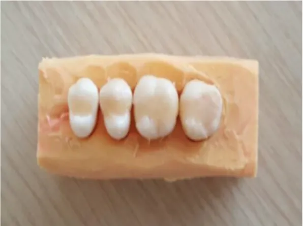
Premolar and molar teeth with approximal contacts were positioned in silicone blocks in a blind manner (Figure 1).
Figure 1. A sample of silicone blocks.
There were 21 blocks that contain four teeth each. Seven different block type was created according to various arrangements of tooth sets. There were three sums for each type of block. Half of the teeth with MOD (n=15) and DO/MO composite filling (n=15) were positioned to be adjacent approximal surfaces with restoration and the other half were located to adjacent approximal surfaces without restoration.
In total, 84 teeth with 24 O, 30 MO/DO and 30 MOD restorations were arranged to simulate various situations in the oral cavity. The number of teeth with O restorations (n=24) was lower than that of the MO/ DO and MOD restorations (n=30). Arrangement of tooth sets and type of blocks are shown in Table 1.
Table 1. Arrangement of tooth sets and block types (O: occlusal, MOD: mesial-occlusal-distal, DO: distal-occlusal, MO: mesial-occlusal).
Type of blocks Arrangement of tooth sets
Block type 1 O-MOD-DO-DO
Block type 2 O-MOD-MO-DO
Block type 3 MOD-O-MOD-DO
Block type 4 MOD-O-MOD-MO
Block type 5 O-MOD-O-MO
Block type 7 DO-MOD-O-O
Image acquisition and evaluation protocols
All blocks were radiographed by ProMax 3Ds CBCT system (Planmeca Oy, Helsinki, Finland). This device was operated at 90 kVp and 6.3 mA with 5×5 cm field of view (FOV) in high resolution settings. The acquired data were reconstructed with 0.48 mm slice thickness. Romexis Viewer (Planmeca Oy, Helsinki, Finland) software was used to evaluate the images in three orthogonal planes. Examples of CBCT images are shown in Figure 2. All images were evaluated by two oral and maxillofacial radiologists on two separate sessions with at least one week interval. The images were displayed on a 19-inch LCD monitor (Dell Inc., Round Rock, TX, USA). The observers were free to modify image algorithms but they did not have access to raw data. The presence or absence of secondary caries was scored using a five-point confidence scale; 1=definitely no caries, 2=probably no caries, 3=questionable, 4=probably caries, 5=definitely caries. Since there must be a final judgement under clinical conditions, scores 3, 4 and 5 were classified as positive diagnosis for caries whereas scores 1 and 2 were considered as negative diagnosis for caries.
Statistical analysis
The Kappa (k) coefficient values of intra- and inter-observer agreements were calculated and the values with higher than 0.7 were denoted as acceptable consistency. SPSS version 19.0 (SPSS Inc., Chicago, IL, USA) was
used for statistical analysis. The area under (Az) the
receiver operating characteristic (ROC) curve was used to evaluate the diagnostic accuracy. The areas under the curves were compared using Z-tests and p values less than 0.05 were considered as statistically significant.
Figure 2. (a) Cupping artifact seen on sagittal section (yellow arrow) (b) Appearance of adhesive system (yellow arrow) and simulated caries lesion (blue arrow) (c) Radiolucent appearance of adhesive system under radiopaque composite (yellow arrow).
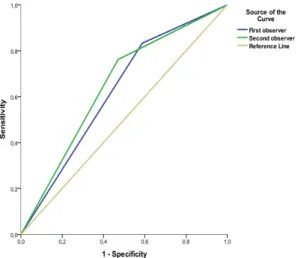
Figure 3. Graphic view of the receiver operating characteristics curves of all specimens.
Results
The k coefficients were 0.89 and 0.79 for intra- and inter-observer agreement, respectively. The areas under the ROC curves (Az values), sensitivity and specificity values of two observers were presented in Figure 3 and Table 2. Az values of the second observer was higher than the first observer. Sensitivity of the first observer was higher than the second observer while specificity
of the second observer was higher than that of the first observer (Table 2). The areas under the ROC curves (Az values), sensitivities and specificities for each observers and cavity types were given in Table 3. The Az values of two observers were the highest for the O restorations, followed by the MOD and DO/MO
restorations, respectively. Az values for MOD and DO/MO restorations were very low and diagnostic accuracy based on sensitivity and specificity values was not statistically significant. In simpler terms, sensitivity for DO/MO restorations and specificity for MOD restorations were found to be the lowest values.
Table 2. The statistical analysis of the areas under the receiver operating characteristics curves (Az values) and the sensitivity and specificity values of two observers.
Observer Area Standard Error p value Sensitivity Specificity
First .623 .066 .068 .833 .412
Second .646 .065 .030* .762 .529
*Statistically significant at p<0.05 level.
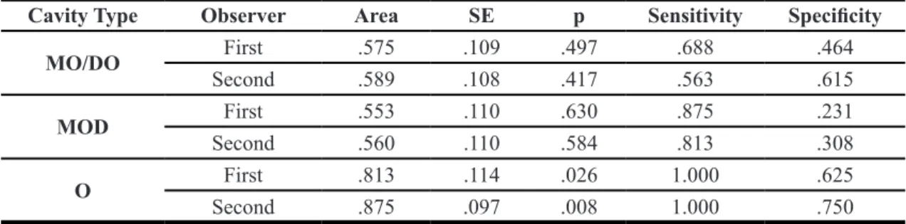
Table 3. The areas under receiver operating characteristics curves (Az values) and sensitivity and specificity values for the cavity types (O: occlusal, MOD: mesial-occlusal-distal, DO: distal-occlusal, MO: mesial-occlusal, SE: standard error).
Cavity Type Observer Area SE p Sensitivity Specificity
MO/DO First .575 .109 .497 .688 .464 Second .589 .108 .417 .563 .615 MOD First .553 .110 .630 .875 .231 Second .560 .110 .584 .813 .308 O First .813 .114 .026 1.000 .625 Second .875 .097 .008 1.000 .750 Discussion
The diagnostic accuracy of CBCT was tested in different types of cavities with composite restorations for secondary caries detection in the present study. The burr with one-mm-diameter was used to simulate secondary caries under restorations in accordance with previous studies (15, 19). The use of small burs has been recommended since larger burs were found to be ineffective for simulation of caries lesions (19). In earlier studies (15, 20), the researchers have standardized the position of artificial caries lesions, however, occurrence of caries in a standard location is unrealistic for clinical situation. Therefore, we created random artificial holes on the base to evaluate the performance of CBCT. In this study, the small voxel size and FOV were selected for CBCT scanning because reducing field size has been reported to increase spatial resolution and image quality (21). It is well known that the beam hardening artifacts occur with amalgam during CBCT scanning (16). Metal artifacts seen in CBCT images restricted the diagnosis of caries under restorations (15). Dark bands around the metallic restorations may look like secondary caries which may lead the clinicians
to reach false positive diagnosis (16, 22). It is also suggested that composite resin fillings could be useful for improving diagnostic accuracy of CBCT (18). But in contrast, recent studies showed that composite fillings were not superior to amalgam in terms of secondary caries detection with CBCT (15, 20). Based on this contradictory findings, it can be stated that the clinical performance of CBCT in the detection of secondary caries under composite resin fillings still remains unclear. In the present study, the diagnostic accuracy of CBCT in the detection of simulated secondary caries under composite resin restorations were found to be highest for the O type of restorations. In addition, its sensitivity for DO/MO restorations and its specificity for MOD restorations were found to be the lowest values. Sensitivity is defined as the percentage of true positive answers while specificity is the percentage of true negatives (23). In other words, the observers were unable to detect true positive cases in DO/MO restorations and also the true negative cases in MOD restorations. This finding could be attributed to the increased intensity of the artifacts which has been found to correlate positively with the restoration size. Nabha et al. (18) assessed artifacts induced by O, MO/DO, MOD
amalgam restorations with CBCT and concluded that increasing amalgam restoration size has caused higher intensity of artifacts on CBCT images. Even though the density of composite resin is lower than that of the amalgam, it is still higher than that of the enamel (24). Thus, it can be stated that composite resin may lead to beam hardening artefacts as well, which makes it harder to detect secondary caries under such fillings. In this context, the findings of the present study are not consistent with those of previous reports which claim that the composite resin improves diagnostic accuracy of CBCT (18). The Az
values of two observers were found to be between 0.813 and 0.875 for O restorations, 0.553 to 0.560 for MOD restorations and 0.589 to 0.575 for DO/MO restorations. In previous studies, accuracy of CBCT were found to be between 75% 97% for occlusal and class 2 cavities respectively in detecting secondary caries lesions restored with composite fillings (16, 20). Such inconsistencies may arise from difference in study designs, evaluation criteria, and experience of the observers as well as observers’ backgrounds.
Conclusion
The in vitro diagnostic performance of CBCT in detecting secondary caries is higher for O composite restorations than for MOD and DO/MO restorations. In addition, CBCT is a reliable imaging method for the detection of secondary caries under O composite restorations. Further clinical studies are needed to determine the accuracy of CBCT for secondary caries detection under different restorative materials.
Source of funding
None declared
Conflict of interest
None declared
References
1. Bianchi S. AS, Castellano S., Rizzi, L., Ragona R. Absorbed doses and risk in implant planning: Comparison between spiral ct and cone beam ct. Dentomaxillofac Radiol. 2001;30:28.
2. Tsiklakis K, Donta C, Gavala S, Karayianni K, Kamenopoulou V, Hourdakis CJ. Dose reduction in maxillofacial imaging using low dose cone beam CT. Eur J Radiol 2005;56(3):413-417.
3. Drage NA, Sivarajasingam V. The use of cone beam computed tomography in the management of isolated orbital floor fractures. Br J Oral Maxillofac Surg 2009;47(1):65-66.
4. Garrett BJ, Caruso JM, Rungcharassaeng K, Farrage JR, Kim JS, Taylor GD. Skeletal effects to the maxilla after rapid maxillary expansion assessed with cone-beam computed tomography. Am J Orthod Dentofacial Orthop 2008;134(1):8-9.
5. Guerrero ME, Jacobs R, Loubele M, Schutyser F, Suetens P, van Steenberghe D. State-of-the-art on cone beam ct imaging for preoperative planning of implant placement. Clin Oral Investig 2006;10(1):1-7.
6. Holberg C, Steinhauser S, Geis P, Rudzki-Janson I. Cone-beam computed tomography in orthodontics: Benefits and limitations. J Orofac Orthop 2005;66(6):434-444.
7. Honda K, Arai Y, Kashima M, Takano Y, Sawada K, Ejima K, Iwai K. Evaluation of the usefulness of the limited cone-beam CT (3DX) in the assessment of the thickness of the roof of the glenoid fossa of the temporomandibular joint. Dentomaxillofac Radiol 2004;33(6):391-395.
8. Misch KA, Yi ES, Sarment DP. Accuracy of cone beam computed tomography for periodontal defect measurements. J Periodontol 2006;77(7):1261-1266.
9. Kayipmaz S, Sezgin OS, Saricaoglu ST, Can G. An in vitro comparison of diagnostic abilities of conventional radiography, storage phosphor, and cone beam computed tomography to determine occlusal and approximal caries. Eur J Radiol 2011;80(2):478-482.
10. Qu X, Li G, Zhang Z, Ma X. Detection accuracy of in vitro approximal caries by cone beam computed tomography images. Eur J Radiol 2011;79(2):e24-27.
11. Senel B, Kamburoglu K, Ucok O, Yuksel SP, Ozen T, Avsever H. Diagnostic accuracy of different imaging modalities in detection of proximal caries. Dentomaxillofac Radiol 2010;39(8):501-511.
12. Tsuchida R, Araki K, Okano T. Evaluation of a limited cone-beam volumetric imaging system: Comparison with film radiography in detecting incipient proximal caries. Oral Surg Oral Med Oral Pathol Oral Radiol Endod 2007;104(3):412-416.
13. Akdeniz BG, Grondahl HG, Magnusson B. Accuracy of proximal caries depth measurements: Comparison between limited cone beam computed tomography, storage phosphor and film radiography. Caries Res 2006;40(3):202-207.
14. American Dental Association Council on Scientific A. The use of cone-beam computed tomography in dentistry: An advisory statement from the american dental association council on scientific affairs. J Am Dent Assoc 2012;143(8):899-902.
15. Murat S, Kamburoglu K, Isayev A, Kursun S, Yuksel S. Visibility of artificial buccal recurrent caries under restorations using different radiographic techniques. Oper Dent 2013;38(2):197-207.
16. Farag S AM. Detection of in vitro secondary caries adjacent to restorations using cone-beam computed tomography. Egypt Dent J 2011;57:907-919.
17. Jaju PP, Jain M, Singh A, Gupta A. Artefacts in cone beam CT. Open J Stomatol 2013;3:292-297. 18. Nabha W, Hong YM, Cho JH, Hwang HS.
Assessment of metal artifacts in three-dimensional dental surface models derived by cone-beam computed tomography. Korean J Orthod 2014;44(5):229-235.
19. Nair MK, Ludlow JB, May KN, Nair UP, Johnson MP, Close JM. Diagnostic accuracy of intraoral film and direct digital images for detection of simulated recurrent decay. Oper Dent 2001;26(3):223-230.
20. Charuakkra A, Prapayasatok S, Janhom A, Pongsiriwet S, Verochana K, Mahasantipiya P. Diagnostic performance of cone-beam computed tomography on detection of mechanically-created artificial secondary caries. Imaging Sci Dent 2011;41(4):143-150.
21. Tohnak S, Mehnert AJ, Mahoney M, Crozier S. Dental ct metal artefact reduction based on sequential substitution. Dentomaxillofac Radiol 2011;40(3):184-190.
22. White SC, Pharoah MJ. The evolution and application of dental maxillofacial imaging modalities. Dent Clin North Am 2008;52(4):689-705.
23. Espelid I, Tveit AB, Erickson RL, Keck SC, Glasspoole EA. Radiopacity of restorations and detection of secondary caries. Dent Mater 1991;7(2):114-117.
24. Emadi N, Safi Y, Akbarzadeh Bagheban A, Asgary S. Comparison of CT-number and gray scale value of different dental materials and hard tissues in CT and CBCT. Iran Endod J 2014;9(4):283-286.
Corresponding Author: Elif YILDIZER KERİŞ
Department of Radiology Çanakkale Dentistry Hospital 17000 Kepez- Çanakkale/Turkey Phone: +90 286 216 00 00 (ext:1108) e-mail: [email protected]