O R I G I N A L A R T I C L E – P A N C R E A T I C T U M O R S
Substaging Nodal Status in Ampullary Carcinomas has Significant
Prognostic Value: Proposed Revised Staging Based on an Analysis
of 313 Well-Characterized Cases
Serdar Balci, MD1, Olca Basturk, MD2, Burcu Saka, MD3, Pelin Bagci, MD1, Lauren M. Postlewait, MD4, Takuma Tajiri, MD5, Kee-Taek Jang, MD6, Nobuyuki Ohike, MD7, Grace E. Kim, MD8, Alyssa Krasinskas, MD1, Hyejeong Choi, MD6, Juan M. Sarmiento, MD9, David A. Kooby, MD4, Bassel F. El-Rayes, MD10, Jessica H. Knight, MPH11, Michael Goodman, MD, PhD11, Gizem Akkas, MD1, Michelle D. Reid, MD1, Shishir K. Maithel, MD4, and Volkan Adsay, MD1
1Department of Pathology and Laboratory Medicine, Emory University, Atlanta, GA;2Department of Pathology, New
York University, New York, NY;3Department of Pathology, I˙stanbul Medipol University, I˙stanbul, Turkey;4Division of Surgical Oncology, Department of Surgery, Emory University, Atlanta, GA;5Department of Pathology, Tokai University Hachiouji Hospital, Tokyo, Japan;6Department of Pathology, Samsung Medical Center, Sungkyunkwan University School of Medicine, Seoul, Korea;7Department of Pathology, Showa University Fujigaoka Hospital, Tokyo, Japan;8Department of Pathology, University of California San Francisco, San Francisco, CA;9Division of General and Gastrointstinal Surgery, Emory University, Atlanta, GA;10Department of Oncology, Emory University, Atlanta, GA;11Department of
Epidemiology, Emory University, Atlanta, GA
ABSTRACT
Background. Current nodal staging (N-staging) of am-pullary carcinoma in the TNM staging system distinguishes between node-negative (N0) and node-positive (N1) dis-ease but does not consider the metastatic lymph node (LN) number.
Methods. Overall, 313 patients who underwent pancre-atoduodenectomy for ampullary adenocarcinoma were categorized as N0, N1 (1–2 metastatic LNs), or N2 (C3 metastatic LNs), as proposed by Kang et al. Clinico-pathological features and overall survival (OS) of the three groups were compared.
Results. The median number of LNs examined was 11, and LN metastasis was present in 142 cases (45 %). When LN-positive cases were re-classified according to the
proposed staging system, 82 were N1 (26 %) and 60 were N2 (19 %). There was a significant correlation between proposed N-stage and lymphovascular invasion, perineural invasion, increased tumor size (each p \ 0.001), and sur-gical margin positivity (p = 0.001). The median OS in negative cases was significantly longer than that in LN-positive cases (107.5 vs. 32 months; p \ 0.001). Patients with N1 and N2 disease had median survivals of 40 and 24.5 months, respectively (p \ 0.0001). In addition, 1-, 3-, and 5-year survivals were 88, 76, 62 %, respectively, for N0; 90, 55, 31.5 %, respectively, for N1; and 68, 34, 30 %, respectively for N2 (p \ 0.001). Even with multivariate modeling, the association between higher proposed N stage and shorter survival persisted (hazard ratio 1.6 for N1 and 1.9 for N2; p = 0.018).
Conclusions. Classification of nodal status in ampullary carcinomas based on the number of metastatic LNs has a significant prognostic value. A revised N-staging classifi-cation system should be incorporated into the TNM staging of ampullary cancers.
The clinicopathologic features of ampullary carcinoma remain poorly characterized for a number of reasons. Ampullary tumors are relatively rare, and the ampulla is This study was presented in part at the annual meeting of the United
States and Canadian Academy of Pathology, San Diego, CA, USA, 1–7 March 2014.
Ó Society of Surgical Oncology 2015 First Received: 9 December 2014; Published Online: 18 March 2015 V. Adsay, MD
e-mail: [email protected] DOI 10.1245/s10434-015-4499-y
anatomically complex.1–3 Tumors from the pancreas, duodenum, and common bile duct are often misdiagnosed as ampullary. There has not been a uniform definition of what qualifies as ‘ampullary cancer’, and often non-inva-sive epithelial lesions have been analyzed, together with invasive carcinomas. Many studies use the expression ‘periampullary cancers’, an imprecise term that could refer to any tumor amenable to resection by pancreatoduo-denectomy,4–13 including tumors of the ampulla and its immediate vicinity,14tumors strictly of the ampulla,15and tumors of the non-ampullary portion of the duodenum.16 Therefore, the results have been highly variable and it is not surprising that some authors continue to question whether ampullary cancer is a distinct entity.17
Recently, the College of American Pathologists pro-posed a more specific classification of ampullary cancer.18 This new classification was further refined in subsequent studies.19,20 Studies following these classifications clearly identify ampullary cancers as a distinct category with vastly different characteristics and outcomes than cancers of neighboring sites.19,21,22
In this highly inconsistent literature, few prognostic factors of ampullary cancers have been identified.15,23–51 Among these, lymph node (LN) status has been identified as an important predictor of survival in multiple stud-ies.15,26–43 In those studies that found no association between LN involvement and prognosis,25 the negative result is likely attributable to the variable definition of ampullary cancer.
Current N staging of ampullary carcinoma in the American Joint Committee on Cancer/Union for Inter-national Cancer Control (AJCC/UICC) TNM classification recognizes only node-negative (N0) and node-positive (N1) disease. However some studies have also shown that among LN-positive cases additional in-formation about prognosis can be gleaned from the number of metastatic LNs.52–58 The prognostic value of the number of positive LNs has been shown for several other cancers and is considered in the TNM staging guidelines for esophageal, gastric, colon, rectal, and breast carcinomas.59 Recently, Kang et al. proposed a new nodal classification for ampullary carcinoma based on analysis of the Surveillance, Epidemiology, and End Results (SEER) database as node-negative (N0), 1–2 LN-positive (N1), and C3 LN-LN-positive (N2); this stratification was found to be a significant factor in the survival ana-lysis. They also verified these findings in their institutional database but limited information was pro-vided regarding the criteria of inclusion.60
The aim of this study was to determine the frequency and clinical significance of LN involvement in ampullary adenocarcinomas through analysis of a well-characterized and pathologically verified cohort.
MATERIALS AND METHODS
This study was conducted in accordance with the re-quirements of the Institutional Review Boards of all institutions involved.
Study Population
A total of 313 cases of invasive ampullary carcinoma (61 % from Emory University, USA; 26 % from the University of California San Francisco, USA; 6 % from Showa University, Japan; 3.5 % from the University of Pittsburgh, USA, and 3.5 % from Marmara University, Turkey) with adequate LN sampling, which met the re-cently established criteria, were included.18,19 Ampullectomy cases were omitted because of the absence of LN sampling. Pancreatic, common bile duct, and non-ampullary duodenal carcinomas were excluded utilizing the purist’s approach, as previously described.61 Only the cases of ampullary origin with convincing invasive ade-nocarcinoma component verified with pathologic re-review by the authors were included. Patients with unusual car-cinoma types such as undifferentiated carcar-cinoma with osteoclast-like giant cells or neuroendocrine neoplasms, and patients who underwent neoadjuvant therapy, were excluded. Demographic, clinical, and survival data were obtained from medical records.
Criteria for Ampullary Origin and the Site-Specific Classification
Five authors (SB, TT, NO, GEK, and VA) re-evaluated the cases to confirm the ampullary origin based on the recently established criteria.18,19 Briefly, a tumor was designated as a primary ampullary carcinoma only if the following criteria were met.
(1) Its epicenter was located in the lumen or walls of the distal ends (intra-ampullary component) of the CBD and/or pancreatic duct, or at the ‘papilla of Vater’ (junction of duodenal and ampullary mucosa, as defined by the College of American Pathologists), or the duodenal surface of the papilla (the duodenal-facing surface of the ampullary protuberance). For the latter, the case was designated as ‘primary ampullary carcinoma’ rather than duodenal, only if the am-pullary orifice was located clearly within this lesion. (2) The epicenter of the tumor or [75 % of the bulk of
the lesion was in the ampulla.
Using these strict definitions, there was consensus on ampullary origin of all 313 cases included in the study. In fact, 68 cases that were originally classified as ampullary
carcinoma were reclassified as carcinomas secondarily in-volving the ampulla (33 from the pancreas, 16 from the duodenum, and 19 from the CBD) and were excluded from the study.
Pathology Evaluation: Histopathologic Parameters and Lymph Node (LN) Assessment
All 313 cases were subjected to pathology re-evaluation by the authors. Pathologic parameters such as tumor size, invasive carcinoma size, typing, and perineural/vascular invasion were reassessed. The cases were classified as in-testinal, pancreatobiliary or ‘other’ based on their resemblance to colonic or pancreatic carcinomas, as de-scribed previously.1–3,62 This classification was supported by immunohistochemical expression of cell-lineage mark-ers, which have also been used for subclassification of pancreatic and biliary intraductal papillary neoplasms in a subset of the cases (n = 59). A representative formalin-fixed paraffin-embedded tissue section of these cases was immunolabeled using the standard avidin–biotin-per-oxidase method with antibodies against the intestinal differentiation markers CK20 (DakoCytomation, Carpin-teria, CA, USA), CDX2 (Biogenex, San Ramon, CA, USA), and MUC2 (Leica Microsystems, Bannockburn, IL, USA) and the pancreatobiliary differentiation markers CK7 (DakoCytomation), MUC1 (Leica Microsystems), and MUC5AC (Leica Microsystems).
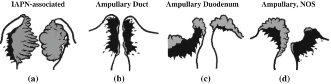
T-stage of the tumors was also re-analyzed. Four site-specific groups comprising the ampullary carcinomas were defined, as recently described (Fig.1).19
Since cases were identified retrospectively at multiple different institutions, the LN sampling method utilized at the time of original assessment was not standardized. The orange-peeling method63,64was used for cases from Emory University.
The revised N-stage protocol proposed by Kang et al.60 was applied to assign patients into node-negative (N0), 1–2
LN-positive (N1), and C3 LN-positive (N2) cohorts. The data were analyzed for all patients regardless of the number of LNs examined. Additionally, subset analysis was per-formed for cases that had C12 LNs examined as 12 is the number of LNs advocated by the AJCC as the minimum needed for accurate staging of periampullary cancers treated with pancreaticoduodenectomy.59
Statistical Analysis
Kaplan–Meier survival curves were constructed and compared across all three LN involvement categories using the log-rank test. Additional analyses compared patients with 1–2 metastatic LNs with those with C3 affected LNs to further assess the relation between the extent of LN involvement and survival. This analysis was repeated after excluding patients with \12 LNs to control for the extent of LN assessment. The cutoff of 12 LNs was used because it was considered to be an indicator of quality performance.59
Hazard ratios (HRs) and the corresponding 95 % con-fidence intervals (CIs) reflecting the association between N stage and survival were calculated using Cox models where patients with no metastatic LNs represented the reference group. The adjusted model included age, sex, lymphovascular invasion, T stage, perineural invasion, surgical margin, and site-specific classification. Propor-tional hazard assumptions were tested by inspecting the log–log curves.
RESULTS
Demographic and Clinicopathologic Data
Overall, 313 patients with invasive ampullary adeno-carcinoma were included in this study. The clinicopathologic characteristics of the study cohort are
IAPN-associated Ampullary Duct Ampullary Duodenum Ampullary, NOS
(a) (b) (c) (d)
FIG. 1 Site-specific classification of ampullary carcinoma. a IAPN-associated carcinomas are characterized by a prominent pre-invasive neoplasm (gray areas) that grows predominantly as an exophytic mass within the distal ends of the common bile duct and main pancreatic duct. b Ampullary duct carcinomas have minimal pre-invasive component, and pre-invasive component (black areas) forms a
plaque-like stricture at the distal ends of the ducts. c Ampullary duodenum carcinomas form ulcerovegetative tumors that grow predominantly on the duodenal surface of the ampulla. d Carcinomas that do not fit in any of these categories are classified as NOS. IAPN intra-ampullary papillary-tubular neoplasm, NOS not otherwise specified
summarized in Table1. Mean age was 64 years (range 27–89), and 59 % were male (n = 183). The mean overall size of the tumors was 2.7 cm, with mean invasion size of
1.9 cm. Perineural invasion was seen in 35 % (n = 110) and lymphovascular invasion was present in 65 % (n = 202) of cases.
TABLE 1 Clinicopathologic features of the study cases (n = 313)
All cases (n = 313) N0 (n = 171, 55 %) N1 (n = 82, 26 %) N2 (n = 60, 19 %) Age (years) Mean 64 ± 12 65 ± 12 65 ± 11 62 ± 12 Median 65 66 67 63 Range 27–89 27–86 38–89 35–86 Gender [n (%)] Female 129 (41.5) 66 (39) 37 (45) 26 (43) Male 183 (58.5) 104 (61) 45 (55) 34 (57)
Overall tumor size (mm)
Mean 27 ± 16 24 ± 15 28 ± 15 33 ± 17
Median 22 20 25 25
Range 3–95 3–95 8–80 12–80
Invasive tumor size (mm)
Mean 19 ± 12 16 ± 11 21 ± 12 26 ± 13
Median 17 15 20 22.5
Range 2–95 2–95 3–60 7–70
T stage (AJCC 2010 staging) [n (%)]
T1 38 (12) 32 (19) 4 (5) 2 (3) T2 128 (41) 81 (47) 34 (42) 13 (22) T3 107 (34) 48 (28) 34 (42) 25 (42) T4 39 (13) 10 (6) 9 (11) 20 (33) Perineural invasion [n (%)] Absent 203 (65) 128 (75) 50 (61) 25 (42) Present 110 (35) 43 (25) 32 (39) 35 (58) Lymphovascular invasion [n (%)] Absent 111 (35.5) 86 (50) 21 (26) 4 (7) Present 202 (64.5) 85 (50) 61 (74) 56 (93) Surgical margin [n (%)] Absent 298 (96) 167 (98) 79 (99) 52 (87) Present 12 (4) 3 (2) 1 (1) 8 (13)
Harvested lymph node number [n (%)]
\12 163 (52) 105 (61) 45 (55) 13 (22) C12 150 (48) 66 (39) 37 (45) 47 (78) Site-specific classification [n (%)] Ampullary, NOS 159 (51) 87 (51) 41 (50) 31 (52) Ampullary duct 60 (19) 25 (15) 18 (22) 17 (28) IAPN-associated 68 (22) 50 (29) 13 (16) 5 (8) Ampullary duodenum 26 (8) 9 (5) 10 (12) 7 (12) Histologic type [n (%)] Pancreatobiliary 172 (55) 95 (56) 45 (55) 32 (53) Intestinal 68 (22) 46 (27) 13 (16) 9 (15) Mixed or other 73 (23) 30 (16) 24 (29) 19 (32) Outcome [n (%)] Alive 155 (50) 97 (58) 33 (40) 25 (43) Dead 153 (50) 71 (42) 49 (60) 33 (57)
Median survival (months) 56 107.5 40 24.5
1-year survival (%) 85 88 90 68
3-year survival (%) 63 76 55 34
5-year survival (%) 49 62 31.5 30
With respect to histologic classification, 55 % of tumors (n = 172) were pancreatobiliary type; 22 % (n = 68) were intestinal type; and 23 % (n = 73) were mixed or other types. Most of the intestinal-type carcinomas revealed CDX2 (90 %) and MUC2 (85 %) expression; however, the specificity of these markers for this phenotype was fairly low (61 and 78 %, respectively). In contrast, all pancreatobil-iary-type carcinomas (100 %) were, at least focally, positive for MUC1 and MUC5AC. The cytokeratin profile was en-tirely non-discriminatory, in that CK7 and CK20 were co-expressed in 53 % of all cases available for immunohisto-chemical staining. More importantly, CK7, which is regarded as a good marker of pancreatobiliary differen-tiation, was expressed in a high proportion (63 %) of intestinal cases, and CK20, which is generally considered a good marker of the intestinal phenotype, was expressed in a substantial number (39 %) of pancreatobiliary cases.
Based on the site-specific classification scheme, 159 cases (51 %) were ampullary, not otherwise specified; 68 (22 %) were intra-ampullary papillary-tubular neoplasm (IAPN)-associated; 60 (19 %) were ampullary duct; and 26 (8 %) were ampullary duodenal.
Median follow-up was 56 months, while overall 1-, 3-, and 5-year survival was 85, 63, and 49 %, respectively.
LN Analysis
A total of 142 cases (45 %) had LN metastasis. The median number of LNs examined was 11 (range 1–61), and the number of metastatic LNs among LN-positive cases ranged from 1 to 19 (median 2). Based on the proposed N-staging protocol, 82 cases (26 %) were N1, and 60 (19 %) were N2. In the analysis restricted to cases with more than 12 LNs examined, 66 cases (44 %) were N0, 37 (25 %) were N1, and 47 (31 %) were N2. The percentage of N2 cases at the Emory University, where the orange-peeling method of LN harvesting was performed,63was 26 %.
There was a statistically significant association between proposed N stage and frequency of aggressive tumor char-acteristics, including lymphovascular invasion (p \ 0.001), perineural invasion (p \ 0.001), invasive tumor size (p \ 0.001), and surgical margin positivity (p = 0.001).
With respect to histologic classification, 55 % of pan-creatobiliary-type carcinomas were N0, 26 % were N1, and 19 % were N2; 68 % of intestinal-type carcinomas were N0, 19 % were N1, 13 % were N2, and, of the mixed or other type carcinomas, 41 % were N0, 33 % were N1, and 0 0.0 0.1 0.2 0.3 0.4 0.5 0.6 0.7 0.8 0.9 1.0 20 p<0.0001 40 60
Time to Event (months) ALL CASES Metastatic LN absent 171 (55%) Metastatic LN present 142 (45%) N (%) Survival Median (months) 1 year 3 year 5 year 88% 76% 62% 81% 47% 31% 107.5 32 Sur vi v al Pr obability 80 100 Absent Present 120 CASES WITH ≥ 12 LNs Metastatic LN Absent 66 (44%) Metastatic LN Present 84 (56%) N (%) Survival Median (months) 1 year 3 year 5 year 85% 76% 64% 85% 57% 45% N/A 46 0 0.0 0.1 0.2 0.3 0.4 0.5 0.6 0.7 0.8 0.9 1.0 20 p=0.04 40 60
Time to Event (months)
Sur v iv a l Pr obability 80 100 Absent Present 120 (a) (b)
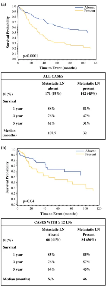
FIG. 2 Comparison of survival between positive and LN-negative cases. a All cases, and b cases with 12 or more LNs sampled only. LN lymph node
26 % were N2. When only pancreatobiliary and intestinal carcinomas were taken into account, the distribution of N stages was not different between pancreatobiliary and in-testinal carcinomas (p = 0.212).
Among the site-specific categories, 74 % of the IAPN-associated carcinoma category were N0, 19 % were N1, and only 7 % were N2. In contrast, the ampullary-ductal group had the highest proportion of N2 cases (28 %).
Survival Analysis
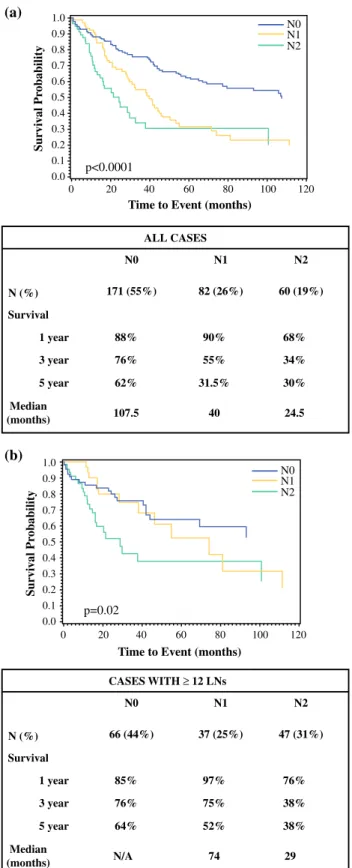
In all patients, the 1-, 3-, and 5-year survival rates were 85, 63, and 49 %, respectively. Median survival of LN-negative cases was significantly longer than that of LN-positive cases (107.5 vs. 32 months; p \ 0.0001), with 1-, 3-, and 5-year survival rates of 88, 76, and 62 % versus 81, 47, and 31 %, respectively (p \ 0.001; Fig. 2a). In the subanalysis of pa-tients who had 12 or more LNs examined, the difference persisted. The 1-, 3-, and 5-year survival rates were 85, 76 %, and 64 %, respectively, for LN-negative cases, and 85, 57, and 45 %, respectively, for LN-positive cases (p = 0.04; Fig.2b). The median survival of patients with N1 disease was 40 months compared with 24.5 months for patients with N2 disease (p \ 0.0001; Fig.3a). The 1-, 3-, and 5-year survival rates were 90, 55, and, 31.5 %, respectively, for the N1 group, and 68, 34, and 30 %, respectively, for the N2 group (p \ 0.001; Fig.3a). The corresponding analyses among patients with 12 or more LNs sampled produced similar results, with median survival estimates of 74 months for N1, and 29 months for N2. The 1-, 3-, and 5-year survival rates among N1- and N2-stage groups with sufficient (C12 LNs) sampling were 97, 75, and 52 %, respectively, and 76, 38, and 38 %, respectively (p = 0.02; Fig.3b).
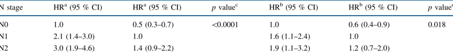
In the multivariable Cox regression model adjusted for age, sex, tumor invasion size, tumor stage, perineural in-vasion, surgical margin, and site-specific classification, the association between higher proposed N stage and shorter survival persisted. Using the N0 group as the reference, the adjusted HRs (95 % CIs) for N1 and N2 were 1.6 (1.1–2.4) and 1.9 (1.1–3.2), respectively (p value for trend = 0.018) [Table2]. In these models, age [HR, 1.3 (1.1–1.5, p = 0.004) for a 10 year increase], lymphovascular inva-sion [HR, 1.7 (1.1–2.5, p = 0.01)], perineural invainva-sion [HR, 1.5 (1.1–2.3, p = 0.04)] and surgical margin posi-tivity [HR, 3.3 (1.6–6.9, p = 0.001)] were also statistically significantly associated with survival.
DISCUSSION
This large, multi-institutional study of ampullary carci-noma examined the prognostic implications of the number ALL CASES N0 171 (55%) N2 60 (19%) N (%) Survival Median (months) 1 year 3 year 5 year 88% 76% 62% 68% 34% 30% 107.5 24.5 N1 82 (26%) 90% 55% 31.5% 40 0 0.0 0.1 0.2 0.3 0.4 0.5 0.6 0.7 0.8 0.9 1.0 20 p<0.0001 40 60
Time to Event (months)
Sur vi v al Pr obability 80 100 N0 N1 120 N2 CASES WITH ≥ 12 LNs N0 66 (44%) N2 47 (31%) N (%) Survival Median (months) 1 year 3 year 5 year 85% 76% 64% 76% 38% 38% N/A 29 N1 37 (25%) 97% 75% 52% 74 0 0.0 0.1 0.2 0.3 0.4 0.5 0.6 0.7 0.8 0.9 1.0 20 p=0.02 40 60
Time to Event (months)
Sur v iv al Pr obability 80 100 N0 N1 120 N2 (a) (b)
FIG. 3 Comparison of survival between proposed N0 (no positive LNs), N1 (1–2 positive LNs), and N2 (C3 positive LNs) cases. a All cases, and b cases with 12 or more LNs sampled only. LNs lymph node, NA not applicable
of positive LNs using recently proposed nodal substages with the primary outcome of survival after pancreatoduo-denectomy for ampullary carcinoma. The proposed N-staging classification was found to stratify patients by survival outcomes in a manner that was clinically and statistically significant. The associations between proposed N stages and survival persisted in a multivariate model that included age, sex, tumor invasion size, tumor stage, per-ineural invasion, surgical margin, and site-specific classification.
Historically, ampullary carcinoma has been ill-defined. Recent studies have proposed guidelines to identify am-pullary carcinoma, which were used in the present study.18,19 Variability in classifying ampullary tumors generated studies with conflicting results, which led some authors to question the validity that ampullary carcinoma is a distinct entity.17 In this study, we found that the
frequency of LN metastasis in ampullary carcinoma was 45 %, which is much lower than the documented LN metastasis rate for pancreatic ductal adenocarcinoma (PDAC) [78 %].22 To account for biases given the retro-spective nature of this study, without the ability to control for the number of LNs sampled, cases with C12 LNs ex-amined were separately analyzed. The rate of LN metastasis was 56 %, which was still lower than that of PDAC. Furthermore, ampullary carcinoma was found to have a better clinical outcome than is typical of PDACs.19,22 These differences support the clinical sig-nificance of the defining criteria employed and reinforce the identity of ampullary carcinoma as a separate clinical entity.
Multiple studies have shown the association between LN metastasis and shorter survival in ampullary carcinoma (Table3).52–58In many studies, the absence or presence of TABLE 2 Comparison of hazard ratios by proposed N stage in all cases
N stage HRa(95 % CI) HRa(95 % CI) p valuec HRb(95 % CI) HRb(95 % CI) p valuec
N0 1.0 0.5 (0.3–0.7) \0.0001 1.0 0.6 (0.4–0.9) 0.018
N1 2.1 (1.4–3.0) 1.0 1.6 (1.1–2.4) 1.0
N2 3.0 (1.9–4.6) 1.4 (0.9–2.2) 1.9 (1.1–3.2) 1.2 (0.7–2.0)
CI confidence interval, HR hazard ratio
a HR based on Cox proportional hazard model only, including N stage
b HR based on Cox proportional hazard model adjusting for age, sex, invasion size, T stage, surgical margin, site specific lassification c p value based on Chi square test (Wald test)
TABLE 3 Different number (cut-off value) of positive lymph nodes deemed to be significant in the literature Author, year No. of
cases
Cut-off values
Comments Risk assessment
determined by
Shroff et al.52 92 0, 1–3, or [3 Disease-free and overall
survival Sakata et al.53 71 0, 1–3, or C4 The number of positive LNs better predicts the
outcome than the LN ratio
Lee et al.54 52 C3 In univariate analysis, the number of positive LNs, and LN ratio and LN location are significantly correlated with survival
In multivariate analysis, the factor of C3 metastatic LNs is the only independent prognostic factor
Recurrence and survival
Choi et al.55 78 0–1 or C2 LN number and presence of metastatic LNs do not affect overall survival
C2 metastatic LNs significantly affects disease-free survival
Recurrence and overall survival
Sommerville et al.56 39 0, 1–3, or [3 [3 vs. 0 metastatic LN hazard ratio is significant Overall survival Sierzega et al.57 111 C4 In univariate analysis, the presence of metastatic LNs, and
their number and ratio are significantly correlated with survival
Overall survival
Sakata et al.58 62 0, 1–3, or C4 The number, not the location, of metastatic LNs independently affects long-term survival
Overall survival
LN metastasis was found to be associated with overall survival38,39,41,42 or disease-free survival15,37,40 in uni-variate analysis. Only some of these associations persisted in multivariate analysis.26–36 As an extension of this, LN ratio was also found to be significant in some studies,44–48 whereas other studies found a different cut-off value of positive LNs to be significant.52–58
The current AJCC staging system for ampullary carci-noma stratifies patients into N-stage groups reflecting only the absence or presence of LN metastasis without consid-eration of the number of positive LNs.59 For many other types of carcinomas, the number of positive LNs had been found to be of prognostic significance and is included in their respective N-stage guidelines.59 For ampullary can-cer, previous studies have found that the number of positive LNs is associated with survival; however, the value dif-fered in studies with [2, [3, and [4 being advocated.52–58 In their analysis of the SEER database, Kang et al. recently determined that stratifying positive LNs as N1 (1 or 2 LNs) versus N2 (3 or more LNs) had significant prognostic value.60Their findings were separately validated in an in-stitutional cohort, although the detailed criteria of patient selection for the latter were not provided. In this study, the value of this nodal substaging was analyzed, and we validated this proposal in a well-characterized, patho-logically-verified cohort, both in univariate and multivariate survival analysis.
In addition to the established relationship to survival, the proposed N substage protocol suggested an association with features observed in aggressive disease, including lymphovascular invasion, perineural invasion, surgical margin positivity, and invasive tumor size. This observa-tion is similar to the results found by Kang et al.60
The prognostic differences in three N-stage groups persist in subset analysis of cases with C12 LNs examined. The rate of LN-positive cases was 56 % in the C12 LN group compared with 45 % in all patients, suggesting that lack of LN sampling could lead to understaging. Further-more, in this subset, the survival curve of N2 category separates more dramatically from the N0 and N1 groups. Intriguingly, also in this subset, the N0 and N1 groups begin to have overlapping survival curves. The fact that N0 and N1 (1–2 metastatic LNs) cases have similar survival in this better-sampled subset with C12 LNs may be at-tributable to various factors, one of which is direct invasion of LN being regarded as ‘metastasis’ by AJCC TNM classification. Some authors believe that direct invasion does not imply the same thing as true LN metastasis as the cells do not yet have the ability to grow and survive within lymphatic channels, and extravasate and form self-sus-taining colonies in LNs.65 Of note, due to anatomic complexity, it may be difficult to determine whether an LN is involved directly or represents true metastasis in the
peri-ampullary region. Another possibility for the similar sur-vivals of N0 and N1 cases in this better-documented group might be that only cases with the ability to metastasize to multiple LNs represent the true biologic aggressiveness of the disease. This may also explain why N2 cases are more common than N1 cases. Regardless, the implication is that an improved LN sampling of pancreatoduodenectomy specimens enhances the prognostic value of separating LN-positive cases into N1 and N2 substages. Of note, LN sampling in the pathology gross room can be augmented by the orange-peeling method yielding improved LN harvest and, consequently, a better assessment of LN status.63,64 This can then have an impact on prognostication, as il-lustrated in the study by Partelli et al.66 In fact, in our study, in cases from the institution where the orange-peeling method is routinely applied, the frequency of N2 cases was higher (26 vs. 13.5 % in the remainder); thus, an orange-peeling approach, or other approach that generates more accurate LN harvest, should be adopted.
We acknowledge the limitation of this study design. This was a retrospective, multi-institutional study, there-fore the gross dissection method of inspecting and sampling for LNs and data for the adjuvant therapy was not available. Additionally, retrospective analysis of data is limited to determining associations between factors and outcomes, and causality cannot be confirmed. Despite these drawbacks, the present study is important because it compares the current N-stage guidelines for ampullary carcinoma with proposed guidelines to create substages within the LN-positive population. The results from the current study validate the prognostic significance of the substaging proposed by Kang et al., which should be in-tegrated into future N-stage guidelines for ampullary carcinoma.60
CONCLUSIONS
In summary, defined with the revised criteria, ampullary carcinoma is a distinct entity. LN status is strongly asso-ciated with survival. Furthermore, substaging N-stage, as proposed by Kang et al., with 1–2 LN-positive (N1) and C3 LN-positive (N2) cases offers improved stratification of survival in ampullary carcinoma. Considering that there are very few established predictors of outcome in ampullary carcinoma, incorporating substaging of LN-positive cases is strongly recommended as a part of the TNM stage of these tumors.
ACKNOWLEDGMENT The authors would like to thank Allyne Manzo and Lorraine Biedrzycki for assistance with the figures. DISCLOSURES None of the authors have no affiliation with, or financial involvement in, any organization with a direct financial in-terest in the subject matter or materials discussed in the manuscript.
REFERENCES
1. Klimstra DS, Albores-Saavedra J, Hruban RH, Zamboni G. Tu-mours of the ampullary region. In: Bosman FT, Carneiro F, Hruban RH, Theise ND, eds. WHO classification of tumours of the digestive system. Lyon: International Agency for Research on Cancer (IARC); 2010. p. 81–94.
2. Adsay NV, Basturk O. Tumors of major and minor ampulla. In: Odze R, Goldblum J, eds. Surgical pathology of the GI tract, liver, biliary tract, and pancreas. Philadelphia: Elsevier; 2014. p. 1120–39.
3. Thompson LDR, Basturk O, Adsay NV. Pancreas. In: Mills SE, ed. Sternberg’s diagnostic surgical pathology. Philadelphia: Lippincott Williams & Wilkins; 2015. p. 1577–662.
4. Shi HY, Wang SN, Lee KT. Temporal trends and volume-out-come associations in periampullary cancer patients: a propensity score-adjusted nationwide population-based study. Am J Surg. 2014;207:512–19.
5. Bronsert P, Kohler I, Werner M, et al. Intestinal-type of differ-entiation predicts favourable overall survival: confirmatory clinicopathological analysis of 198 periampullary adenocarcino-mas of pancreatic, biliary, ampullary and duodenal origin. BMC Cancer. 2013;13:428.
6. Kumari N, Prabha K, Singh RK, Baitha DK, Krishnani N. In-testinal and pancreatobiliary differentiation in periampullary carcinoma: the role of immunohistochemistry. Hum Pathol. 2013;44:2213–9.
7. Lee SR, Kim HO, Park YL, Shin JH. Lymph node ratio predicts local recurrence for periampullary tumours. ANZ J Surg. 2014;84:353–8.
8. Bornstein-Quevedo L, Gamboa-Dominguez A. Carcinoid tumors of the duodenum and ampulla of vater: a clinicomorphologic, immunohistochemical, and cell kinetic comparison. Hum Pathol. 2001;32:1252–6.
9. Gutierrez JC, Franceschi D, Koniaris LG. How many lymph nodes properly stage a periampullary malignancy? J Gastrointest Surg. 2008;12:77–85.
10. Kim K, Chie EK, Jang JY, et al. Prognostic significance of tumour location after adjuvant chemoradiotherapy for periampullary ade-nocarcinoma. Clin Transl Oncol. 2012;14:391–5.
11. Maithel SK, Khalili K, Dixon E, et al. Impact of regional lymph node evaluation in staging patients with periampullary tumors. Ann Surg Oncol. 2007;14:202–10.
12. Westgaard A, Pomianowska E, Clausen OP, Gladhaug IP. In-testinal-type and pancreatobiliary-type adenocarcinomas: how does ampullary carcinoma differ from other periampullary ma-lignancies? Ann Surg Oncol. 2013;20:430–9.
13. Riall TS, Cameron JL, Lillemoe KD, et al. Resected peri-ampullary adenocarcinoma: 5-year survivors and their 6- to 10-year follow-up. Surgery. 2006;140:764–72.
14. Ahn DH, Bekaii-Saab T. Ampullary cancer: an overview. Am Soc Clin Oncol Educ Book. 2014; 112–115.
15. Williams JA, Cubilla A, Maclean BJ, Fortner JG. Twenty-two year experience with periampullary carcinoma at Memorial Sloan-Kettering Cancer Center. Am J Surg. 1979;138:662–5. 16. Cubilla AL, Fitzgerald PJ. Surgical pathology aspects of cancer
of the ampulla-head-of-pancreas region. Monogr Pathol. 1980;21:67–81.
17. Perysinakis I, Margaris I, Kouraklis G. Ampullary cancer: a separate clinical entity? Histopathology. 2014;64:759–68. 18. Washington K, Berlin J, Branton P; for the College of American
Pathologists (CAP). Protocol for the examination of specimens from patients with carcinoma of the ampulla of Vater. 2012. Available at: http://www.cap.org/apps/docs/committees/cancer/
cancer_protocols/2012/Ampulla_12protocol_3101.pdf. Accessed 24 Jan 2014.
19. Adsay V, Ohike N, Tajiri T, et al. Ampullary region carcinomas: definition and site specific classification with delineation of four clinicopathologically and prognostically distinct subsets in an analysis of 249 cases. Am J Surg Pathol. 2012;36:1592–608. 20. Ohike N, Kim GE, Tajiri T, et al. Intra-ampullary
papillary-tubular neoplasm (IAPN): characterization of tumoral intraep-ithelial neoplasia occurring within the ampulla: a clinicopathologic analysis of 82 cases. Am J Surg Pathol. 2010;34:1731–48.
21. Tajiri T, Basturk O, Krasinskas A, et al. Prognostic differences between ampullary carcinomas and pancreatic ductal adenocarci-nomas: the importance of size of invasive component [abstract]. Mod Pathol. 2009;22:323A.
22. Saka B, Tajiri T, Ohike N, et al. Clinicopathologic comparison of ampullary versus pancreatic carcinoma: preinvasive component, size of invasion, stage, resectability and histologic phenotype are the factors for the significantly favorable outcome of ampullary carcinoma [abstract]. Mod Pathol. 2013;26:429A.
23. Mori K, Ikei S, Yamane T, et al. Pathological factors influencing survival of carcinoma of the ampulla of Vater. Eur J Surg Oncol. 1990;16:183–8.
24. Shyr YM, Su CH, Wu LH, Li AF, Chiu JH, Lui WY. DNA ploidy as a major prognostic factor in resectable ampulla of Vater can-cers. J Surg Oncol. 1993;53:220–5.
25. Woo SM, Ryu JK, Lee SH, et al. Recurrence and prognostic factors of ampullary carcinoma after radical resection: compar-ison with distal extrahepatic cholangiocarcinoma. Ann Surg Oncol. 2007;14:3195–201.
26. Zhou J, Zhang Q, Li P, Shan Y, Zhao D, Cai J. Prognostic factors of carcinoma of the ampulla of Vater after surgery. Tumour Biol. 2014;35(2):1143–8.
27. Kim WS, Choi DW, Choi SH, Heo JS, You DD, Lee HG. Clinical significance of pathologic subtype in curatively resected ampulla of vater cancer. J Surg Oncol. 2012;105:266–72.
28. Inoue Y, Hayashi M, Hirokawa F, Egashira Y, Tanigawa N. Clinicopathological and operative factors for prognosis of carcinoma of the ampulla of vater. Hepatogastroenterology. 2012;59:1573–6. 29. Kohler I, Jacob D, Budzies J, et al. Phenotypic and genotypic
characterization of carcinomas of the papilla of Vater has prog-nostic and putative therapeutic implications. Am J Clin Pathol. 2011;135:202–11.
30. Ohike N, Coban I, Kim GE, et al. Tumor budding as a strong prognostic indicator in invasive ampullary adenocarcinomas. Am J Surg Pathol. 2010;34:1417–24.
31. Hsu HP, Shan YS, Jin YT, Lai MD, Lin PW. Loss of E-cadherin and beta-catenin is correlated with poor prognosis of ampullary neoplasms. J Surg Oncol. 2010;101:356–62.
32. de Paiva Haddad LB, Patzina RA, Penteado S, et al. Lymph node involvement and not the histophatologic subtype is correlated with outcome after resection of adenocarcinoma of the ampulla of vater. J Gastrointest Surg. 2010;14:719–28.
33. Lowe MC, Coban I, Adsay NV, et al. Important prognostic fac-tors in adenocarcinoma of the ampulla of Vater. Am Surg. 2009;75:754–60; discussion 761.
34. Yao HS, Wang Q, Wang WJ, Hu ZQ. Intraoperative allogeneic red blood cell transfusion in ampullary cancer outcome after curative pancreatoduodenectomy: a clinical study and meta-ana-lysis. World J Surg. 2008;32:2038–46.
35. Shimizu Y, Kimura F, Shimizu H, Yoshidome H, Ohtsuka M, Miyazaki M. The morbidity, mortality, and prognostic factors for ampullary carcinoma and distal cholangiocarcinoma. Hepato-gastroenterology. 2008;55:699–703.
36. Qiao QL, Zhao YG, Ye ML, et al. Carcinoma of the ampulla of Vater: factors influencing long-term survival of 127 patients with resection. World J Surg. 2007;31:137–43; discussion 144-136. 37. Chen J, Cai S, Dong J. Predictors of recurrence after
pancreati-coduodenectomy for carcinoma of the ampulla of Vater [in Chinese]. Nan Fang Yi Ke Da Xue Xue Bao 2012;32:1242–4. 38. Showalter TN, Zhan T, Anne PR, et al. The influence of
prog-nostic factors and adjuvant chemoradiation on survival after pancreaticoduodenectomy for ampullary carcinoma. J Gastroin-test Surg. 2011;15:1411–6.
39. Casaretto E, Andrada DG, Granero LE. Results of cephalic pancreaticoduodenectomy for ampullary carcinoma. Analysis of 18 consecutive cases [in Spanish]. Acta Gastroenterol Latinoam. 2010;40:22–31.
40. Sudo T, Murakami Y, Uemura K, et al. Prognostic impact of perineural invasion following pancreatoduodenectomy with lymphadenectomy for ampullary carcinoma. Dig Dis Sci. 2008;53:2281–6.
41. Barauskas G, Gulbinas A, Pranys D, Dambrauskas Z, Pundzius J. Tumor-related factors and patient’s age influence survival after resection for ampullary adenocarcinoma. J Hepatobiliary Pan-creat Surg. 2008;15:423–8.
42. Sessa F, Furlan D, Zampatti C, Carnevali I, Franzi F, Capella C. Prognostic factors for ampullary adenocarcinomas: tumor stage, tumor histology, tumor location, immunohistochemistry and mi-crosatellite instability. Virchows Arch. 2007;451:649–57. 43. Dahl S, Bendixen M, Fristrup CW, Mortensen MB. Treatment
outcomes for patients with papilla of Vater cancer [in Danish]. Ugeskr Laeger. 2010;172:1361–5.
44. Pomianowska E, Westgaard A, Mathisen O, Clausen OP, Glad-haug IP. Prognostic relevance of number and ratio of metastatic lymph nodes in resected pancreatic, ampullary, and distal bile duct carcinomas. Ann Surg Oncol. 2013;20:233–41.
45. Roland CL, Katz MH, Gonzalez GM, et al. A high positive lymph node ratio is associated with distant recurrence after surgical re-section of ampullary carcinoma. J Gastrointest Surg. 2012;16:2056–63.
46. Hurtuk MG, Hughes C, Shoup M, Aranha GV. Does lymph node ratio impact survival in resected periampullary malignancies? Am J Surg. 2009;197:348–52.
47. Zhou J, Zhang Q, Li P, Shan Y, Zhao D, Cai J. Prognostic relevance of number and ratio of metastatic lymph nodes in re-sected carcinoma of the ampulla of Vater. Chin J Cancer Res. 2013;25:735–42.
48. Falconi M, Crippa S, Dominguez I, et al. Prognostic relevance of lymph node ratio and number of resected nodes after curative resection of ampulla of Vater carcinoma. Ann Surg Oncol. 2008;15:3178–86.
49. Bogoevski D, Chayeb H, Cataldegirmen G, et al. Nodal mi-croinvolvement in patients with carcinoma of the papilla of vater receiving no adjuvant chemotherapy. J Gastrointest Surg. 2008;12:1830–7; discussion 1837-1838.
50. van der Gaag NA, ten Kate FJ, Lagarde SM, Busch OR, van Gulik TM, Gouma DJ. Prognostic significance of extracapsular lymph node involvement in patients with adenocarcinoma of the ampulla of Vater. Br J Surg. 2008;95:735–43.
51. Hornick JR, Johnston FM, Simon PO, et al. A single-institution review of 157 patients presenting with benign and malignant
tumors of the ampulla of Vater: management and outcomes. Surgery. 2011;150:169–76.
52. Shroff S, Overman MJ, Rashid A, et al. The expression of PTEN is associated with improved prognosis in patients with ampullary adenocarcinoma after pancreaticoduodenectomy. Arch Pathol Lab Med. 2013;137:1619–26.
53. Sakata J, Shirai Y, Wakai T, Ajioka Y, Akazawa K, Hatakeyama K. Assessment of the nodal status in ampullary carcinoma: the number of positive lymph nodes versus the lymph node ratio. World J Surg. 2011;35:2118–24.
54. Lee JH, Lee KG, Ha TK, et al. Pattern analysis of lymph node metastasis and the prognostic importance of number of metastatic nodes in ampullary adenocarcinoma. Am Surg. 2011;77:322–9. 55. Choi SB, Kim WB, Song TJ, Suh SO, Kim YC, Choi SY.
Sur-gical outcomes and prognostic factors for ampulla of Vater cancer. Scand J Surg. 2011;100:92–8.
56. Sommerville CA, Limongelli P, Pai M, et al. Survival analysis after pancreatic resection for ampullary and pancreatic head carcinoma: an analysis of clinicopathological factors. J Surg Oncol. 2009;100:651–6.
57. Sierzega M, Nowak K, Kulig J, Matyja A, Nowak W, Popiela T. Lymph node involvement in ampullary cancer: the importance of the number, ratio, and location of metastatic nodes. J Surg Oncol. 2009;100:19–24.
58. Sakata J, Shirai Y, Wakai T, et al. Number of positive lymph nodes independently affects long-term survival after resection in patients with ampullary carcinoma. Eur J Surg Oncol. 2007;33:346–51. 59. Edge SB, American Joint Committee on Cancer. AJCC cancer
staging manual. 7th ed. New York: Springer; 2010.
60. Kang HJ, Eo SH, Kim SC, et al. Increased number of metastatic lymph nodes in adenocarcinoma of the ampulla of Vater as a prognostic factor: a proposal of new nodal classification. Surgery. 2014;155(1):74–84.
61. Saka B, Bagci P, Krasinskas A, et al. Duodenal carcinomas of non-ampullary origin are significantly more aggressive than ampullary carcinomas. Mod Pathol. 2013;26:176A–177A. 62. Balci S, Kim GE, Ohike N, et al. Applicability and prognostic
relevance of ampullary carcinoma histologic typing as pancre-atobiliary versus intestinal [abstract]. Mod Pathol. 2010;23:350A. 63. Adsay NV, Basturk O, Altinel D, et al. The number of lymph nodes identified in a simple pancreatoduodenectomy specimen: comparison of conventional vs orange-peeling approach in pathologic assessment. Mod Pathol. 2009;22:107–12.
64. Adsay NV, Basturk O, Saka B, et al. Whipple made simple for surgical pathologists: orientation, dissection, and sampling of pancreaticoduodenectomy specimens for a more practical and accurate evaluation of pancreatic, distal common bile duct, and ampullary tumors. Am J Surg Pathol. 2014;38(4):480–93. 65. Pai RK, Beck AH, Mitchem J, et al. Pattern of lymph node
in-volvement and prognosis in pancreatic adenocarcinoma: direct lymph node invasion has similar survival to node-negative dis-ease. Am J Surg Pathol. 2011;35:228–34.
66. Partelli S, Crippa S, Capelli P, et al. Adequacy of lymph node retrieval for ampullary cancer and its association with improved staging and survival. World J Surg. 2013;37:1397–404.