389
CASE REPORT
50-year-old man was admitted to our neurosurgery depart-ment with the chief complaint of difficulty in standing and walk-ing due to leg weakness for nearly 2 days. He also described back pain while sitting and standing. Neurological examination dem-onstrated paraparesis with impaired anal sphincter tonus. He had no history of trauma or any systemic illness.
Thoracic magnetic resonance imaging (MRI) and computer-ized tomography (CT) revealed expansile mass lesion that was compressing the spinal cord at T9 level. Homogenously enhanc-ing mass lesion was found to originate from the anterior portion of the spinous process and both laminas of T9 vertebra (Fig. 1). Routine blood tests were uneventful except a calcium value of 14.3 mg/dL (8.4–10.2). As blood parathormone (PTH) test also revealed a very high value of 547.45 pg/mL (15–68.3), endocri-nology consultation was ordered to rule out primary hyperpara-thyroidism. Neck sonography demonstrated hyperplasia and multiple nodules on the thyroid gland. However, any cystic or sol-id pathologic lesion compatible with adenoma were not seen on the parathyroid regions. Parathyroid scintigraphy with Tc-99m
INTRODUCTION
Brown tumors (BTs), also called as osteoclastomas, are rare nonneoplastic lesions that arise in the setting of hyperparathy-roidism3,19). Hyperparathyroidism is overactivity of the parathy-roid glands resulting in excess production of parathyparathy-roid hor-mone (PTH). PTH regulates calcium and phosphate metabolism of the body3). Excessive PTH secretion may be due to problems in the parathyroid glands such as adenomas, hyperplasia or car-cinoma. This is known as primary hyperparathyroidism3,19). How-ever, it may also occur in response to low calcium levels in such conditions as vitamin D deficiency or chronic renal disease that is known as secondary hyperparathyroidism3,19). BTs can arise as solitary or multiple lesions of any bone, more common in extremi-ties, clavicle, ribs and pelvis. Spinal involvement is very rare1-20).
In this study, we report a rare thoracic BT case that occured in the setting of primary hyperthyroidism. Tumor was treated suc-cesfully by cooperation of endocrinology, general surgery and neurosurgery departments.
Brown Tumor of the Thoracic Spine : First Manifestation
of Primary Hyperparathyroidism
Erkin Sonmez, M.D.,1 Tugan Tezcaner, M.D.,2 Ilker Coven, M.D.,3 Aysen Terzi, M.D.4
Departments of Neurosurgery,1 General Surgery,2 Pathology,4 Baskent University School of Medicine, Ankara, Turkey
Department of Neurosurgery,3 Baskent University Konya Training and Research Hospital, Konya, Turkey
Brown tumors also called as osteoclastomas, are rare nonneoplastic lesions that arise in the setting of primary or secondary hyperparathyroidism. Parathyroid adenomas or hyperplasia constitute the major Brown tumor source in primary hyperparathyroidism while chronic renal failure is the leading cause in secondary hyperparathyroidism. Most of the patients with the diagnosis of primary hyperparathyroidism present with kidney stones or isolated hypercalcemia. However, nearly one third of patients are asymptomatic and hypercalcemia is found incidentally. Skeletal involvement such as generalized osteopenia, bone resorption, bone cysts and Brown tumors are seen on the late phase of hyperparathyroidism. The symptoms include axial pain, radiculopathy, myelopathy and myeloradiculopathy according to their locations. Plasmocytoma, lymphoma, giant cell tumors and metastates should be ruled out in the differential diagnosis of Brown tumors. Treatment of Brown tumors involve both the management of hyper-parathyroidism and neural decompression. The authors report a very rare spinal Brown tumor case, arisen as the initial manifestation of primary hy-perparathyroidism that leads to acute paraparesis.
Key Words : Brown tumor · Primary hyperparathyroidism · Spine · Treatment. Case Report
•Received : October 29, 2014 •Revised : March 20, 2015 •Accepted : March 23, 2015 •Address for reprints : Erkin Sonmez, M.D.
Department of Neurosurgery, Baskent University School of Medicine, Ankara, Turkey Tel : +90 3122126868/1080, Fax : +90 3122237333, E-mail : [email protected]
•This is an Open Access article distributed under the terms of the Creative Commons Attribution Non-Commercial License (http://creativecommons.org/licenses/by-nc/3.0)
which permits unrestricted non-commercial use, distribution, and reproduction in any medium, provided the original work is properly cited. J Korean Neurosurg Soc 58 (4) : 389-392, 2015
http://dx.doi.org/10.3340/jkns.2015.58.4.389
Copyright © 2015 The Korean Neurosurgical Society
PrintISSN 2005-3711 On-line ISSN 1598-7876
390 J Korean Neurosurg Soc 58 |October 2015
MIBI revealed focal activity retention on the inferior portion of the right thyroid lobe. Abdomen and thorax CT were uneventful. Endocrinology department insisted for the urgent parathyroid-ectomy in order to minimize hypercalcemia-related complications. So, patient with the diagnosis of primary hyperparathyroidism underwent surgery for parathyroidectomy first. Pathological parathyroid tissue was found and excised. Total thyroidectomy was also performed since frozen examination of a suspected thy-roid nodule was reported as micropapillary carcinoma. The day after parathyroid and thyroid surgery blood calcium and PTH levels decreased to 10.2 mg/dL and 10.42 pg/mL, respectively. Patient underwent spine surgery. Tumor was excised in piece-meal fashion. Wide posterior decompression (T8 partial/T9 to-tal laminectomy+bilateral T9 transvers process and pedicle ex-cision) was followed by T8–10 posterior instrumentation and
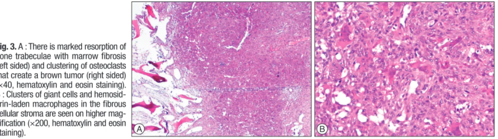
fusion (Fig. 2). Paraparesis resolved postoperatively. Patient was mobilised with a brace on postoperative day 1. Absolute pathol-ogy reports were consistent with the parathyroid adenoma and spinal Brown tumor (Fig. 3).
DISCUSSION
Brown tumors occur due to increased osteoclastic activity in the setting of either primary or secondary hyperparathyroid-ism3,8,19). Parathyroid adenomas or hyperplasia constitute the ma-jor BT source in primary hyperparathyroidism while chronic renal failure is the leading cause in secondary hyperparathyroid-ism3). BTs are localized form of osteitis fibrosa cystica (OFC) that is commonly associated with hyperparathyroidism. OFC is a process that is characterized by hyperstimulation of osteoclastic proliferation via longstanding excessive PTH production caus-ing bone resorption and bone marrow fibrosis3,7,10,14).
Histopathologically, BTs are composed of osteoclast-like giant cells and hemosiderin with a fibrovascular stroma. The name Brown tumor comes from the accumulation of hemosiderin that gives the surrounding stroma a brown color3,8). BTs look like sim-ilar histologically to other giant cell lesions such as giant cell tu-mor, aneursymal bone cyst and reparative giant cell granuloma8). Only the clinical manifestation and endocrinologic status help to differentiate BTs from other giant cell lesions.
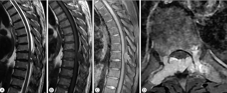
BTs usually develop in the third or fourth decades of life8). In the beginning, BTs that develop in primary hyperparathyroid-ism were seen much more common. However, they are seen more often recently as a result of secondary hyperparathyroidism that is closely related with increased life expectancy of chronic renal disease patients who require hemodialysis3,8). Most of the patients with the diagnosis of primary hyperparathyroidism present with kidney stones or isolated hypercalcemia3). However, nearly one third of patients are asymptomatic and hypercalcemia is Fig. 1. Sagittal T2W (A), sagittal T1W (B), sagittal T1W postcontrast (C) and axial T2W MR (D) images showing an expansile mass lesion that was com-pressing the spinal cord at T9 level. Homogenously enhancing mass lesion was found to originate from the anterior portion of the spinous process and both laminas of T9 vertebra.
A B C D
Fig. 2. AP (A) and lateral (B) X-Ray images demonstrate a short segment spinal instrumentation and fusion with transpedicular screws and rods at the level of T8–10.
391
Brown Tumor of the Thoracic Spine |E Sonmez, et al.
found incidentally. Skeletal involvement such as bone resorption, bone cysts, BTs and osteopenia is seen on the late phase of hy-perparathyroidism3,11).
The symptoms of spinal BTs include axial pain, radiculopathy, myelopathy and myeloradiculopathy according to their loca-tions1,3,12-20). Radiographically, BTs do not have a pathognomon-ic appearance. It can mimpathognomon-ic multiple myeloma, metastases, sar-comas and other giant cell lesions6,7,10,19). X-Rays demonstrate solitary or multiple, lytic, expansile lesions. Bone cortex might
be thinned and expanded but it is not penetrated by the tumor frequently. On CT scan, relatively well demarcated soft tissue mass with local bone erosion and expansion is observed. MRI is the diagnosis modality of choice in evaluating the location, ex-tent of spinal tumor and neural compression. On MRI, BTs are commonly seen hypointense on T1-weighted images, hypoin-tense or hyperinhypoin-tense on T2-weighted images and homogenous enhancement is noted after contrast injection. Intratumoral hem-orrhages lead to fluid-fluid level appearance on MRI3,8,19).
Table 1. Literature review of spinal Brown tumors seen in primary hyperparathyroidism
Authors (year) Sex Age Affected spinal level Symptoms Treatment
Shaw and Davies (1968)15) F 58 T10 pedicle Paraparesis, urinary retention Resection of lesion and parathyroidectomy
Shuangshoti et al. (1972)16) M 32 L4 posterior element Paraparesis, radicular pain Resection of lesion and parathyroidectomy
Sundaram and Scholz (1977)18) F 63 T10 body and pedicle Paraparesis, urinary retention Resection of lesion and parathyroidectomy
Siu et al. (1977)17) F 64 T10 Paraplegia, urinary retention Resection of lesion and parathyroidectomy
Ganesh et al. (1981)4) M 40 T2 body and pedicle Paraparesis, radicular pain Parathyroid adenoma excision only
Yokota et al. (1989)20) F 58 T5 pedicle Paraparesis, numbness Resection of lesion and parathyroidectomy
Daras et al. (1990)2) F 54 T9 pedicle Paraparesis Resection of lesion
Kashkari et al. (1990)7) F 51 T6/T7 body Paraparesis Resection of lesion, instrumentation, fusion
and parathyroidectomy
Sarda et al. (1993)14) F 23 T3/T4 Paraparesis, radicular pain Resection of lesion and parathyroidectomy
Motateanu et al. (1994)12) M 57 L4–5 facet joint Radiculopathy Resection of lesion, instrumentation and
fusion
Mustonen et al. (2004)13) M 28 L2 posterior elements Radiculopathy, numbness Resection of parathyroid adenoma only
Haddad et al. (2007)5) F 26 T2/T3 posterior
elements, T4 body Paraparesis, numbness Resection of lesion and parathyroidectomy Khalil et al. (2007)9) M 69 T2 body and pedicle Radiculopathy Resection of lesion
Altan et al. (2007)1) F 44 Left lateral side of S2 Radiculopathy, low back pain Resection of lesion and parathyroidectomy
Hoshi et al. (2008)6) F 23 Sacrum Radicular pain Resection of parathyroid adenoma only
Lee et al. (2013)10) M 65 L2 body and L1
posterior elements Low back pain, radicular pain Resection of lesion, instrumentation, fusion and parathyroidectomy Khalatbari and Moharamzad
(2014)8) M 16 L2 pedicle and posterior elements Paraparesis, sphincter dysfunction Resection of lesion, instrumentation, fusion and parathyroidectomy
Khalatbari and Moharamzad
(2014)8) F 46 L3 posterior elements Paraparesis, low back pain Resection of lesion and parathyroid adenoma
Khalatbari and Moharamzad
(2014)8) F 52 C6 lamina and pedicle C6 radiculopathy, neck pain Resection of lesion, instrumentation, fusion and parathyroidectomy
Khalatbari and Moharamzad
(2014)8) M 38 T7 body, pedicle and lamina Paraparesis, sphincter dysfunction Resection of lesion, instrumentation, fusion and parathyroidectomy
Sonmez et al. (present study) M 50 T9 pedicle and
posterior elements Paraparesis, sphincter dysfunction Resection of lesion, instrumentation, fusion and parathyroidectomy Fig. 3. A : There is marked resorption of
bone trabeculae with marrow fibrosis (left sided) and clustering of osteoclasts that create a brown tumor (right sided) (×40, hematoxylin and eosin staining). B : Clusters of giant cells and hemosid-erin-laden macrophages in the fibrous cellular stroma are seen on higher mag-nification (×200, hematoxylin and eosin
392 J Korean Neurosurg Soc 58 |October 2015
Plasmocytoma, lymphoma, giant cell tumors and metastates should be ruled out in the differential diagnosis of BTs3,6,8,19). When a solitary lesion that correspond to BT is found on the spine, dif-ferential diagnosis might include true giant cell tumor, aneurys-mal bone cyst and giant cell reparative granuloma. Also, hyper-parathyroidism associated bone changes such as loss of lamina dura of the teeth roots and generalized demineralization of the medullary bone of the jaw could help the surgeon to differentiate BT from other giant cell lesions8).
Treatment of BTs involve both the management of hyperthyroidism and neural decompression. Total or subtotal para-thyroidectomy is the gold standard for the treatment of primary hyperparathyroidism3,7,11,19). Parathyroid surgery rapidly decreases the excessive amount of PTH thus achieving complete regres-sion of the leregres-sions with remineralization3,4,6). Parathyroidectomy and medical treatment may also be used together. Neurological status is the main determinant in case of neural compression in the treatment of BTs3,7). Immediate surgical decompression could be necessary for achieving good outcome. Unless the decom-pression procedure does lead to instability, patients can be mo-bilized in braces without spinal fixation. However, spinal instru-mentation and fusion is needed if the tumor is too large, affects multiple levels or pathological spinal fractures exist7,10,12,19).
Our search in English literature demonstrated 20 spinal BT patients with primary hyperparathyroidism (Table 1). 12 of 20 patients (60%) were women. The patients’ age ranged from 16 to 69 with a mean of 45.3 years. Thoracic spine was the most af-fected part of the spine (55%) followed by lumbar, sacral and cer-vical regions. Almost all the patients were presented with major neurologic deficit. 15 patients (75%) underwent double surger-ies for both removing the mass and to treat the primary hyperpara-thyroidism. 6 patients (35%) underwent instrumentation and fusion surgery whilst 11 patients (65%) underwent only decom-pression surgery. After surgery, improvement of symptoms were observed in all patients.
CONCLUSION
BTs should be kept in mind in the differential diagnosis of lytic, expansile spinal tumors, especially with the histopatholog-ical diagnosis of giant cell tumors. Treatment consists of both treating the underlying primary pathology and decompression of the neural structures, if necessary. Urgent surgery can be needed to preserve neural function and stabilize the spine. References
1. Altan L, Kurtoğlu Z, Yalçinkaya U, Aydinli U, Ertürk E : Brown tumor of
the sacral spine in a patient with low-back pain. Rheumatol Int 28 : 77-81, 2007
2. Daras M, Georgakopoulos T, Avdelidis D, Gravani A, Tuchman AJ : Spi-nal cord compression in primary hyperparathyroidism. Report of a case and review of the literature. Spine (Phila Pa 1976) 15 : 238-240, 1990 3. Fargen KM, Lin CS, Jeung JA, Yachnis AT, Jacob RP, Velat GJ : Vertebral
brown tumors causing neurologic compromise. World Neurosurg 79 : 208.e1-e6, 2013
4. Ganesh A, Kurian S, John L : Complete recovery of spinal cord compres-sion following parathyroidectomy. Postgrad Med J 57 : 652-623, 1981 5. Haddad FH, Malkawi OM, Sharbaji AA, Jbara IF, Rihani HR : Primary
hyperparathyroidism. A rare cause of spinal cord compression. Saudi Med J 28 : 783-786, 2007
6. Hoshi M, Takami M, Kajikawa M, Teramura K, Okamoto T, Yanagida I, et al. : A case of multiple skeletal lesions of brown tumors, mimicking carcinoma metastases. Arch Orthop Trauma Surg 128 : 149-154, 2008 7. Kashkari S, Kelly TR, Bethem D, Pepe RG : Osteitis fibrosa cystica (brown
tumor) of the spine with cord compression : report of a case with needle aspiration biopsy findings. Diagn Cytopathol 6 : 349-353, 1990 8. Khalatbari MR, Moharamzad Y : Brown tumor of the spine in patients
with primary hyperparathyroidism. Spine (Phila Pa 1976) 39 : E1073-E1079, 2014
9. Khalil PN, Heining SM, Huss R, Ihrler S, Siebeck M, Hallfeldt K, et al. : Natural history and surgical treatment of brown tumor lesions at vari-ous sites in refractory primary hyperparathyroidism. Eur J Med Res 12 : 222-230, 2007
10. Lee JH, Chung SM, Kim HS : Osteitis fibrosa cystica mistaken for malig-nant disease. Clin Exp Otorhinolaryngol 6 : 110-113, 2013
11. Marcocci C, Cianferotti L, Cetani F : Bone disease in primary hyperpara-thyrodism. Ther Adv Musculoskelet Dis 4 : 357-368, 2012
12. Motateanu M, Déruaz JP, Fankhauser H : Spinal tumour due to primary hyperparathyroidism causing sciatica : case report. Neuroradiology 36 : 134-136, 1994
13. Mustonen AO, Kiuru MJ, Stahls A, Bohling T, Kivioja A, Koskinen SK : Radicular lower extremity pain as the first symptom of primary hyper-parathyroidism. Skeletal Radiol 33 : 467-472, 2004
14. Sarda AK, Arunabh, Vijayaraghavan M, Kapur M : Paraplegia due to os-teitis fibrosa secondary to primary hyperparathyroidism : report of a case. Surg Today 23 : 1003-1005, 1993
15. Shaw MT, Davies M : Primary hyperparathyroidism presenting as spinal cord compression. Br Med J 4 : 230-231, 1968
16. Shuangshoti S, Hongsaprabhas C, Chandraprasert S, Rajatapiti B : Para-thyroid adenoma, brown tumor and cauda equina compression. J Med Assoc Thai 55 : 251-258, 1972
17. Siu K, Sundaram M, Schultz C, Kirwan L : Primary hyperparathyroidism presenting as spinal cord compression : report of a case. Aust N Z J Surg 47 : 668-672, 1977
18. Sundaram M, Scholz C : Primary hyperparathyroidism presenting with acute paraplegia. AJR Am J Roentgenol 128 : 674-676, 1977
19. Tayfun H, Metin O, Hakan S, Zafer B, Vardar AF : Brown tumor as an unusual but preventable cause of spinal cord compression : case report and review of the literature. Asian J Neurosurg 9 : 40-44, 2014 20. Yokota N, Kuribayashi T, Nagamine M, Tanaka M, Matsukura S,
Wakisa-ka S : Paraplegia caused by brown tumor in primary hyperparathyroidism. Case report. J Neurosurg 71 : 446-448, 1989