Bosn J Basic Med Sci 2012; 12 (4): 213-218
Abstract
Insulin-like Growth Factor- (IGF-) is one of the signifi cant substances aff ecting the growth and development of cartilage tissue in the body. Th e aim of this study is to evaluate the possible histopathological eff ects of local IGF- injection on the viability of rabbit auricular cartilage
autografts.
To this end, the single-piece and sliced cartilage tissues obtained from albino rabbits’ auricula were implanted in the subcutaneous pock-ets created on the back skins of the experimental animals. Every two weeks IGF- ( mg/ml) injections were performed on the autograft implants of one group and normal saline (.) injections were performed on the other group. Experimental animals were sacrifi ced at the end of the third month. A total of tissue samples obtained after dissection were evaluated and scored histopathologically according to their cartilage viability, environmental reaction, and regenerative activities.
Th e intergroup evaluation carried out for the single-piece and sliced cartilage grafts revealed that there was statistically more cartilage viability and less foreign-body reaction in the IGF- group than the normal saline group (p<.).
While there was a statistically signifi cant diff erence between the groups for single-piece grafts regarding regenerative activity (p<.), there was no signifi cant diff erence for sliced grafts. Th e IGF- group, however, showed more activity.
Th e results we obtained point out to the fact that IGF- increases the tissue viability of the implanted auricular autograft and it suppresses
im-mune modulation eff ect. © Association of Basic Medical Sciences of FBIH. All rights reserved
KEY WORDS: auricle, rabbit, cartilage, autograft, IGF-.
Growth Factor-1 (IGF-1) injection on rabbit
auricular cartilage autograft viability
Guclu Kaan Beriat1*, Sefi k Halit Akmansu1, Cem Dogan1, Hande Ezerarslan1,
Unsal Han2, Mehmet Saglam3, Oytun Okan Senel3, Sinan Kocaturk1
1 Department of Otorhinolaryngology, Medical School, Ufuk University, Mevlana Avenue No: 84, 06520 Ankara, Turkey. 2 Department of
Pathology, Yıldırım Beyazıt Training and Research Hospital, İrfan Baştuğ Street, 06110 Ankara, Turkey. 3 Department of Surgery, Veterinary
Medicine School, Ankara University, İrfan Baştuğ Street, 06110 Ankara, Turkey.
INTRODUCTION
Cartilage grafts are frequently needed in cosmetic and recon-structive surgery of the head and neck area. Th e autogeneous cartilage tissue that is used to this end is typically preferred because they are easily obtainable, shapeable, and they have high histocompatibility []. On the other hand, inflamma-tory tissue response and shortages in tissue nutrition make it hard for the graft tissue to survive in the implanted area. When the fact that the auricular cartilage tissue is structurally
thin and labile is added to these eff ects, it all together causes loss of volume and tissue resistance in the implant [, ]. Insulin-like Growth Factor- (IGF-), also known as Somato-medin-C, which is a growth factor having a protein structure
and coded to IGF- gene in humans [,]. Th e IGF- molecule includes amino acids with a molecular weight of Dalton. It has disulphide bonds in a single chain []. IGF- produced by many tissues during embryogenesis starts to be produced by the liver at the end of embryogenesis. Th e main source of IGF- for adults is the liver []. Th e Insulin-like Growth Factors (IGFs) are carried through the bind-ing proteins in blood and form their eff ects on the tissues by way of Insulin-like Growth Factor (IGF) receptors [, ]. IGF- is an important factor in the growth and differen-tiation of many tissues in the body, especially in the growth and differentiation of the skeleton []. All the cells re-sponsible for bone modeling (osteoprogenitor cells, osteo-blasts, osteocytes, and osteoclasts) can produce IGF []. Studies carried out with osteoblast cell cultures demon-strated that IGFs increase the proliferation, diff erentiation, matrix production, and mineralization functions of the chon-droblasts at every stage of development [, ]. Th ere are many studies proving the anabolic and chondrogenic eff ects of IGF- on the cartilage tissue by increasing the type
col-* Corresponding author: Guclu Kaan Beriat,
Department of Otorhinolaryngology, Medical School, Ufuk University, Mevlana Avenue No: 84 Balgat, 06520 Ankara, Turkey Phone: +90 312 204 41 84; Fax: +90 312 284 40 88
e-mail: [email protected]
Bosn J Basic Med Sci 2012; 12 (4): 214-218lagen mRNA expression and proteoglycan synthesis [, ]. Due to its mentioned characteristics, we have therefore, eval-uated the local eff ects of IGF- on rabbits’ auricular cartilage autograft viability.
MATERIALS AND METHODS
The study was carried out at Ankara University, Fac-ulty of Veterinary Medicine’s Animal Research Labo-ratory with the approval of the same university’s ani-mal research ethics board. All the procedures were performed in compliance with the rules set by An-kara University Board of Animal Research Ethics.
Animals
Twenty albino rabbits weighing between .- kg were used in the study as experimental animals. The animals were kept in single cages of their own with water and food pres-ent all the time at humidity and a temperature of oC.
Procedures
The surgical procedures performed on the animals were performed under general anaesthesia in sterile operation room conditions. Intraperitoneal ketamine (mg/kg) (KE-TALAR, Pfizer- Pharmaceutical Company) and Xylazine (mg/kg) (Rompun, Bayer) were used for general anaesthesia. Th e hair on the ear and dorsal areas of all the animals were shaved with a shaving machine under anesthesia. The ex-posed skin tissues were cleansed with an iodinated disinfec-tant solution. One auricle of each animal was excised with an electrocautery leaving some cartilage tissue around the external ear canal. After peeling the skin layer of the excised auricular tissue, the auricular cartilage was cut into x cm square shaped parts protecting the pericondrium on the car-tilage tissues. One of these parts was further cut into smaller parts measuring - mm by a scalpel. Two cm skin incisions
with - cm distance among them were performed on the rabbits’ dorsal area. Th e skin and the subcutaneous tissues were cut and pockets were formed with some elevation later-ally from the incision line in this area. Utmost care was paid to prevent bleeding and surrounding tissue damage. Th e car-tilage tissues were placed in these pockets and the incision line was sutured with . prolene suture material. Th e skin covering the implantation area was marked with drawing ink. IGF- (Octreotide), . mg/ml (Sandostatin LAR Vial No-vartis) for the experiment group, and . normal saline/ml (Adeka ampoule) for the control group were subcutaneously injected into the pocket where the implant was placed, the fi rst being injected following the implantation. Th e same pro-cedure was repeated every two weeks for a total of months. The hair on the application area was shaved and cleansed with alcohol before each injection when necessary. Three animals died during the study due to various rea-sons. The remaining experimental animals were sacri-fi ced at the end of three months by administrating a high dose of ketamine and intramuscular muscle relaxant. The skin and the subcutaneous tissue were penetrated with an incision on the implantation area. Th e implant and the surrounding soft tissue were taken out in a single piece. A total of tissue samples were obtained having taken cartilage graft samples from each group ( sliced, single-pieced) from animals from the IGF- group and from the control group. Each group was further categorized into sub-groups of single-pieced and sliced samples. Th e obtained tis-sue samples were fi xed in buff ered formaldehyde in sepa-rate storage boxes and they were labelled and enumesepa-rated.
Histopathological evaluation
Th e histopathological evaluation of the obtained samples was carried out at Ankara Social Security Administration Educa-tion and Research Hospital’s Pathology Clinic Laboratory. All the tissue samples were stained in hematoxylin eosin after
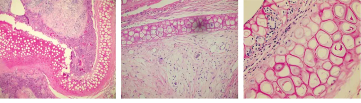
FIGURE 1. Totally empty lacunas and defi -nite foreign body (environmental reaction) in sliced cartilage group (H&E x100).
FIGURE 2. Viable cartilage tissue and mini-mal environmental reaction in single-piece cartilage group (H&E x200).
FIGURE 3. Cytoplasmic vacuolization and regenerative activity fi ndings observed on the mid-line in sliced cartilage group (H&E x400).
Bosn J Basic Med Sci 2012; 12 (4): 215-218
taking sections of microns thickness following the routine tissue follow-up stages and the stained preparations were an-alyzed with Olympus Bx model light microscope. Th e fol-lowing pathological evaluation criteria were formed accord-ing to the observed tissue changes.
Cartilage viability, foreign body reaction (environment reaction), regenerative activity and cyto-plasmic changes were scored as , , , . (Figure -), (Table ).
RESULTS
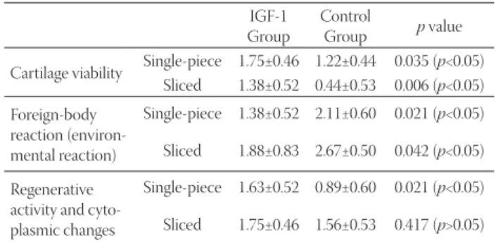
The histopathological scoring results according to cartilage vi-ability, environmental reaction, cytoplasmic changes, and regen-erative activities of single-piece and sliced samples of all car-tilage tissue samples, of which belonged to the IGF- group and of which belonged to the control group were evaluated. The scoring results of the study were evaluated with Shapiro-Wilk’s normality test. The vari-ables were seen to have a normal range. Intergroup differences were statistically analyzed by us-ing the Mann-Whitney U test. The intergroup evaluation of all the cartilages revealed that the IGF- group had more car-tilage viability and cytoplasmic activity while it expressed less environmental reaction than the control group. The differ-ences between these values were found to be statistically signifi cant (p<.), (Table , Graphs , )
Th e intergroup evaluation of the sliced cartilages revealed that the IGF- group had more cartilage viability while it showed less environmental reaction than the control group (Graph ). The differences between these values
Cartilage viability (Score) Foreign-body reaction (Environmental reac-tion) (Score) Regenerative activity and cytoplasmic changes
(Score) Completely Dead (0) None (0) No changes, similar to the literature (0) Partially Viable (1) Focal (1)
Increase in the cytoplas-mic vacuolization and
eosinophilia (1) Viable
(2)
Present (2)
Binucleation and multi-nucleation (2) Widespread and
destructive (3)
TABLE 1. Histopathological evaluation criterias
GRAPH 1. Comparison of histopathologic evaluation scores of the sliced cartilage groups.
GRAPH 2. Comparison of the histopathologic evaluation scores belongs to the one pieced cartilage groups.
IGF-1 Group
Control
Group p value Cartilage viability Single-piece 1.75±0.46 1.22±0.44 0.035 (p<0.05)
Sliced 1.38±0.52 0.44±0.53 0.006 (p<0.05) Foreign-body reaction (environ-mental reaction) Single-piece 1.38±0.52 2.11±0.60 0.021 (p<0.05) Sliced 1.88±0.83 2.67±0.50 0.042 (p<0.05) Regenerative
activity and cyto-plasmic changes
Single-piece 1.63±0.52 0.89±0.60 0.021 (p<0.05) Sliced 1.75±0.46 1.56±0.53 0.417 (p>0.05)
TABLE 2. The mean ± standard deviation and p values of histo-pathological evaluation. Scores for all groups.
Bosn J Basic Med Sci 2012; 12 (4): 216-218were found to be statistically signifi cant (p<.). Th ere was, however, no statistically signifi cant diff erence between the groups regarding cytoplasmic activity (p<.), but there was more activity in the IGF- group (Table ), (Graph ). There was no significant difference regarding all three his-topathological parameters for the sliced and single-piece cartilage groups of the IGF- group (p>.), (Graph ).
DISCUSSION AND CONCLUSION
One of the substances used in order to repro-duce and augment cartilage tissue is IGF-. An am-ple amount of studies have been conducted until now evaluating the effects of this factor on cartilage tissue have either been done only with IGF- or in addi-tion to that in combinaaddi-tion with different growth factors. One of the fi rst studies advocating that IGF- has positive eff ects on cartilage tissue was conducted by Jennische et al. who demonstrated that perichondrial cells were expressed a maximum level of hypertrophy at week two by using hu-man IGF- following freeze-thaw injury in rats’ ears []. Van Osch et al. [] compare the chondrogenic
capac-ity of auricular and nasal perichondrium from young and adult rabbits, using serum containing IGF- ( ng/mL) plus transforming growth factor-beta (TGF-beta) ( ng/ mL) and serum-free culture conditions. When serum was replaced, however, by IGF- plus TGF-beta an increased glycosaminoglycan production and induction of collagen type II was observed, especially in cells isolated from ear perichondrium. Cells derived from perichondrium of young rabbits showed larger chondrogenic potential than cells from perichondrium of adult rabbits. Moreover, stimula-tion of both glycosaminoglycan synthesis and collagen type II production was about fi ve times higher in cells isolated
from the ear perichondrium of young rabbits than of adult rabbits. In a study by Bos et al. [] using a rabbit auricular cartilage wound model, immunohistochemical staining of IGF- was used to de-fine growth factor expression at the cartilage wound sites. Th e au-thors ascertained that this increase was related to the regeneration capacity and that the expression reached maximum limits in days but then decreased gradually. Another study holds that IGF-I possessed promotional ef-fects on proliferation, while the combination of fibroblast growth factor (FGF-) with insulin or IGF-I synergisti-cally enhanced auricular chondrocyte proliferation []. To summarize, all the above mentioned studies have clearly demonstrated that IGF- plays a crucial role in cartilage tissue regeneration and the proliferation of chondrocytes. Following these results, scientists began to conduct stud-ies on how to obtain implantable cartilage tissue using in
vitro tissue engineering techniques and/or by way of in vivo
reproduction of graft material. Studies using IGF- in or-der to achieve this did not get the same successful results. Within the framework of a study by Kaplan et al. [] con-ducted for this purpose, a portion of the chondrocytes obtained from a - week old rabbit was implanted to the back of a donor rabbit after having been in vitro exposed to IGF- ( ng/mL) in a tissue culture. This cartilage tis-sue was evaluated regarding its histological and biome-chanical characteristics weeks later and no difference was found between the study group and the control group. Another study by Westreich et al. [] dealt with chon-drocytes obtained from New Zealand white rabbits’ ears treated with b-FGF and IGF- in order to enable elastic-ity in cartilage tissues obtained in vitro and to prevent volume losses in cartilage tissues obtained in vivo. The cells were injected to the subcutaneous rabbit tissue with tissue seal fibrin adhesive material and were left in the implanted area for months. The results showed that IGF- decreases the efficiency of the chondrocytes. In another study by Liu et al. [] conducted on New Zealand rabbits, a pedunculated fl ap was formed from the perichon-drium in rabbits’ ears and the demineralized ZMK bone ma-trix tissue was buried there. Th e rd day after the operation, the rabbits were injected with growth factors [TGF-β, IGF- and Bone Morphogenic Protein- (BMP-)] every days. Grafts were collected in the rd and th week after the ZMK
Bosn J Basic Med Sci 2012; 12 (4): 217-218
bone matrix implantation, and the silvers made from them were stained for the presence of collagen II, collagen I, and macrophages, and analyzed morphometrically. It was found that the application of growth factors only slightly intensifi ed the synthesis of collagen II, and had no eff ect on the degree of macrophage infi ltration or collagen I contents, while the numerous injections exerted a negative impact on the archi-tecture of the newly-formed tissue and contributed to an in-creased number of complications (haematomas, infections). The results of this study which are in contradiction with our information about the effects of IGF- on the preser-vation of viability of the cartilage tissue and its reproduc-tion directed us to conduct our own study on the subject. In our study, the auricular cartilage tissue, which was subcu-taneously treated for three months with IGF- (. mg/ ml), was evaluated at the end of three months. Th e cartilage tissue was evaluated regarding the cytoplasmic changes in the chon-drocytes, regenerative activity, cartilage viability, and foreign-body reaction and was histopathologically scored. Th ese eval-uations were carried out by the pathologist who had done the analysis during the microscopic analysis of the graft tissue samples based on the changes that he thought was related to tissue viability. Th e reason why the evaluation was carried out in this manner was the fact that the changes that would happen in the cartilage graft and the surrounding tissues was not known beforehand and that there was no known stan-dard histopathological evaluation method on the subject. Single-piece cartilage graft intergroup comparison re-vealed that there was statistically more cartilage viability and cytoplasmic activity, and statistically less foreign-body reaction in the IGF- group than in the control group. Sliced cartilage intergroup evaluation, too, revealed that there was statistically more cartilage viability and sta-tistically less foreign-body reaction in the IGF- group than in the control group. There was, however, no sta-tistically significant difference between the groups, but the IGF- group showed more cytoplasmic activity. Th ese results demonstrated that IGF- increased tissue viabil-ity and repressed the former foreign-body reaction indepen-dent of (not eff ected by) the fact that whether the implant-ed auricular cartilage tissue was in a single-piece or slicimplant-ed. Th e results of our study support other studies which argue that IGF- has positive eff ects on the viability and development of cartilage tissue [- ]. On the other hand, it is clear that the results are in contradiction with the views that IGF- has no positive eff ects on the implanted cartilage tissue [] and further it decreases the effi ciency of the chondrocytes []. We believe that it is of utmost importance to cleanse the hair on the skin thoroughly without damaging the skin in order to harvest and implant the cartilage tissue under sterile condi-tions, to use sterile disposable injectors with appropriate
nee-dle diameter during the subcutaneous injections, to clean the skin with a disinfectant agent, and to have a careful skin care process following the implantation in order to reduce the risk of infections and tissue rejection. Further, we also think that in order to prevent haematoma, infection, granulation tis-sue, and tissue rejection, it is again of utmost importance to dissect the skin in a way that will not upset the tissue plans and integrity of the area where the tissue is implanted during the implantation and performing a subcutaneous injection in an appropriate plan without giving way to tissue destruc-tion. We are of the opinion that such complications seen in other studies [] might be related to such application mis-takes and that it is not right to connect these eff ects to IGF-. Th e results of our study demonstrate that when local subcu-taneous IGF- injections are carried out under appropriate conditions using appropriate surgical techniques, they in-crease the viability of the auricular cartilage in the implant-ed area and rimplant-educe the risk of tissue rejection. On the other hand, the fact that there is a limited number of in vivo studies and that the results of these studies are in contradiction, ne-cessitates novel studies to be conducted on the subject. We believe that comprehensive studies that will be conducted especially to elucidate the dose, duration, and combinations with other agents of IGF- applications to gain maximum benefi t, will contribute to clear the confusion on the subject. The positive results that might be obtained by these studies can give us the chance to have a practical method to sustain the viability, to extend the lifes-pan, and even to increase the tissue volumes of the car-tilage grafts in the area where they were implanted.
DECLARATION OF INTEREST
There is no financial support received for this pres-ent study for all authors, no financial involve-ment of any kind or affiliation with any organiza-tion whose financial interests may be affected by material in the manuscript or which may potentially bias it.
REFERENCES
[] van Osch GJ, van der Veen SW, Verwoerd-Verhoef HL. In vitro rediff erentiation of culture-expanded rabbit and human auricu-lar chondrocytes for cartilage reconstruction. Plast Reconstr Surg ; ():-.
[] Westreich R, Kaufman M, Gannon P, Lawson W. Validating the subcutaneous model of injectable autologous cartilage using a fi -brin glue scaff old. Laryngoscope ; :-.
[] Höppener JW, de Pagter-Holthuizen P, Geurts van Kessel AH, Jan-sen M, Kittur SD, Antonarakis SE, Lips CJ, SusJan-senbach JS. Th e hu-man gene encoding insulin-like growth factor I is located on chro-mosome . Hum Genet. ; ():-.
[] Jansen M, Van Schaik FM, Ricker AT, Bullock B, Woods DE, Gab-bay KH, et al. Sequence of cDNA encoding human insulin-like growth factor I precursor. Nature ; ():-.
Bosn J Basic Med Sci 2012; 12 (4): 218-218[] Allan GJ, Flint DJ, Patel K. Insulin-like growth factor axis during embryonic development. Reproduction ;():-. [] Stratikopoulos E, Szabolcs M, Dragatsis I, Klinakis A, Efstratiadis A.
Th e hormonal action of IGF in postnatal mouse growth. Proc Natl Acad Sci U S A. ;():-.
[] Florini JR, Ewton DZ, Coolican SA. Growth hormone and the insulin-like growth factor system in myogenesis. Endocr Rev ;():-
[] Fu Z, Kubo T, Noguchi T, Kato H. Developmental changes in the mRNA levels of IGF-I and its related genes in the reproductive or-gans of Japanese quail (Coturnix coturnix japonica). Growth Horm IGF Res. ;():-.
[] Rosen CJ. Insulin-like growth factor- and calcium balance: evolv-ing concepts of an volutionary process. Endocrinology. ; ():-.
[] Kawai M, Rosen CJ. Th e IGF-I regulatory system and its impact on skeletal and energy homeostasis. J Cell Biochem. ; ():-.
[] Jonsson KB, Wiberg K, Ljunghall S, Ljunggren O. Insulin-like growth factor I does not stimulate bone resorption in cultured neo-natal mouse calvarial bones. Calcif Tissue Int. ; (): -. [] Tsukazaki T, Usa T, Matsumoto T, Enomoto H, Ohtsuru A,
Nam-ba H, et al. Eff ect of transforming growth factor-beta on the insu-lin-like growth factor- autocrine / paracrine axis in cultured rat articular chondrocytes. Exp Cell Res. ; ():–.
[] Sah RL, Chen AC, Grodzinsky AJ, Trippel SB. Diff erential eff ects of bFGF and IGF-I on matrix metabolism in calf and adult bovine
cartilage explants. Arch Biochem Biophys ; ():-. [] Jennische E, Skottner A, Hansson HA. Dynamic changes in
insulin-like growth factor I immunoreactivity correlate to repair events in rat ear after freeze-thaw injury. Exp Mol Pathol. ; ():-.
[] van Osch GJ, van der Veen SW, Burger EH, Verwoerd-Verhoef HL. Chondrogenic potential of in vitro multiplied rabbit perichondri-um cells cultured in alginate beads in defi ned mediperichondri-um. Tissue Eng. ; (): -.
[] Bos PK, van Osch GJ, Frenz DA, Verhaar JA, Verwoerd-Verhoef HL. Growth factor expression in cartilage wound healing: tempo-ral and spatial immunolocalization in a rabbit auricular cartilage wound model. Osteoarthritis Cartilage ; ():-. [] Takahashi T, Ogasawara T, Kishimoto J, Liu G, Asato H,
Nakat-suka T, et al. Synergistic eff ects of FGF- with insulin or IGF-I on the proliferation of human auricular chondrocytes. Cell Transplant ; ():-.
[] Kaplan BA, Gorman CR, Gupta AK, Taylor SR, Iezzoni JC, Park SS. Eff ects of transforming growth factor Beta and insulinlike growth factor on the biomechanical and histologic properties of tissue-engineered cartilage. Arch Facial Plast Surg ; ():-. [] Westreich R, Kaufman M, Gannon P, Lawson W. Validating the
subcutaneous model of injectable autologous cartilage using a fi -brin glue scaff old. Laryngoscope ; (): -. [] Liu X, Sun H, Yan D, Zhang L, Lv X, Liu T, et al. In vivo ectopic
chondrogenesis of BMSCs directed by mature chondrocytes. Bio-materials ; ():-.