Paediatric orthopaedics through paintings
Y. Camurcu
1H. Sofu
2H. Ucpunar
3S. Duman
4A. Cobden
5 AbstractPurpose Some famous artistic representations created throughout the centuries can reveal a hidden or mysterious diagnosis of some diseases and these paintings have always drawn the attention of physicians interested in art. Artistic illustration of a child with a malformation or disability can reflect the characteristic appearance of a disease and its his-toric perspective. Some articles have revealed the definite di-agnosis of a child with achondroplasia through portraits of dwarfs and some studies have discussed the secret diagnosis of a crippled child with Pes Equinovarus or poliomyelitis. In this study, we aim to introduce some paintings that reveal musculoskeletal diseases related to paediatric orthopaedics. Methods Paintings painted since the Renaissance were re-viewed and collected via web searches. Artistic paintings depicting children with suspected paediatric orthopaedic diseases were analyzed in this study.
Results Paintings in which artists have depicted children with achondroplasia, poliomyelitis and clubfoot were found. Conclusion The investigation of a drawing depicting a disabled child may encourage an orthopaedic surgeon to introduce an analytical approach using visual cues. These paintings may also enhance the observational skills of paediatric orthopaedic surgeons, give information about the historical process of a disease and demonstrate the impact of the disease at the time the painting was painted.
1 Department of Orthopaedics and Traumatology, Erzincan University Faculty of Medicine, Erzincan, Turkey
2 Department of Orthopaedics and Traumatology, Bahcelievler Medicalpark, Istinye University, Erzincan, Turkey
3 Department of Orthopaedics and Traumatology, Erzincan University Faculty of Medicine, Basbaglar Mah, Erzincan, Turkey 4 Department of Orthopaedics and Traumatology, Selahaddin Eyyubi State Hospital, Diyarbakir, Turkey
5 Department of Orthopaedics and Traumatology, Sivas Numune Hospital, Sivas, Turkey
Correspondence should be sent to Yalkin Camurcu MD,
Department of Orthopaedics and Traumatology, Erzincan University Faculty of Medicine, Basbaglar Mah, 24100, Erzincan, Turkey. E-mail: [email protected]
Cite this article: Camurcu Y, Sofu H, Ucpunar H, Duman S, Cobden A. Paediatric orthopaedics through paintings. J Child Orthop 2018;12:647-651. DOI: 10.1302/1863-2548.12.180141 Keywords: paediatric; orthopaedics; paintings; achondroplasia; poliomyelitis
Introduction
Some famous artistic representations rendered through-out the last few centuries can reveal a hidden or mysteri-ous diagnosis of some diseases and these paintings have always drawn the attention of physicians interested in art. An artistic illustration of a child with a malformation or dis-ability can reflect the characteristic appearance of a disease and its historic perspective. Some articles have revealed the definite diagnoses of a child, such as achondroplasia through portraits of dwarfs or some studies which have discussed the secret diagnosis of a crippled child diag-nosed as having Pes Equinovarus or poliomyelitis.1-4
The investigation of a drawing depicting a disabled baby or child may encourage an orthopaedic surgeon to introduce an analytical approach using visual cues.5 These paintings may also enhance observational skills of paedi-atric orthopaedic surgeons, give information about the historical process of a disease and demonstrate the impact of the disease at the time the painting was created. In this study, we aim to introduce some paintings that reveal mus-culoskeletal diseases related to paediatric orthopaedics.
Dwarfism and achondroplasia
Achondroplasia is the most common form of skeletal dysplasia, which is a type of rhizomelic dwarfism, and it occurs in one between 15 000 and 40 000 live births.6 Patients are of short stature and have spinal anomalies, neurologic and craniofacial abnormalities and limb defor-mities.7 Genu varum is a common deformity of the lower limb.7 A mutation in the fibroblast growth factor recep-tor-3 gene on the short arm of chromosome 4 causes achondroplasia by affecting the maturation of chondro-cytes in the growth plate.6
Dwarfs have been described and portrayed in paintings throughout the ages, and most of these have been diag-nosed as having achondroplasia, according to their charac-teristic appearance with short limbs, a big head, prominent forehead and low nasal bridge.1 Spanish painter Diego Velasquez (1599 to 1660) included a number of dwarfs in
his paintings.3 In his oil-on-canvas painting titled Francisco
Lezcano ‘El Nino de Vallecas’ (1643 to 1645), Velasquez
painted a boy who was a jester at the court of Philip IV of Spain8 (Fig. 1). In this portrait, the boy with a green dress is holding cards in his hands. He has short arms, short fin-gers, a large head and a depressed nasal bridge. His right foot is small and his right leg is bowed.9
In another oil-on-canvas painting by Velasquez, Prince
Baltasar Carlos with a Dwarf (1631) (Fig. 2), a dwarf child
is seen, who served as jester and companion of the little prince. This painting presents the contrast of social stature between a prince and a dwarf. The prince is portrayed as a commander, holding a sword in his left hand and a com-mander’s baton at his right side. However, the dwarf jester is portrayed as a child holding an apple and a rattle, ready to entertain the little prince. Both paintings tell us that children with achondroplasia were employed as jesters of kings and their families because of their physical appearance.
Sometimes, our point of view may lead us to a wrong diagnosis. As an example, in their study, Benedicenti and Superti-Furga reported the multiple faces of artwork diag-nosis.10 The Wedding Room of Mantua by Andrea Man-tegna has an interesting history. In this painting, a woman dwarf is depicted within the group of the wedding party, and was diagnosed as neurofibromatosis type 1 because of dark spots on her face. After the restoration of
the painting, the skin spots, caused by surface dirt, could not be seen anymore. Thus, the final correct diagnosis was achondroplasia.
Poliomyelitis
Poliomyelitis (or polio), also called ‘infantile paralysis’, is a highly infectious and incurable viral disease.11 Polio was first introduced as a disease of a separate clinical entity by an English physician named Dr. Michael Underwood in 1789, and the first effective vaccination was developed by Dr. John Salk in 1952.12 Polio has not been eradicated completely despite successful worldwide eradication pro-grammes, thus paediatric orthopaedic surgeons should be aware of the risk of resurgence of the disease.11 It is possible that this endemic disease may have historic origins. Phil-lippe Hernigou previously reported on the possible diag-nosis of poliomyelitis cases through medieval paintings.13
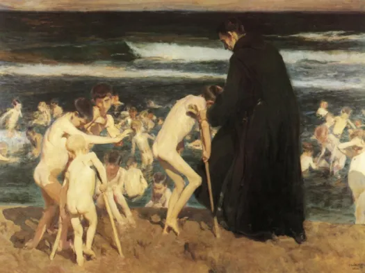
Sad Inheritance (1899) is the most famous and
remark-able painting by the Spanish painter Joaquín Sorolla y Bastida (1863 to 1923) (Fig. 3). The painting shows a group of children bathing in the sea under the watchful eye of a monk in Valencia. The painting seems to describe the polio epidemic which we know occurred in Valencia.14 We can see two boys with crutches with a probable diagnosis of the sequel of poliomyelitis. The crutch is designed as
Fig. 2 Prince Baltasar Carlos with a Dwarf. By Velasquez, Diego
Rodriguez De Silva Y (1599 to 1660). (The Museum of Fine Arts Boston, United States).
Fig. 1 El Nino de Vallecas. By Velasquez, Diego Rodriguez De
Silva Y (1599 to 1660). (Museo Nacional del Prado, Madrid, Spain).
one long stick appropriate to the height of the child, with an axillary part bound to the stick. The artist has empha-sized the lower limb disability by painting both children as lifting their right legs.
One of José Gallegos y Arnosa’s (1857 to 1917) earliest paintings is named Crippled Boy (year unknown) (Fig. 4). It portrays a begging boy with a crutch on his left side. He has an atrophic left leg and leg-length discrepancy; polio-myelitis is the most suitable diagnosis for this boy accord-ing to his appearance.
Beggars have commonly been painted by artists to show human charity. Some of them display no remark-able disability and let us think that they are imitating dis-ability for their own benefit. The poor in these paintings provided an opportunity for the prudent and beneficent wealthy to display their charity. Sir William Beechey’s (1753 to 1839) painting Portrait of Sir Francis Ford’s
Chil-dren Giving a Coin to a Beggar Boy serves as an example.
Two well-dressed children and a beggar boy, who is dressed in tatty clothes, are shown. The girl is giving a coin to the beggar boy, who seems to have no apparent orthopaedic disability.
Clubfoot
Archeological studies of ancient Egyptian tombs and mummies by Smith and Warren report the first know depictions of clubfoot.15 Pes equinovarus or clubfoot,
Fig. 3 Sad Inheritance. By Joaquín Sorolla y Bastida (1863 to 1923). (Private Collection).
Fig. 4 Crippled Boy. By José Gallegos y Arnosa (1857 to 1917).
occurs in 0.3 to 7.8 per 1000 births and is one of the most common musculoskeletal abnormalities.16 Clubfoot may be associated with myelodysplasia, arthrogryposis or mul-tiple congenital anomalies. However, the majority of cases are idiopathic.
The most famous illustration of a child with clubfoot was painted by Spanish artist Jusepe de Ribera (1591 to 1652). In The Clubfoot (1642), a young beggar boy is portrayed with a right unilateral clubfoot and a bending right hand (Fig. 5). The boy’s smile demonstrates that he is happy to be portrayed by the artist. Additionally, although this painting evokes charitable feelings at first view, the boy is showing us that he can handle his condition. He is carrying his crutch like a soldier and standing straight with confidence and dignity. The artist’s low viewpoint from ankle level also supports this by making the boy appear much taller.
Stahl et al17 analyzed Ribera’s painting and mentioned that hemiplegia and arthrogryposis are the most suitable diagnosis for this child. The authors reported that the boy holds a begging note enscribed ‘DA MIHI ELIMOSINAM
PROPTER AMOREM DEI’ (Give me alms for the love of God),
suggesting some difficulty in speaking. This suggests that his condition may have been caused by cerebral palsy, which consists of a brain injury in the left hemisphere
responsible for right hemiplegia and speech distur-bance. On the other hand, Ramachandran and Aronson18 reported that the diagnosis is more likely to be arthrogry-posis because of three-limb involvement rather than right-side involvement. These authors mentioned: ‘Although the latter could be as a result of shouldering the crutch, there appears to be flexion deformity of the wrist with an adducted thumb and extended elbow’. Recently, Abul et al19 analyzed the painting and the authors proposed a different diagnosis: poliomyelitis. The diagnosis of this young boy is still unknown although three centuries have passed.
Conclusion
Artists painted children with bone malformations or con-sequences of diseases that affected the musculoskeletal system in the historical periods when paintings and draw-ings were the most common means of portraying humans and reflecting social life. Works of art can be used for edu-cational purposes and the development of observational skills. Physicians can learn about the manifestations of the orthopaedic conditions depicted while discussing the painting and suggesting a diagnosis. Works of art may also reflect social environment, attitude and emotions towards sick disabled people or events. However, it is dif-ficult for a physician to conclude on the exact diagnosis of the underlying medical condition shown in paintings of past centuries; the diseases depicted in these paint-ings hold their mystery. We appreciate all artists who had excellent observation skills and captured many signs of various diseases.
Received 15 August 2018; accepted after revision 07 September 2018.
COMPLIANCE WITH ETHICAL STANDARDS FUNDING STATEMENT
No benefits in any form have been received or will be received from a commercial party related directly or indirectly to the subject of this article.
OA LICENCE TEXT
This article is distributed under the terms of the Creative Commons Attribution-Non Commercial 4.0 International (CC BY-NC 4.0) licence (https://creativecommons.org/ licenses/by-nc/4.0/) which permits non-commercial use, reproduction and distribu-tion of the work without further permission provided the original work is attributed.
ETHICAL STATEMENT
Ethical approval: This article does not contain any studies with human participants
or animals performed by any of the authors.
Informed consent: Not required for this work.
ICMJE CONFLICT OF INTEREST STATEMENT
All authors declare that they have no conflict of interest.
Fig. 5 The Clubfoot. By Jusepe de Ribera (1591 to 1652). (Louvre
REFERENCES
1. Haworth J, Chudley A. Dwarfs in art. Clin Genet 2001;59:84-87. 2. Foote JA. Evidence of rickets prior to 1650. Am J Dis Child 1927;34:443-452. 3. Schlesinger BE. Paediatrics in classical art. BMJ 1962;2:1671-1677.
4. Levitas AS, Reid CS. An angel with Down syndrome in a sixteenth century Flemish Nativity painting. Am J Med Genet A 2003;116A:399-405.
5. Friedlaender GE, Friedlaender LK. Art in science: enhancing observational skills. Clin Orthop Relat Res 2013;471:2065-2067.
6. Engberts AC, Jacobs WC, Castelijns SJ, Castelein RM,
Vleggeert-Lankamp CL. The prevalence of thoracolumbar kyphosis in
achondroplasia: a systematic review. J Child Orthop 2012;6:69-73.
7. Ain MC, Shirley ED, Pirouzmanesh A, Skolasky RL, Leet AI. Genu varum in achondroplasia. J Pediatr Orthop 2006;26:375-379.
8. Ravin JG, Fried RI. Picasso, Velasquez, and dwarfs. JAMA 1974;228: 1671-1672.
9. Harris JC. Portrait of Francisco Lezcano-the ‘Niño de Vallecas’. Arch Gen Psychiatry 2011;68:229.
10. Benedicenti F, Superti-Furga A. The multiple faces of artwork diagnoses.
Lancet Neurol 2017;16:417.
11. Joseph B, Watts H. Polio revisited: reviving knowledge and skills to meet the challenge of resurgence. J Child Orthop 2015;9:325-338.
12. Horstmann DM. The poliomyelitis story: a scientific hegira. Yale J Biol Med 1985;58:79-90.
13. Hernigou P. Crutch art painting in the middle age as orthopaedic heritage (part I: the lepers, the poliomyelitis, the cripples). Int Orthop 2014;38:1329-1335.
14. Cano de la Cuerda R, Collado-Vazquez S. Deficiency, disability, neurology and art. Rev Neurol 2010;51:108-116.
15. Hernigou P, Huys M, Pariat J, Jammal S. History of clubfoot treatment, part I: from manipulation in antiquity to splint and plaster in Renaissance before tenotomy. Int Orthop 2017;41:1693-1704.
16. Shyy W, Wang K, Sheffield VC, Morcuende JA. Evaluation of embryonic and perinatal myosin gene mutations and the etiology of congenital idiopathic clubfoot. J Pediatr Orthop 2010;30:231-234.
17. Stahl A, Tourame P, Montjean D. The clubfoot painted by Jusepe de Ribera: a controversial diagnosis. J Matern Fetal Neonatal Med 2016;29:1308-1310. 18. Ramachandran M, Aronson JK. The diagnosis of art: arthrogryposis and Ribera’s The Clubfoot. J R Soc Med 2006;99:321-322.
19. Abul K, Misir A, Buyuk AF. Art in science: Jusepe de Ribera’s Puzzle in The Clubfoot. Clin Orthop Relat Res 2018;476:942-945.