BONE MINERAL DENSITY IN FEMALE ELITE HANDBALL PLAYERS AND SEDANTERIES
Bilgehan BAYDİL
Kastamonu University, School of Physical Education and Sports, Kastamonu, Turkey.
İlk Kayıt Tarihi: 13.06.2013 Yayına Kabul Tarihi: 18.09.2013 Abstract
In this study, it was aimed to assess bone mineral densities (BMD) in female athletes and sedanteries. Regional bone mineral densities were measured by a bone densitometer using dual-energy x-ray absorptiometry (DXA). It was found that there was significant differences in bone mineral density values of the lumbar spine and the right femoral regions between athletes and sedanteries (P˂0.01). As result; it can be said that the young women who participate in sports exhibit the great associated gains in bone health.
Key Words : BMD, athlete, sedantery
BAYAN ELİT HENTBOL OYUNCULARINDA VE SEDANTERLERDE KEMİK MİNERAL YOĞUNLUKLARI Özet
Bu çalışmada bayan sporcularda ve sedanterlerde kemik mineral yoğunluklarının (KMY) değerlendirilmesi amaçlandı. Bölgesel kemik mineral yoğunlukları dual energy x-ray (DXA) absorbsiyometri kullanılarak kemik dansitometresi ile ölçüldü. Sporcular ve sedanterler arasında lumbar omurga ve sağ femur bölgelerinin kemik mineral yoğunluklarında anlamlı farklılıklar bulundu (P˂0.01). Sonuç olarak genç bayanlarda spora katılımla kemik sağlığında faydalar arasında büyük ilişki olduğu söylenebilir.
Anahtar Kelimeler : KMY, sporcu, sedanter 1. Introduction
Involvement in athletics can promote healthy lifestyle behaviors and decrease the risk for a number of health problems. In particular, weight-bearing exercise in female athletes increases bone mineral density (BMD) and lean body mass, which may help to prevent stress fractures and osteoporosis later in life (Lanay, 2007).
Bone is a unique, metabolically active tissue that undergoes a continuous remo-delling throughout its life cycle (Rita, 2012). A great number of factors influence the
accumulation of bone mineral in humans. Some are endogenic, such as heredity, eth-nicity, gender, or endocrine status; the others – exogenic, such as nutrition or physical activity (Račienė, 2012, Długołęcka,2011).
Sports participation during the years that peak bone mass is being acquired may lead to adaptive changes that improve bone architecture through increased density and enhanced geometric properties (Tenforde,2011). The variable mechanical stress indu-ced through physical activity is the stimulus for the related increase in bone formation. The skeletal response to exercise is therefore dependent on the nature of the loading forces associated with the activity (Chang, 2013)
The aim of this study was to assess bone mineral densities of the lumbar spine and the femur in female athletes and sedanteries.
2. Materials and Methods
Female elite level handball players (n : 10, age: 21±1,49 years, height: 170,50±7,66 cm, body weight : 64,80±8,02 kg) and a sedantery group (n : 10, age: 20,90±1,44 years, height: 161,90±5,98 cm, body weight :54,10±7,48kg) volunteered to be part of the study. The sedantery group consisted of girls at the same age, who were not involved in sports
Information on sporting career including length of training and weekly training loads was collected by way of a questionnaire completed by subjects.
Under anthropometric assessment height and weight recordings were done on the same day on which bone mineral density was measured. The weight of subjects were meausered by an electronic scale without shoes and wearing only short and t shirt. The height measurements were determined with metric scale. Regional bone mineral density was measured by a bone densitometer (GE / LUNAR DPX PRO, USA) using dual-energy x-ray absorptiometry. The measured regions were lumbar spine (L1, L2, L3, L4) and the right femoral regions neck, trochanter, wards, shaft and total.
Statistical evaluation was conducted using SPSS (Statistical Package for the So-cial Sciences) 13 software program. The differences between athlete and sedantery groups results were determined by Mann-Whitney U test method at a significance level of P<0.01.
3. Results
Table 1 : Descriptive Statistic of the Subjects
Groups n Age (year) Height (cm) Weight (kg)Body Length of training (year) Weekly training load (hour) Sedantery 10 20,90±1,44 161,90±5,98 54,10±7,48 - -Athletes 10 21±1,49 170,50±7,66 64,80±8,02 9,90±1,44 11,20±1,03
Table 1 shows the average age, height, body weight, length of training and we-ekly training load values of the subjects. The athletes values were determined to be 21±1,49 years, 170,50±7,66 cm. and 64,80±8,02 kg., 9,90±1,44 years and 11,20±1,03
hours, respectively. The sedanteries values 20,90±1,44 years, 161,90±5,98 cm. and 54,10±7,48 kg. respectively.
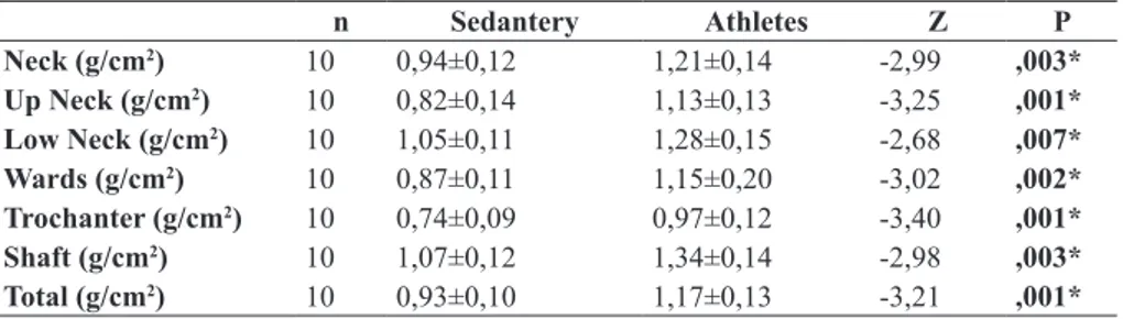
Table 2 : Bone Mineral Density Values of Femoral Regions
n Sedantery Athletes Z P Neck (g/cm2) 10 0,94±0,12 1,21±0,14 -2,99 ,003* Up Neck (g/cm2) 10 0,82±0,14 1,13±0,13 -3,25 ,001* Low Neck (g/cm2) 10 1,05±0,11 1,28±0,15 -2,68 ,007* Wards (g/cm2) 10 0,87±0,11 1,15±0,20 -3,02 ,002* Trochanter (g/cm2) 10 0,74±0,09 0,97±0,12 -3,40 ,001* Shaft (g/cm2) 10 1,07±0,12 1,34±0,14 -2,98 ,003* Total (g/cm2) 10 0,93±0,10 1,17±0,13 -3,21 ,001* *P˂0,01
Table 2 shows the comparisons in bone mineral density values of femoral regions. It can clearly be seen from the table that athletes neck, wards, trochanter, shaft and total bone mineral density values higher than sedanteries with statistically significant extent (P<0.01).
Table 3 : Bone Mineral Density Values of Lumbar Spine Regions
n Sedantery Athletes Z P L1 (g/cm2) 10 1,03±0,10 1,24±0,11 -3,21 ,001* L2 (g/cm2) 10 1,10±0,13 1,32±0,14 -3,02 ,002* L3 (g/cm2) 10 1,15±0,13 1,37±0,12 -2,87 ,004* L4 (g/cm2) 10 1,07±0,11 1,30±0,10 -3,25 ,001* L1 - L4 (g/cm2) 10 1,09±0,11 1,31±0,11 -3,14 ,002* *P˂0,01
Table 3 shows the comparisons in bone mineral density values of lumbar spine regions. According to findings, L1, L2, L3, L4 and L1-L4 BMD values of athletes are higher (P<0.01).
4. Discussion and Conclusion
It is so far unknown, which component of stress has the strongest anabolic ef-fects on bones in humans: kind of stress, intensity, frequency or duration? Experiments with animals revealed that new bone formation depends less on duration of mechanical stress but more on its magnitude and rate: especially strains of high rate and magnitude stimulated new bone formation. One of the most important factors influencing bone metabolism in humans probably is impact force that causes compression (e.g. spine) or deflection (e.g. proximal femur) of bones (Hinrichs, 2010). Strength-based and high impact sports seem to be associated with higher BMD, whereas non–weight-bearing sports have a neutral or negative relationship (Lanay, 2007).
In our study was found significant diffrences in bone mineral density levels betwe-en athletes and sedanteries (P<0.01). The all BMD values of the athletes were higher
than sedanteries. In parallel with findings, large number of studies indicated that regular physical activity contributes significantly to gain in BMD.
Marwaha et al. investigated the effects of sports training & nutrition on bone mine-ral density in young Indian healthy females. They found that sportswomen who have undergone at least three years of regular physical training, had significantly higher total BMD and BMD at femur, and lumbar (L1-L4) skeletal sites when compared to sedan-tery controls (Marwaha, 2011).
Conroy et al examined the relationship of bone mineral density to muscular strength in highly trained young male athletes in order to gain insights concerning the influence of heavy resistance training on BMD. Twenty-five elite junior weightlifters (age, 17.4 +/- 1.4 yr) and 11 age-matched controls (16.9 +/- 1.1 yr) volunteered for this investiga-tion. Measurements of BMD (g.cm-2) utilizing dual energy x-ray absorptiometry were obtained for the lumbar spine (L2-4) and the proximal femur (neck; trochanter, Ward’s triangle). The BMD values for the junior lifters were found to be significantly greater at all sites for the junior weightlifters compared with their age-matched control group (Conroy, 1993).
Düppe et al examined the BMD active female junior (age : 13-17, n : 62) and senior (age : 18-28, n : 34) football players and their respective controls. Body compositions and total body, lumbar spine and proximal femur BMD were measured with dual energy x-ray absorptiometry. They found that football players had significantly greater BMD than controls at all sites measured. Further, differences were greater for seniors than for junior players (Düppe,1996).
The effects of sporting activity and of menstrual status on the bone mineral density of the femoral mid-shaft were investigated by Wolman et al. The cohort consisted of 67 elite, female athletes comprising 21 runners, 36 rowers, and 10 dancers. The mean bone mineral content in the runners was 1.51 (1.47 to 1.55) g/cm2, which was significantly higher than in the rowers, dancers, and sedentary controls whose values were 1.43 (1.40 to 1.47), 1.39 (1-33 to 1.45), and 1.40 (1.34 to 1.45) g/cm2 respectively (Wolman, 1991).
Fehlings and colleagues study demonstrated that a group of female athletes who engaged in a sport that loads the skeletal system with high magnitude, short duration stimuli (volleyball or gymnastics) had greater BMD than athletes who participated in a sport that actively taxes their muscular system (swimming). They found that the group of swimmers in the study were not significantly different from the nonathletic controls (Fehling, 1995).
Heinonen measured anatomic sites were at the lumbar spine, femoral neck, distal femur, patella, proximal tibia, calcaneus, and distal radius in Finnish female athletes, physically active referents (they reported an average of five various types of exercise sessions per week) and sedentary referents . The athlete group consisted of aerobic dan-cers (N = 27), squash players (N = 18), and speed skaters (N = 14). The squash players had the highest values for weight-adjusted bone mineral density (BMD) at the lumbar spine (13.8% P < 0.001 as compared with the sedentary reference group), femoral neck (16.8%, P < 0.00l), proximal tibia (12.6%, P < 0.001) and calcaneus (l&5%, P < 0.001). Aerobic dancers and speed skaters also had significantly higher BMD values at the loa-ded sites than the sedentary reference group, the difference ranging from 5.3% to 13.5%. The physically active referents’ BMD values did not differ from those of the sedentary
referents at any site. The results support the concept that training, including high strain rates in versatile movements and high peak forces, is more effective in bone formation than training with a large number of low-force repetitions (Heinonen, 1995).
Taaffe et al examined the role of athletic activity, specifically gymnastics, on bone mineral density accretion, they monitored longitudinal changes (8-12 mounths) in re-gional and whole body BMD in collegiate women gymnasts and competitive athletes whose skeletons are exposed to differential loading patterns: runners and swimmers. Lumbar spine (L2–4), femoral neck, and whole body BMD (g/cm2) were assessed by dual-energy X-ray absorptiometry. The percent change in lumbar spine BMD was signi-ficantly greater in the gymnasts than in the runners, swimmers or controls. An increase in femoral neck BMD in gymnasts was also greater than runners, swimmers and appro-ached significance compared with controls (Taaffe, 1997).
In the study conducted by Egan et al, whole-body, lumbar spine (L1–4), and total left hip areal bone mineral density were measured according to standard operating procedu-res, using a dual-energy X-ray absorptiometry in club and university level female Rugby Union football players (n = 30, age: 21.4 ± 1.9 years), netball players (n = 20, 20.7 ± 1.3 years), distance runners (n = 11, 21.5 ± 2.6 years) and sedentary controls (n = 25, 21.4 ± 1.1 years). Bone mineral density was significantly higher (P b 0.05) in the Rugby Union players compared with sedentary controls at all sites, regions, and segments. In the netball players, BMD was significantly higher at the ribs, spine, pelvis, legs, femoral neck, and inter-trochanter region than in the sedentary controls. Differences between the runners and the controls were significant for the legs and the femoral neck, trochanter, and inter-trochanter regions of the proximal femur (P ˂ 0.05) (Egan, 2006).
There are a lot of studies in the literature like mentioned aboves supporting to our results (Creighton, 2001; Fiore, 1996; Hinrichs, 2010; Chang 2013; Multani, 2011; Pre-lack, 2012). In these studies there is a critical point : type of sport. These studies have shown that weight bearing and high impact loading sports are associated with greater levels of BMD in comparison to non-active control groups. High bone strain rates and magnitude are produced during handball participation. Physical requirements during practice and competition are anaerobic in nature requiring repeated bone loading activi-ties such as jumping, pushing, pulling and turning. These movements may have power-ful osteogenic effects.
The mechanical stress in the form of sporting activity may be a major factor in bone mineralization, though the physiological mechanisms involved in the response of bone cells to mechanical stress are still unclear. A possible explanation may be that osteocytes, acting as mechanoreceptors, respond and release chemical factor capable for promoting osteoblast proliferation at the local bone site (Multani, 2011).
The results of this study indicate that regular training in sports producing high strain rates, such as handball, may positively contribute to the bone health of adolescent girls. 5. References
Chang R.P.Y., Briffa K.N., Edmondston S.J. (2013). Bone mineral density and body composition in elite female golf and netball players. European Journal of Sport Science, 13(2), 183-190.
Conroy B.P., Kraemer W.J., Maresh C.M., Fleck S.J., Stone M.H., Fry A.C. (1993). Bone mine-ral density in elite junior olympic weightlifters, Medicine and Science in Sports and Exerci-se, 25(10), 1103-1109.
Creighton D.L., Morgan A.L., Boardley D., Brolinson P.G. (2001). Weightbearing exercise and markers of bone turnover in female athletes. J Appl Physiol, 90(2), 565-570.
Długołęcka B., Czeczelewski J., Raczyńska B. (2011). Bone mineral content and bone mineral density in female swimmers during the time of peak bone mass attainment. Biology of Sport, 28(1), 69-74.
Düppe H., Gärdsell P., Johnell O., Ornstein E. (1996). Bone mineral density in female junior, senior and former football players. Osteoporosis International, 6(6), 437-441.
Egan E., Reilly T., Giacomoni M, Redmond L., Turner C. (2006). Bone mineral density among female sports participants. Bone, 38, 227–233.
Fehling P.C., Alekel L., Clasey J., Rector A., Stillman R. J. (1995). A Comparison of Bone Mineral Densities Among Female Athletes in Impact Loading and Active Loading Sports. Bone, 17(3), 205-210.
Fiore C.E., Dieli M., Vintaloro G. (1996). Body composition and bone mineral density in competi-tive athletes in different sports. Int J Tissue React-Exp Clin Asp, 18(4-6), 121-124.
Heinonen A., Oja P., Kannus P., Sievanen H., Haapasalo H., Manttari A., Vuori I. (1995). Bone Mi-neral Density in Female Athletes Representing Sports With Different Loading Characteristics of the Skeleton. Bone, 17(3), 197-203.
Hinrichs T., Chae E.H., Lehmann R., Allolio B., Platen P. (2010). Bone mineral density in athletes of different disciplines: a cross-sectional study. The Open Sports Sciences Journal, 3, 129-133. Lanay M.M., Fornetti W., Pivarnik J.M. (2007). Bone mineral density in collegiate female athletes:
Comparisons among sports. Journal of Athletic Training 42(3), 403–408.
Marwaha R.K., Puri S., Tandon N., Dhir S., Agarwal N., Bhadra K., et al. (2011). Effects of sports training & nutrition on bone mineral density in young ındian healthy females. Indian J. Med. Res., 134, 307-313.
Multani N.K., Kaur H., Chahal A. (2011). Impact of Sporting activities on Bone mineral density. Journal of Exercise Science and Physiotherapy, 7(2), 103-109.
Prelack K., Dwyer J., Ziegler P., Kehayias J. J. (2012). Bone mineral density in elite adolescent female figure skaters. Journal of the International Society of Sports Nutrition, 9(1), 57. Račienė R.G., Jurimae J., Saar M., Cicchella A., Stefanelli C., Passariello C. (2012). Bone
mine-ral density and hormonal status in adolescent athletic girls. Acta Kinesiologiae Universitatis. Tartuensis, 18, 56–67.
Taaffe D. R., Robinson T. L., Snow C. M., Marcus R. (1997). High-Impact Exercise Promotes Bone Gain In Well-Trained Female Athletes. Journal of Bone and Mineral Research, 12(2), 255-260.
Tenforde A. D., Fredericson M. (2011). Influence of sports participation on bone health in the young athlete: A review of the literature. PM R, 3, 861-867.
Wolman R.L., Faulmann L., Clark P., Hesp R., Harries M.G. (1991). Different training patterns and bone mineral density of the femoral shaft in elite, female atheletes. Annals of the Rheumatic Diseases, 50, 487-489.