Global Impact of the COVID-19 Pandemic
on Cytopathology Practice: Results From an International
Survey of Laboratories in 23 Countries
Elena Vigliar, MD, PhD 1; Rima Cepurnaite, MD1; Eduardo Alcaraz-Mateos, MD, PhD2;
Syed Z. Ali, MD, FRCPath, FIAC3; Zubair W. Baloch, MD4; Claudio Bellevicine, MD, PhD 1;
Massimo Bongiovanni, MD, MIAC 5; Pavlina Botsun, MD6; Dario Bruzzese, PhD1; Lukas Bubendorf, MD 7;
Reinhard Büttner, MD, PhD8; Sule Canberk, MD 9; Arrigo Capitanio, MD10; Chiara Casadio, MD11;
Eugeniu Cazacu, MD12; Beatrix Cochand-Priollet, MD, MIAC, PhD13; Alessandro D’Amuri, MD, PhD14;
Catarina Eloy, MD, PhD9; Marianne Engels, MD, FIAC8; Guido Fadda, MD15; Gabriella Fontanini, MD16;
Franco Fulciniti, MD, PhD17; Paul Hofman, MD, PhD 18; Antonino Iaccarino, PhD1; Antonio Ieni, MD, PhD15;
Xiaoyin Sara Jiang, MD, FCAP19; Kennichi Kakudo, MD, PhD 20; Izidor Kern, MD21; Ivana Kholova, MD, PhD22;
Chinhua Liu, CT23; Anandi Lobo, MD24; Maria D. Lozano, MD, PhD25; Umberto Malapelle, PhD1;
Zahra Maleki, MD, FCAP, MIAC 3; Pamela Michelow, MD26; Jamal Musayev, MD, MIAC27; Gonca Özgün, MD28;
Meltem Oznur, MD29; Francisca Maria Peiró Marqués, MD30; Pasquale Pisapia, MD 1;
David Poller, MD, FRCPath31; Michal Pyzlak, MD, PhD32; Betsy Robinson, CT19;
Esther Diana Rossi, MD, PhD, MIAC 33; Sinchita Roy-Chowdhuri, MD, PhD 23;
Mauro Saieg, MD, PhD, FIAC 34; Spasenija Savic Prince, MD7; Fernando C. Schmitt, MD, PhD9;
Francisco Javier Seguí Iváñez, MD30; Tajana Štoos-Veić, MD, PhD35; Oksana Sulaieva, MD, PhD6;
Brenda J. Sweeney, MD36; Giovanni Tuccari, MD15; Marie-Louise van Velthuysen, MD, PhD37;
Paul A. VanderLaan, MD, PhD38; Philippe Vielh, MD, PhD39; Patrizia Viola, MD40; Rinus Voorham, PhD41;
Birgit Weynand, MD42; Pio Zeppa, MD, PhD 43; William C. Faquin, MD, PhD36; Martha Bishop Pitman, MD36;
and Giancarlo Troncone, MD, PhD 1
Corresponding Author: Giancarlo Troncone, MD, Department of Public Health, University of Naples Federico II, Via Sergio Pansini 5, 80131 Naples, Italy ([email protected]).
1 Department of Public Health, University of Naples Federico II, Naples, Italy; 2 Pathology Department, Jose M. Morales Meseguer University General Hospital,
Murcia, Spain; 3 Division of Cytopathology, Department of Pathology, The Johns Hopkins Hospital, Baltimore, Maryland; 4 Department of Pathology, University
of Pennsylvania, Philadelphia, Pennsylvania; 5 Cytopathology, SYNLAB Suisse SA, Lausanne, Switzerland; 6 Laboratory of Pathology CSD, Kyiv, Ukraine; 7 Institute
of Pathology, University Hospital Basel, Basel, Switzerland; 8 Department of Pathology, University Hospital Cologne, Cologne, Germany; 9 Medical Faculty,
IPATIMUP, Institute of Molecular Pathology and Immunology, University of Porto, Porto, Portugal; 10 Department of Pathology, Linkoping University Hospital,
Linkoping, Sweden; 11 Division of Pathology and Laboratory Medicine, European Institute of Oncology, Milan, Italy; 12 Department of Morphopathology, Nicolae
Testemitanu State University of Medicine and Pharmacy, Chisinau, Moldova; 13 Cochin Hospital, APHP University Medical Center of Paris, Paris, France; 14 Anatomic
Pathology Unit, A. Perrino Hospital, ASL Brindisi, Brindisi, Italy; 15 Section of Pathological Anatomy, Department of Human Pathology “Gaetano Barresi”, A.O.U.
Polyclinic G. Martino, Messina, Italy; 16 Department of Surgical, Medical and Molecular Pathology and Critical Care Medicine, University of Pisa, Pisa, Italy; 17 Clinical Cytopathology Service, Histopathology Service, Cantonal Institute of Pathology, Locarno Cantonal Hospital, Locarno, Switzerland; 18 Laboratory of
Clinical and Experimental Pathology, Hospital-related Biobank (BB-0033-00025), FHU OncoAge, Pasteur Hospital, Nice, France; 19 Department of Pathology, Duke
University Medical Center, Durham, North Carolina; 20 Department of Pathology, Izumi City General Hospital, Izumi, Japan; 21 University Clinic Golnik, Golnik,
Slovenia; 22 Department of Pathology, Fimlab Laboratories, Faculty of Medicine and Health Technology, Tampere University, Tampere, Finland; 23 Department
of Pathology, The University of Texas MD Anderson Cancer Center, Houston, Texas; 24 Department of Pathology, Kapoor Path Laboratories, Raipur, India; 25 Department of Pathology, University Clinic of Navarra, Pamplona, Spain; 26 Department of Anatomical Pathology, University of the Witwatersrand, National
Health Laboratory Service, Johannesburg, South Africa; 27 Baku Pathology Center, Baku, Azerbaijan; 28 Department of Pathology, Baskent University Faculty of
Medicine, Ankara, Turkey; 29 Department of Pathology, Tekirdag Namik Kemal University, Suleymanpaşa/Tekirdag, Turkey; 30 Department of Pathology, General
University Hospital of Alicante, Alicante, Spain; 31 Department of Pathology and Cytology, Queen Alexandra Hospital, Portsmouth, United Kingdom; 32 Center for
Diagnostic Pathology, Warsaw, Poland; 33 Division of Anatomic Pathology and Histology, Catholic University–University Polyclinic Foundation “A. Gemini,” Rome,
Italy; 34 Department of Pathology, A.C. Camargo Cancer Center, Santa Casa Medical School, Sao Paulo, Brazil; 35 Department of Pathology and Cytology, University
Hospital Dubrava, Zagreb, Croatia; 36 Department of Pathology, Massachusetts General Hospital, Harvard Medical School, Boston, Massachusetts; 37 Department
of Pathology, Erasmus Medical Centre, Rotterdam, The Netherlands; 38 Department of Pathology, Beth Israel Deaconess Medical Center, Harvard Medical School,
Boston, Massachusetts; 39 Medipath, American Hospital of Paris, Paris, France; 40 North West London Pathology, Imperial College Healthcare, NHS Trust Charing
Cross Hospital, London, United Kingdom; 41 Quirinus JM Voorham, PALGA Foundation, Houten, The Netherlands; 42 Department of Pathology, University Hospitals
Leuven, Leuven, Belgium; 43 Department of Medicine and Surgery, University of Salerno, Fisciano, Italy
The last 3 authors contributed equally to this article.
Received: August 11, 2020; Revised: September 14, 2020; Accepted: September 16, 2020 Published online October 27, 2020 in Wiley Online Library (wileyonlinelibrary.com) DOI: 10.1002/cncy.22373, wileyonlinelibrary.com
BACKGROUND: To the authors’ knowledge, the impact of the coronavirus disease 2019 (COVID-19) pandemic on cytopathology practices worldwide has not been investigated formally. In the current study, data from 41 respondents from 23 countries were reported. METHODS: Data regarding the activity of each cytopathology laboratory during 4 weeks of COVID-19 lockdown were col-lected and compared with those obtained during the corresponding period in 2019. The overall number and percentage of exfoliative and fine-needle aspiration cytology samples from each anatomic site were recorded. Differences in the malignancy and suspicious rates between the 2 periods were analyzed using a meta-analytical approach. RESULTS: Overall, the sample volume was lower compared with 2019 (104,319 samples vs 190,225 samples), with an average volume reduction of 45.3% (range, 0.1%-98.0%). The percentage of samples from the cervicovaginal tract, thyroid, and anorectal region was significantly reduced (P < .05). Conversely, the percentage of samples from the urinary tract, serous cavities, breast, lymph nodes, respiratory tract, salivary glands, central nerv-ous system, gastrointestinal tract, pancreas, liver, and biliary tract increased (P < .05). An overall increase of 5.56% (95% CI, 3.77%-7.35%) in the malignancy rate in nongynecological samples during the COVID-19 pandemic was observed. When the suspicious category was included, the overall increase was 6.95% (95% CI, 4.63%-9.27%). CONCLUSIONS: The COVID-19 pandemic resulted in a drastic reduction in the total number of cytology specimens regardless of anatomic site or specimen type. The rate of malignancy increased, reflecting the prioritization of patients with cancer who were considered to be at high risk. Prospective monitoring of the effect of delays in access to health services during the lockdown period is warranted. Cancer Cytopathol 2020;128:886-894. © 2020 American Cancer Society.
KEY WORDS: coronavirus disease 2019 (COVID-19); cytopathology; fine-needle aspiration; malignancy rate.
INTRODUCTION
During the recent coronavirus disease (COVID-19) pan-demic outbreak caused by the severe acute respiratory syndrome coronavirus 2 (SARS-CoV-2),1 several national health organizations and different pathology scientific societies recommended reductions in routine health main-tenance due to the health emergency, which also affected cytopathological practices around the world.2-5 Normally, cytological specimens are obtained routinely regardless of whether the procedure is a screening procedure for more invasive histopathological examinations or a complete di-agnostic, prognostic, and predictive evaluation. However, during the COVID-19 pandemic, procedures leading to a cytological sample needed to be carefully evaluated with respect to the risks and benefits to the patient as well as the health care provider. Indeed, to maintain the effi-ciency of health systems and to reduce the risk of infec-tion for patients and medical staff, screening procedures were minimized or postponed until the “flattening of the curve” could be accomplished.3,4 However, cytopatholo-gists still were asked to ensure timely malignancy-related diagnoses because any delay could lead to an increase in cancer-related mortality.6
Clinical recommendations may be difficult to apply in routine practice, and to our knowledge it is unclear how these guidelines were implemented. The perception of reduced cytological activity during the COVID-19 pandemic has not yet been studied via real-world, prac-tice-based evidence generated from different laboratories worldwide. Currently, the only available data have been reported by single institutions, and demonstrate a decrease
in cytological workload.7,8 It is interesting to note that,
despite the reduced activity, the rate of malignant diag-noses significantly increased.8 However, single-institution
reports are not robust enough to draw reliable conclu-sions on a global scale or for assessment of the effect of the prioritization of cytological samples from patients considered to be at high risk of malignancy. Because the implementation of a nationwide network and registry of cytopathology diagnoses still is limited, worldwide data are difficult to gather without a collective effort. To fill this knowledge gap, a large number of cytopathologists from different countries reviewed their clinical reports to assess how cytological practices were impacted during the COVID-19 pandemic worldwide.
MATERIALS AND METHODS
Survey
Data regarding the activity of cytopathology laboratories were collected through an international survey.
An Excel questionnaire template was distributed through email to members of the CytoESP Working Group (cytopathologists from the European Society of Pathology) (https://www.esp-patho logy.org/worki ng-group s/esp-worki ng-ng-group s/cytop athol ogy.html) and to cytopathologists who have taken part in 1 of the 9 Annual National Molecular Cytopathology meetings in Naples, Italy (https://www.molec ularc ytopa tholo gy.com/), ac-counting for a total of 65 invited participants. Only a single email and no reminders were sent. Participants were asked to provide data regarding their cytopathology
practice during the first 4 weeks of the COVID-19 national lockdown.
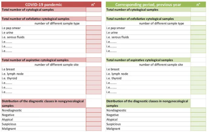
The study period was individualized for each in-stitution due to the variability of the lockdown among countries. In countries in which lockdown did not take place, cytopathologists were asked to provide data from the first 4 weeks of the peak infection spread. To assess changes, if any, in cytopathological practice, participants were asked to provide the same data compared with the corresponding period in 2019. Questions included in the survey are listed in Figure 1. Specifically, participants were asked to report on the total number of processed cytolog-ical samples, the total number of exfoliative samples spec-ifying the number of different specimen types, the total number of fine-needle aspiration (FNA) samples specify-ing the different samplspecify-ing sites, and the distribution of diagnostic classes in nongynecological samples (nondiag-nostic, negative, atypical, suspicious, and malignant). Statistical Analyses
All analyses were performed using the R statistical plat-form (version 4.0.2).
Differences between the 2 periods with respect to the ratio of exfoliative-to-FNA samples and to the ma-lignancy rate were summarized using a meta-analytical approach, treating each institution as a different study. In the first analysis, the odds ratio (OR) was used as sum-mary measure. The random effects model of DerSimonian and Laird was a priori selected due to the anticipated het-erogeneity among institutions. Statistical hethet-erogeneity between institutions was assessed using the I2 statistic (ie, the percentage of total variability across institutions not due to sampling error). Standard thresholds were considered for the determination of I2: ≤25% for low
heterogeneity, 26% to 50% for moderate heterogeneity, and >50% for high heterogeneity. Results were shown using forest plots.
Global differences between the 2 periods with re-spect to the percentage of samples for each single ana-tomic site were assessed using the Fisher exact test and the corresponding P values were adjusted for multiplicity using the Benjamini-Hochberg correction procedure.
The ratio between exfoliative and FNA specimens and the sample site distribution were evaluated taking
into account results from respondents who provided both exfoliative and FNA data (39 respondents). The sample site list was built considering sites provided from all par-ticipants; sites accounting for <30 samples in the refer-ence period (2019) and sites reported as “other” all were grouped in the “other sites” category.
RESULTS
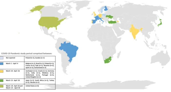
A total of 41 of 65 respondents (63%) from 23 countries worldwide (Azerbaijan [1 respondent], Belgium [1 re-spondent], Brazil [1 rere-spondent], Croatia [1 rere-spondent], Finland [1 respondent], France [2 respondents], Germany [1 respondent], India [1 respondent], Italy [7 respond-ents], Japan [1 respondent], Moldova [1 respondent], the Netherlands [1 respondent], Poland [1 respondent], Portugal [1 respondent], Slovenia [1 respondent], South Africa [1 respondent], Spain [3 respondents], Sweden [1 respondent], Switzerland [3 respondents], Turkey [2 re-spondents], Ukraine [1 respondent], the United Kingdom [2 respondents], and the United States [6 respondents]) joined the survey (Fig. 2). For the most part, data reflected single-institution activity (39 of 41 respondents; 95.1%), except in 2 instances (4.9%) in which multi-institutional data were provided, namely from the Pathological National Automated Archive (PALGA) Public Pathology Database of the Netherlands (https://www.palga.nl/
en/publi c-patho logy-datab ase/) and from the National Health Laboratory Service of South Africa. Since the tim-ing of COVID-19 lockdown differed among countries, as reported in Figure 2, each institution selected a 4-week time frame between March 1 and April 30, 2020, as the most significant health emergency period.
A total of 36 of the 41 respondents (87.8%) com-pleted all required fields; in 2 instances (4.9%) only data relative to gynecological samples (Papanicolaou tests) were provided. With regard to the distribution of diag-nostic classes, in 1 case (2.4%) suspicious and malignant diagnoses were merged; in another, only malignant diag-noses were reported; and, finally, in 1 case data were not reported.
Overall, data relative to 294,544 cytological sam-ples, including 104,319 cytological specimens from the COVID-19 pandemic period and 190,225 cytological samples from the corresponding period in 2019, were provided, with an overall workload reduction of 45.3% (range, 0.1%-98.0%). Data for each single respondent are reported in Table 1. Data were anonymized and a number randomly was assigned to each respondent.
Because changes in cytological practice could mod-ify the ratio between exfoliative versus FNA samples, a detailed analysis was performed; although the pooled analysis did not demonstrate a significant variation in the
ratio of exfoliative to FNA samples between the COVID-19 pandemic and the reference period (OR, 0.89; 95% CI, 0.74-1.08), a very high heterogeneity among the in-stitutions was observed (I2 of 95%) (Fig. 3).
For any single anatomic site, an absolute reduction in the total number of cases was observed consistently; this reduction was more evident (>50%) in samples from the cervicovaginal tract, urinary tract, breast, thy-roid, salivary gland, soft tissue, anorectal region, and bone marrow, whereas it was less pronounced (<50%) in samples from 8 sites (serous cavities, lymph nodes,
respiratory tract, central nervous system, gastrointes-tinal tract, pancreas, liver, and biliary tract); data are summarized in Table 2. Considering the contribution of any single anatomic site to the overall activity, signif-icant decreases were observed in samples from the cer-vicovaginal tract, thyroid, anorectal region, and bone marrow (P < .05) during the COVID-19 pandemic compared with the corresponding period in 2019. Conversely, the percentage of samples from the urinary tract, serous cavities, breast, lymph nodes, respiratory tract, salivary gland, central nervous system, gastroin-testinal tract, pancreas, liver, and biliary tract increased (P < .05). No significant variation in the percentage of soft-tissue samples was observed (Table 2).
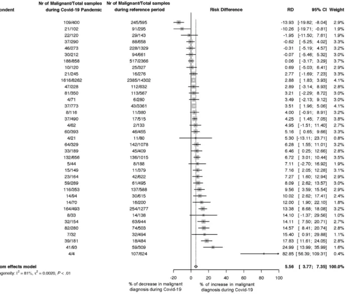
With regard to the distribution of diagnostic classes in nongynecological samples, an overall increase of 5.56% (95% CI, 3.77%-7.35%) in the malignancy rate during the COVID-19 pandemic compared with the corre-sponding period in 2019 was observed (Fig. 4). When the suspicious category also was added to the malignant cat-egory, the overall increase was 6.95% (95% CI, 4.63%-9.27%) (Fig. 5). The heterogeneity among institutions was found to be very high in both analyses (I2 of 81% and I2 of 87%, respectively).
DISCUSSION
To our knowledge to date, the effect of COVID-19 on cytological practice has been documented by only a few reports reflecting single-institution experience.7,8 The results of the current study demonstrated that the COVID-19 pandemic impacted cytology practices around the world by dramatically reducing the cytolog-ical specimen volume across specimen types by 45.3%. There are a number of explanations. First, screening programs were suspended or widely reduced according to recommendations issued by pathology scientific soci-eties.3,4 During the COVID-19 pandemic, the number
of cervicovaginal tract samples obtained was dramati-cally reduced, both in absolute terms (−74.6%) and in proportion to the overall cytological sample volume (53.7% vs 68.61%; P < .001). Because it was recom-mended that cervical cancer screening activities be post-poned rather than cancelled, future investigations once the health emergency is over are warranted to assess to what extent women have returned to cervical cancer screening programs.
TABLE 1. Total Number of Cytological Samples
During 4 Weeks of the COVID-19 Pandemic Compared With the Corresponding Period in 2019 for Each Respondent
Respondent COVID-19 Pandemic Corresponding Period in 2019 Difference, % 1 440 1361 −67.7 2 164 622 −73.6 3 547 1001 −45.4 4 185 983 −81.2 5 1207 3402 −64.5 6 173 858 −79.8 7 50 475 −89.5 8 456 736 −38.0 9 273 1329 −79.5 10 436 2576 −83.1 11 292 2532 −88.5 12 289 495 −41.6 13 704 2856 −75.4 14 102 295 −65.4 15 120 143 −16.1 16 702 2784 −74.8 17 858 2366 −63.7 18 384 677 −43.3 19 526 3099 −83.0 20 87 353 −75.4 21 94 615 −84.7 22 2352 6214 −62.1 23 398 1237 −67.8 24 39 128 −69.5 25 162 806 −79.9 26 212 661 −67.9 27 190 962 −80.2 28 400 595 −32.8 29 248 828 −70.0 30 1615 1616 −0.1 31 3531 8658 −59.2 32 4 1783 −99.8 33 3479 12,680 −72.6 34 13,345 38,824 −65.6 35 456 508 −10.2 36 263 635 −58.6 37 126 1191 −89.4 38 10 500 −98.0 39 198 697 −71.6 40 68,429 79,116 −13.5 41 773 3361 −77.0 Total 104,319 190,558 −45.3
Compared with the reduction in pap smears, the percentage of other exfoliative specimens, such as serous fluid, urine, and cerebrospinal fluid, demon-strated a significant increase, thereby explaining why there was no significant difference noted with regard to exfoliative versus FNA samples during the COVID-19 pandemic compared with the corresponding period in 2019.
A second explanation for the reduction in cytolog-ical samples lies in the fact that FNA specimens were
limited to patients in whom a diagnosis rendered by the cytopathologist would immediately affect manage-ment. As an example, the current survey demonstrated an overall reduction in thyroid FNA samples both in absolute number (−78.9%) and, considering the over-all cytological sample volume, in percentage (3.26% vs 5.02%; P < .001). It is interesting to note that the ma-jority of asymptomatic thyroid nodules are not medi-cally urgent9,10; in addition, most differentiated thyroid
cancers have an indolent clinical course, thus explaining
FIGURE 3. Meta-analysis forest plot demonstrating the differences between the coronavirus disease 2019 (COVID-19) pandemic and the corresponding period in 2019 (reference period) with respect to the ratio of exfoliative–to–fine-needle aspiration (FNA) samples. For each institution, the odds ratio (OR) between the exfoliative compared with the FNA samples observed during the COVID-19 pandemic and the reference period is shown. ORs with corresponding 95% CIs were graphically represented. ORs <1 indicate a reduction in the odds of exfoliative samples during the COVID-19 pandemic whereas ORs >1 demonstrate an increase in the exfoliative-to-FNA ratio during the COVID-19 pandemic. The pooled OR was obtained through a random effect model and is shown in bold. Nr indicates number.
the recommendation to postpone thyroid FNAs to the end of the health emergency, taking into account that a long and undefined “waiting time” between an endo-crinologist’s referral and performance of FNA generates anxiety for patients. Ideally, the decision to postpone the FNA should be taken by a multidisciplinary board, based on nodule location, ultrasound features, and clin-ical pathology laboratory data, in particular serum thy-rotropin and calcitonin levels.9,11 Dedicated guidelines, also addressing medicolegal issues, could be useful to assist the interventional cytopathologist in deciding to delay a thyroid FNA.
Compared with the reduction in thyroid FNAs, the overall reduction in cytology volume was less ev-ident for specimens for which the rate of malignancy usually is higher. In fact, lymph node, respiratory tract, breast, and salivary gland specimens were reduced in absolute terms but, considering the overall cytological sample volume, their percentage was significantly in-creased compared with 2019. As an example, respiratory tract cytological specimens demonstrated a reduction of 49.9% but a relative increase in percentage compared with 2019 (6.43% vs 4.16%; P < .001). Moreover,
respiratory tract FNA samples showed a slight relative increase (18% vs 13% in 2019) with respect to exfoli-ative specimens (82% vs 87% in 2019) (Table 2), sug-gesting a prioritization of FNA procedures that directly sample a suspicious lesion. The data from the current study indicate that, despite biosafety issues,12-14 which are especially relevant in the handling of specimens from the upper and lower airways, lung cytopathology still was relatively robust during the health emergency. A more focused survey could shed light on how cyto-pathologists applied the recommendations to limit the practice of rapid on-site evaluation to avoid smears air-drying before Romanowsky staining15 and to mod-ify the alcohol content of liquid-based cytology collec-tion medium.16
The overall data from the current study demonstrated a remarkable reduction in cytological workload across lab-oratory practices around the world, and also indicated that patients at high oncological risk were prioritized. The re-sults also demonstrated an overall increase in the relative malignancy rate among nongynecological samples during the COVID-19 pandemic compared with the correspond-ing period in 2019 (+5.56%). This is even more evident TABLE 2. Overall Number and Percentage of Samples From Each Anatomic Site During the COVID-19 Pandemic
and the Corresponding Period in 2019 (Global Volume Resulting From 39 Respondents Who Provided Both Exfoliative and FNA Data)
Site
Overall No. Percentage
COVID-19 Pandemic Corresponding Period in 2019 Difference, % COVID-19 Pandemic, % Corresponding Period in 2019, % Adjusted P Cervicovaginal tract 19,269 75,884 −74.6 53.7 68.61 <.001 Urinary tract 3778 8379 −54.9 10.53 7.58 <.001 Serous cavities 3101 4626 −33.0 8.64 4.18 <.001 Breast 980 2248 −56.4 2.73 2.03 <.001 Lymph node 2850 4651 −38.7 7.94 4.2 <.001 Thyroid 1169 5551 −78.9 3.26 5.02 <.001 Respiratory tract 2308 4606 −49.9 6.43 4.16 <.001 Exfoliative samples (n = 1892) (82%) Exfoliative samples (n = 4007) (87%) FNA sample (n = 416) (18%) FNA sample (n = 599) (13%) Salivary gland 195 482 −59.5 0.54 0.44 .021 Soft tissue 143 386 −63.0 0.4 0.35 .172 CNS 901 1309 −31.2 2.51 1.18 <.001 Gastrointestinal tract 81 161 −49.7 0.23 0.15 .005 Pancreas 378 518 −27.0 1.05 0.47 <.001 Liver 98 158 −38.0 0.27 0.14 <.001 Biliary tract 54 94 −42.6 0.15 0.08 .004 Anorectal region 6 183 −96.7 0.02 0.17 <.001 Bone marrow 41 220 −81.4 0.1 0.2 .003 Other sites 528 1153 −54.2 1.5 1.04 <.001 Total 35,880 110,609 100% 100%
when the suspicious category also is taken into account (+6.95%), which is conceivable considering that the risk of malignancy of this category is not negligible, generally ranging from 50% to 100%.17 The significance of this “relative” increase in the percentage of malignant and sus-picious diagnoses should be investigated further, bearing in mind that the majority of laboratories had a dramatic decrease in the “absolute” volumes of many specimens.
Although the current study has provided robust data reflecting an international collective effort, several
limitations should be highlighted. First, the study period was limited to 4 weeks between March 1 and April 30, 2020. This period of time does not necessarily correspond to the peak of the COVID-19 pandemic in countries still facing the health emergency at the time of this writing (eg, Brazil, India, and South Africa). Second, the data were analyzed globally, which may conceal differences among institutional practices. In this setting, further in-vestigations may be warranted when also taking into con-sideration that certain continents are better represented
FIGURE 4. Meta-analysis forest plot demonstrating the difference between the coronavirus disease 2019 (COVID-19) pandemic and the corresponding period in 2019 (reference period) with respect to the malignancy rate. Participants who reported only data relative to gynecological samples (2 participants), did not report nongynecological diagnostic classes (1 participant), or who reported merged suspicious and malignant diagnoses (1 participant) were not included. For each institution, the malignancy rate observed during the COVID-19 pandemic and the reference period is shown. Rate differences (RDs) with corresponding 95% CIs were graphically represented. Negative RDs (RD <0) indicate a reduction in the malignancy rate during the COVID-19 pandemic whereas positive RDs (RD >0) represent an increase in the malignancy rate during the COVID-19 pandemic. The pooled RD was obtained through a random effect model and is shown in bold. Nr indicates number.
than others and additional data from Asia and Oceania would make the survey more complete.
Conclusions
The COVID-19 pandemic dramatically impacted health systems and the activity of cytopathology laboratories world-wide. Laboratories universally experienced a dramatic reduc-tion in overall cytological specimen volume across specimen types, which resulted in a higher malignancy rate overall. Although the increase in the percentage of malignant cases demonstrates the efficacy of prioritizing high-risk patients with cancer despite the pandemic, prospective monitoring of the effect of delays in access to health services during the COVID-19 pandemic warrants further investigation.
FUNDING SUPPORT
Supported by Monitoraggio ambientale, studio ed approfondi-mento della salute della popolazione residente in aree a rischio–In attuazione della D.G.R. Campania n.180/2019; POR Campania FESR 2014-2020 Progetto “Sviluppo di Approcci Terapeutici Innovativi per patologie Neoplastiche resistenti ai trattamenti (SATIN)”; and Campania Region for the Investigation of the Molecular Biology of Thyroid Cancer (grant LR n.24 29/12/2005).
CONFLICT OF INTEREST DISCLOSURES
Umberto Malapelle has received personal fees for acting as a member of the Speakers’ Bureau or as an advisor from Boehringer Ingelheim, AstraZeneca, Roche, MSD, Amgen, Merck, Eli Lilly, and Diaceutics for work performed outside of the current study. Paul A. VanderLaan has acted as a paid consultant for Gala Therapeutics, Foundation Medicine, Caris Life Sciences, Flatiron Health, Intuitive Surgical, and Clearview Healthcare Partners for work performed outside of FIGURE 5. Meta-analysis forest plot demonstrating the difference between the coronavirus disease 2019 (COVID-19) pandemic and the corresponding period in 2019 (reference period) with respect to the malignancy and suspicious rates. Participants who reported only data relative to gynecological samples (2 participants), did not report nongynecological diagnostic classes (1 participant), or reported only malignant diagnoses (1 participant) were not included. For each institution, the malignancy and suspicious rates observed during the COVID-19 pandemic and reference period are shown. Rate differences (RDs) with corresponding 95% CIs are graphically represented. Negative RDs (RD <0) indicate a reduction in the malignancy and suspicious rates during the COVID-19 pandemic whereas positive RDs (RD >0) indicate an increase in the malignancy and suspicious rates during the COVID-19 pandemic. The pooled RD was obtained through a random effect model and is shown in bold. Nr indicates number.
the current study. Giancarlo Troncone has received personal fees as a member of the Speakers’ Bureau or as an advisor from Roche, MSD, Pfizer, and Bayer for work performed outside of the current study. The other authors made no disclosures.
AUTHOR CONTRIBUTIONS
Conceptualization: Elena Vigliar, William C. Faquin, Martha
Bishop Pitman, and Giancarlo Troncone. Methodology: All authors. Software and formal analysis: Elena Vigliar and Dario
Bruzzese. Validation: All authors. Investigation: All authors. Resources: All authors. Data curation: All authors. Writing–original draft: Elena Vigliar and Giancarlo Troncone. Writing–review and editing: All authors. Visualization: All authors. Supervision:
Elena Vigliar, William C. Faquin, Martha Bishop Pitman, and Giancarlo Troncone. Project administration: Elena Vigliar,
William C. Faquin, Martha Bishop Pitman, and Giancarlo
Troncone. Funding acquisition: Giancarlo Troncone.
REFERENCES
1. Zhu N, Zhang D, Wang W, et al; China Novel Coronavirus Investigating and Research Team. A novel coronavirus from patients with pneumonia in China, 2019. N Engl J Med. 2020;382:727-733. doi:10.1056/NEJMo a2001017
2. Centers for Disease Control and Prevention. Interim laboratory bio-safety guidelines for handling and processing specimens associated with Coronavirus Disease 2019 (COVID-19). Accessed June 15, 2020. https://www.cdc.gov/coron aviru s/2019-ncov/lab/lab-biosa fe-ty-guide lines.html
3. Royal College of Pathologists, Institute of Biomedical Science, the Association of Clinical Biochemistry and Laboratory Medicine, Association of Clinical Pathologists. Recommendations from RCPath and Professional Bodies (IBMS, ACP and ACB): Prioritisation/ Deferral of Pathology Laboratory Work (in Light of SARS-CoV-2 (COVID19) Epidemic). Accessed September 21, 2020. https://www. rcpath.org/uploa ds/asset s/f5123 842-950f-49c5-bf69e d866a 7ca3d a/ Prior itisa tion-defer ral-of-patho logy-labor atory -work.pdf
4. College of American Pathologists. Cytopathology laboratory con-siderations during the COVID-19 pandemic: College of American Pathologists Cytopathology Committee. Accessed July 29, 2020. https://www.cap.org/labor atory -impro vemen t/news-and-updat es/cytop athol ogy-labor atory -consi derat ions-durin g-the-covid -19-pandemic 5. COVID-19 - World Health Organization. Laboratory biosafety
guid-ance related to coronavirus disease (COVID-19). https://www.who. int/
6. Garassino MC, Whisenant JG, Huang LC, et al; TERAVOLT in-vestigators. COVID-19 in patients with thoracic malignancies (TERAVOLT): first results of an international, registry-based, co-hort study. Lancet Oncol. 2020;21:914-922. doi:10.1016/S1470 -2045(20)30314 -4
7. de Pelsemaeker MC, Guiot Y, Vanderveken J, Galant C,Van Bockstal MR. The impact of the COVID-19 pandemic and the associated Belgian governmental measures on cancer screening, surgical pathol-ogy and cytopatholpathol-ogy. Pathobiolpathol-ogy. 2020:1-10. doi:10.1159/00050 9546
8. Vigliar E, Iaccarino A, Bruzzese D, Malapelle U, Bellevicine C, Troncone G. Cytology in the time of coronavirus disease (Covid-19): an Italian perspective. J Clin Pathol. 2020:jclinpath-2020-206614. doi:10.1136/jclin path-2020-206614
9. Li M, Marquez RC, Vinales KL, et al. Considerations for thyroid fine needle aspiration (FNA) biopsies during the COVID-19 pan-demic. Clin Thyroidol. Published online April 2020. doi:10.1089/ CT.2020;32.156-158
10. Durante C, Grani G, Lamartina L, Filetti S, Mandel SJ, Cooper DS. The diagnosis and management of thyroid nodules: a review. JAMA. 2018;319:919-924. doi:10.1001/jama.2018.0898
11. Troncone G. Thyroid cytology in the times of coronavirus. Diagn
Cytopathol. Published online June 1, 2020. doi:10.1002/dc.24510
12. Barbareschi M, Ascoli V, Bonoldi E, et al. Biosafety in surgical pathol-ogy in the era of SARS-Cov2 pandemia. A statement of the Italian Society of Surgical Pathology and Cytology. Pathologica. 2020;112:59-63. doi:10.32074/ 1591-951X-14-20
13. Chen CC, Chi CY. Biosafety in the preparation and processing of cytology specimens with potential coronavirus (COVID-19) in-fection: perspectives from Taiwan. Cancer Cytopathol. 2020;128: 309-316. doi:10.1002/cncy.22280
14. Miller JM, Astles R, Baszler T, et al; Biosafety Blue Ribbon Panel; Centers for Disease Control and Prevention (CDC). Guidelines for safe work practices in human and animal medical diagnostic laborato-ries. Recommendations of a CDC-convened, Biosafety Blue Ribbon Panel. MMWR Suppl. 2012;61:1-102.
15. Pambuccian SE. The COVID-19 pandemic: implications for the cytology laboratory. J Am Soc Cytopathol. 2020;9:202-211. doi:10.1016/j.jasc.2020.03.001
16. Rossi ED, Fadda G, Mule A, Zannoni GF, Rindi G. Cytologic and his-tologic samples from patients infected by the novel coronavirus 2019 SARS-CoV-2: an Italian institutional experience focusing on biosafety procedures. Cancer Cytopathol. 2020;128:317-320. doi:10.1002/ cncy.22281
17. Poller DN, Schmitt F. Should uncertainty concerning the risk of malignancy be included in diagnostic (nongynecologic) cytopa-thology reports? Cancer Cytopathol. Published online July 10, 2020. doi:10.1002/cncy.22322