Original Paper
Med Princ Pract 2004;13:260–266 DOI: 10.1159/000079524
Vitamin D Receptor Alleles, Bone Mineral
Density and Turnover in Postmenopausal
Osteoporotic and Healthy Women
Belgin Süsleyici Duman
aRefik Tanakol
bNevin Erensoy
cMelek Öztürk
cSelma Yılmazer
caDepartment of Medical Biology and Genetics, Kadir Has University Medical Faculty, Gayrettepe-Istanbul, and Departments of bEndocrinology and Metabolism and cMedical Biology, Istanbul University Faculty of Medicine, Çapa-Istanbul, Istanbul, Turkey
Received: January 20, 2003 Revised: June 28, 2003
Key Words
Bone mineral densityW Vitamin D receptor gene
polymorphismW OsteoporosisW Menopause
Abstract
Objective: Vitamin D receptor (VDR) gene polymor-phisms and bone metabolic markers were investigated as potential genetic markers for osteoporosis in post-menopausal Turkish women. The relationship between their VDR gene polymorphisms and bone states was determined. Materials and Methods: Restriction frag-ment length polymorphisms at the VDR gene locus (i.e., for BsmI, ApaI, and TaqI) was investigated in 75 post-menopausal osteoporotic (53.16 B 1.31 years) and 66 healthy (52.62 B 1.69 years) Turkish women and the genotypes were related to bone mineral density (BMD) at femoral neck (FN), lumbar spine (L1–4), trochanter, Ward’s triangle (Ward’s) and metabolic parameters of bone turnover. Results: In osteoporotic women, TaqI genotype-related differences of the VDR gene were found to be significant at all BMD sites; TT genotype had higher L1–4 BMD values than Tt and tt (p ! 0.05); tt geno-type had significantly lower BMD at FN (p ! 0.05), tro-chanter (p ! 0.01), and Ward’s (p ! 0.05) compared to TT
genotype. The tt genotype was found to be associated with higher (p ! 0.05) serum osteocalcin levels compared to Tt and TT genotypes in the osteoporotic women, whereas no such association was found for the healthy women. Conclusion: Our data showed an association between VDR TaqI genotype and BMD at the FN, L1–4, trochanter and Ward’s triangle in nonobese postmeno-pausal osteoporotic women. Thus the VDR gene Taql polymorphism modulates differences in BMD in the postmenopausal osteoporotic women.
Copyright © 2004 S. Karger AG, Basel
Introduction
Osteoporosis is characterized by low bone mineral density (BMD) and microarchitectural deterioration of bone leading to increased bone fragility and a high risk of fracture. BMD is influenced by genetic factors [1] and sev-eral twin studies suggest that genetic factors account for as much as 80% of total variance of BMD, the major predic-tor of osteoporosis and fragility fractures [1–7].
Although studies performed by Morrison et al. [8, 9] have focused attention on the possibility that polymor-phisms in the vitamin D receptor (VDR) gene may
account for a significant part of this variation, the distri-bution of BMD in the population strongly suggests a poly-genic inheritance [10, 11]. Several groups have studied the relationship between VDR and bone mass as well as bone turnover in different populations, and obtained discor-dant results [8, 12–23]. In Australian women of English-Irish descent, after adjustment for age and environmental factors, subjects with BB or the tt genotype were found to have higher serum osteocalcin levels and lower BMD than those with the bb or TT genotypes. Heterozygotes (Bb/Tt) had intermediate values [8]. Furthermore, the BB geno-type is associated with a higher rate of bone loss in preme-nopausal Japanese women [12] and in postmepreme-nopausal Caucasian women [13, 14]. These findings show an active role of the VDR genotype in determining bone density in postmenopausal women. Other studies have found a cor-relation between the BMD and VDR genotype [15–17], while others still have failed to detect such a correlation [18–23]. In general, the studies that have detected lower BMD found smaller differences between groups than that reported by Morrison et al. [9]. The ethnic differences among the study populations may explain this variability to some extent but only a few studies have focused on this issue [22, 24, 25].
No data have yet been published examining the preva-lence and relevance of the various VDR polymorphisms in Turkish women. We therefore studied VDR genotypes in Turkish postmenopausal osteoporotic women and ex-amined the relationship between the genotypes defined by the polymorphisms ApaI, BsmI, and TaqI at the L1–4, the femoral neck (FN), trochanter and Ward’s triangle in healthy and osteoporotic women. The major aim of this study was to define the possible associations of VDR gene alleles and bone turnover markers with BMD.
Materials and Methods
Subjects and Bone Density Measurements
Seventy-five postmenopausal osteoporotic (53.16 B 1.31 years) and 66 healthy (52.62 B 1.69 years) women were included in the study. All osteoporotic women had a BMD of at least 2.5 SD below the mean value of healthy premenopausal women in either the hip or spine. The control group consisted of healthy, age- and risk factor-matched subjects with BMD B1 SD T score. A detailed medical his-tory was obtained and physical examination was performed by one of the investigators (R.T.). Patients were excluded from the study on the basis of the following criteria: presence of concomitant disease (disor-ders of calcium metabolism, renal, thyroid, hepatic dysfunction, Paget’s disease, Cushing’s syndrome, sarcoidosis, rheumatoid arthri-tis, malignancy, malabsorption, and malnutrition), previous use of oral/transdermal hormone replacement therapy or any other osteo-porosis treatment, thyroid hormone replacement, glucocorticoid or
anticonvulsant drug, biochemical evidence of osteomalacia and severe osteoarthritis. Osteoporosis risk factors, such as cigarette smoking, premenstrual irregularity of menses, insufficient sun expo-sure and calcium intake as well as sodium, protein, coffee and alcohol consumption, and physical activity, were assessed by a question-naire. Lateral radiographs of the spine were examined, and the com-pression fracture was defined as 1 20% reduction in anterior height. BMD of the lumbar spine and proximal femur in grams per square centimeter was measured by dual energy X-ray absorptiometry (Ho-logic QDR 1000 Plus, USA).
Measurement of Bone Turnover Markers
Following an overnight fast, urine samples for the measurement of 24-hour calcium and hydroxyproline [26] were collected and mea-sured. Serum calcium and osteocalcin were also meamea-sured. Body mass index was calculated as an estimate of obesity [weight (kg)/ height (m2)].
Genotype Assignment
Genomic DNA was prepared from 10 ml of EDTA-treated blood with a simple salting-out procedure [27]. The DNA sequences were amplified by polymerase chain reaction (PCR) [28]. Detection of the
BsmI site in intron 8 was performed by PCR amplification of a
region carrying the BsmI site with primers originating in exon 7 (primer 1: 5)-CAA CCAAGACTACAAGTACCGCGTCAGTGA-3)) and intron 8 (primer 2: 5)-AACCAGCGGGAAGAGGTCAAGGG-3)) producing an 825-basepair (bp) fragment [9]. To amplify the VDR DNA sequence carrying ApaI and TaqI sites, primers in intron 8 CAGAGCATGGACAGGGAGCAAG-3)) and exon 9 (5)-GCAACTCCTCATGGCTGAGGTCTCA-3)) were used which pro-duced a 740-bp fragment [8]. PCR products were generated in a 60-Ìl reaction volume containing 100–200 ng of DNA, 0.5 ÌM of each primer, 200 ÌM of dNTPs, 50 mM KCl, 10 mM Tris-HCl (pH = 9.0), 1.5 mM MgCl2, 0.1% Triton X-100, and 0.8 U of Taq DNA
poly-merase.
To determine the presence of a restriction site within an ampli-fied product, a 10-Ìl aliquot was digested with 5 U of endonuclease
BsmI at 65° C, ApaI at 37 ° C, or TaqI at 65 ° C for 1 h. Digestion products were electrophoresed in a 2% agarose gel containing ethid-ium bromide (50 Ìg/ml). DNA fragments were visualized by ultra-violet illumination and fragment sizes estimated by comparison to the 50-bp ladder run on the same gel. The genotype detection proto-col was repeated 2 times for the heterozygous samples to eliminate the probability of partial digestion. The presence of the BsmI restric-tion site generates 175- and 650-bp fragments, whereas the absence of this site yields a 825-bp fragment. The homozygous absence of the
TaqI site yields bands of 245 and 495 bp, whereas the homozygous
presence of this site yields fragments of 205, 245 and 290 bp. Hetero-zygotes for the TaqI site exhibit fragments of 490, 290, 245 and 205 bp. Digestion of the 740-bp PCR product with ApaI gives fragments of 220 and 520 bp for the presence of the restriction site, whereas the absence of the restriction site leaves the PCR product undigested. The presence of the restriction enzyme site is indicated by a lower-case letter and the absence of the site by an upperlower-case letter.
Statistical Analysis
Statistical analyses were conducted using Unistat 5.1 software. Rare genotypes (n ! 10 in either control or osteoporotic women) were excluded from the analysis. Data were considered significant at p ! 0.05. The frequency distributions of VDR genotypes in osteoporotic
Table 1. Main clinical and biochemical characteristics, BMD, and bone turnover parameters in osteoporotic and control patients
Osteoporosis (n = 75)
Control (n = 66)
Age, years 53.16B1.31 52.62B 1.69
Menopause age, years 42.09B0.72 44.42B 0.85
Weight, kg 62.28B1.06** 67.56B 1.18
Height, m 1.55B0.01** 1.59B 0.01
Body mass index, kg/m2 25.67B0.44 26.66B 0.59
Serum calcium, mg/dl 9.41B0.17 9.69B 0.06
Urinary calcium mg/day 131.49B7.49* 166.32B13.98
Osteocalcin, ng/ml 5.03B0.56*** 2.61B 0.55 Hydroxyproline, mmol/mol Cr 15.87B1.34 14.75B 1.53 L1–4 BMD, g/cm2 0.80B0.02*** 0.93B 0.02 FN BMD, g/cm2 0.68B0.01* 0.76B 0.02 Trochanter BMD, g/cm2 0.57B0.01* 0.66B 0.02 Ward’s BMD, g/cm2 0.53B0.01*** 0.65B 0.03 Total BMD, g/cm2 0.76B0.03* 0.89B 0.09
Values are means B SE.
* p ^ 0.05; ** p ^ 0.01; *** p ^ 0.001.
Table 2. Allele and genotype frequencies of the BsmI, ApaI, and
TaqI polymorphisms of the VDR gene
VDR genotype Osteoporotic women observed n frequency % Control women observed n frequency % BsmI Bb54 72 42 63.6 BB 18 24 17 25.8 bb 3 4 7 10.6 ApaI Aa 56 74.8 45 68.2 AA 13 17.2 15 22.7 aa 6 8 6 9.1 Taq Tt 42 56 28 42.4 TT 23 30.8 23 34.9 tt 10 13.2 15 22.7
Table 3. Frequency of VDR-combined genotypes Combined genotype Osteoporotic women (n = 75) n % Control women (n = 66) n % BbAaTt 31 41.3 17 25.8 BbAatt 15 20 10 15.15 bbAaTT 6 8 10 15.15 bbaaTt 5 6.76 – – BbaaTt 3 4 5 7.57 BBAAtt 3 4 3 4.54 Bbaatt 2 2.66 5 7.57 bbAaTt 2 2.66 5 7.57
and normal groups were compared using the ¯2 test. Both for the
osteoporotic and control subjects, BMD levels and bone turnover parameters for the three genotype classes were compared with analy-sis of variance (ANOVA). Student’s t test and the Mann-Whitney test were applied for statistical analysis between the two groups. All data are shown as mean B standard error.
Results
The demographic and major clinical and biochemical values for both osteoporotic and control subjects are given in table 1. The osteoporotic women’s height, weight, uri-nary calcium and all BMD values were significantly lower than those of the control women (p ^ 0.01 and 0.05). Although not significant, the bone resorption marker, hydroxyproline, was higher in the osteoporotic than the control group. Osteoporotic women had significantly higher (p ! 0.001) serum osteocalcin levels compared to the control group. After amplification of genomic DNA with VDR-specific primers, followed by digestion with
ApaI, BsmI, or TaqI and agarose gel electrophoresis,
restriction fragment length polymorphisms (RFLPs) were determined. No different result was obtained from partial digestion testing. The genotypic frequencies observed for these polymorphisms both in osteoporotic patients and control subjects are shown in table 2. The genotype
fre-quencies in this population were 24% for BB, 72% for Bb, 4% for bb, 17.2% for AA, 74.8% for Aa, 8% for aa, 30.8% for TT, 56% for Tt, 13.2% for tt in the osteoporotic wom-en, and 25.8% for BB, 63.6% for Bb, 10.6% for bb, 22.7% for AA, 68.2% for Aa, 9.1% for aa, 34.9% for TT, 42.4% for Tt and 22.7% for tt in the control women. The combi-nation of genotypes defined by all three RFLPs is shown in table 3. The BbAaTt, BbAatt, and bbAaTT define the most frequently combined ones (69.3% in osteoporotic women, 56.1% in control women). The most common
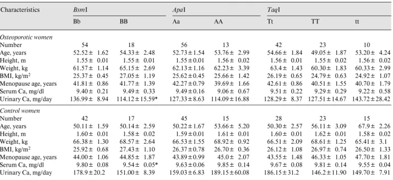
Table 4. Selected characteristics of unrelated osteoporotic and healthy postmenopausal women in relation to VDR gene alleles Characteristics BsmI BbBB ApaI Aa AA TaqI Tt TT tt Osteoporotic women Number 54 18 56 13 42 23 10 Age, years 52.52B 1.62 54.33B 2.48 52.73B1.54 53.76B 2.99 54.66B 1.84 49.05B 1.87 53.20B 4.24 Height, m 1.55B 0.01 1.55B 0.01 1.55B0.01 1.56B 0.02 1.56B 0.01 1.55B 0.02 1.56B 0.02 Weight, kg 61.57B 1.14 65.15B 2.69 62.13B1.16 62.23B 3.39 63.4B 1.43 60.30B 1.83 60.33B 2.99 BMI, kg/m2 25.37B 0.45 27.05B 1.19 25.62B0.45 25.66B 1.42 26.19B 0.65 24.79B 0.63 24.92B 1.07 Menopause age, years 41.81B 0.86 41.77B 1.39 42.27B0.79 39.69B 1.66 42.61B 0.86 40.51B 1.55 40.70B 1.79
Serum Ca, mg/dl 9.40B 0.21 9.49B 0.33 9.49B0.16 9.06B 0.67 9.51B 0.22 9.29B 0.29 9.22B 0.58
Urinary Ca, mg/day 136.99B 8.94 114.12B15.59* 127.33B8.63 114.09B16.88 128.29B 8.37 127.51B14.67 143.72B28.42
Control women Number 42 17 45 15 28 23 15 Age, years 50.11B 1.59 50.14B 2.59 50.22B1.67 53.66B 5.20 50.30B 2.57 56.11B 3.09 67.9B 2.26 Height, m 1.60B 0.01 1.58B 0.02 1.59B0.01 1.61B 0.01 1.60B 0.01 1.62B 0.01 1.58B 0.02 Weight, kg 66.38B 1.30 68.57B 2.64 66.53B1.55 68.92B 0.92 66.51B 2.09 68.61B 1.25 65.41B 3.1 BMI, kg/m2 25.92B 0.68 27.43B 1.10 26.37B0.78 26.70B 0.36 26.12B 1.08 26.97B 0.74 26.50B 1.33 Menopause age, years 44.00B 1.06 44.85B 1.87 43.89B0.99 45.0B 2.07 43.55B 1.48 46.33B 1.05 47.70B 1.81
Serum Ca, mg/dl 9.80B 0.08 9.54B 0.05* 9.63B0.06 9.85B 0.14 9.67B 0.08 9.81B 0.14 9.55B 0.04
Urinary Ca, mg/day 178.9B20.2 151.00B 8.39 159.03B6.83 189.15B60.08 186.15B31.2 146.2B11.90 149.70B 7.91 Values are means B SE.
* p ! 0.05. Student’s t test was used to compare the characteristics between the alleles of the VDR genes (BsmI, ApaI), whereas ANOVA was used to test for the difference across the TaqI genotypes. BMI = Body mass index.
genotype combination in postmenopausal osteoporotic and control women was BbAaTt.
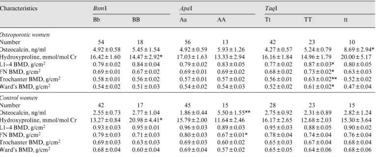
The relationship between VDR gene alleles and select-ed characteristics of osteoporotic and control subjects is shown in table 4. In osteoporotic and control women, there were no significant differences in age, age at meno-pause, height, weight or body mass index among the com-mon genotypes in BsmI, ApaI, and TaqI RFLPs. Urinary calcium concentration was to be found lower in the osteo-porotic women with the BB genotype than in those carry-ing Bb (p ! 0.05). Table 5 demonstrates BMD and bone turnover markers with respect to VDR gene alleles in osteoporotic and control subjects. Hydroxyproline levels were significantly lower in BB than in the Bb genotype in osteoporotic women (p ! 0.05); in contrast to osteoporotic women, hydroxyproline levels in the control group were significantly different with respect to genotype, in which lower values were found in Bb than BB. Although serum osteocalcin levels were not significantly different accord-ing to BsmI and ApaI genotypes, higher concentrations were observed in the BB and AA for osteoporotic women. For the control women, osteocalcin levels were found to differ according to the ApaI genotype, where AA carriers had higher levels compared to Aa (p ! 0.01), but no such
difference was observed between BsmI genotypes. In osteoporotic women serum osteocalcin levels were found to be highest in the tt genotype and lowest in the Tt, and intermediate in the TT genotype (p ! 0.05).
There is some evidence of a relationship with higher BMD levels associated with the T allele at the FN. Our data showed significant TaqI-related differences in the osteoporotic women in FN as well as L1–4, trochanter and Ward’s BMD. The osteoporotic group with the TT genotype had significantly higher FN (p ! 0.05), trochan-ter (p ! 0.01), L1–4 (p ! 0.05) and Ward’s (p ! 0.05) BMD values with respect to the tt genotype.
Discussion
In the last two decades, compelling data have been obtained indicating that BMD is, at least in part, geneti-cally determined [15, 16, 18, 19]. In the present study, we analyzed ApaI, BsmI, and TaqI VDR polymorphisms and addressed the question whether BMD was affected by the VDR gene polymorphisms in postmenopausal osteopo-rotic women as well as healthy control subjects. The observed VDR genotype distributions were similar to
pre-Table 5. BMD and bone turnover markers according to different VDR gene alleles in unrelated osteoporotic and healthy postmenopausal women Characteristics BsmI BbBB ApaI Aa AA TaqI Tt TT tt Osteoporotic women Number 54 18 56 13 42 23 10 Osteocalcin, ng/ml 4.92B0.58 5.45B1.54 4.92B0.59 5.93B1.26 4.27B0.57 5.24B0.79 8.69B2.94* Hydroxyproline, mmol/mol Cr L1–4 BMD, g/cm2 16.42B1.60 0.79B0.02 14.47B2.92* 0.84B0.04 17.03B1.63 0.79B0.02 13.33B2.94 0.83B0.05 16.16B1.84 0.77B0.02 14.96B1.79 0.87B0.03* 20.00B5.17 0.80B0.05 FN BMD, g/cm2 0.69B0.01 0.67B0.02 0.69B0.01 0.69B0.02 0.68B0.02 0.73B0.02* 0.63B0.03 Trochanter BMD, g/cm2 0.58B0.01 0.56B0.02 0.57B0.01 0.57B0.02 0.56B0.01 0.63B0.02** 0.52B0.02 Ward’s BMD, g/cm2 0.54B0.02 0.51B0.03 0.54B0.02 0.54B0.03 0.52B0.02 0.61B0.02* 0.47B0.04 Control women Number 42 17 45 15 28 23 15 Osteocalcin, ng/ml Hydroxyproline, mmol/mol Cr L1–4 BMD, g/cm2 2.55B0.73 13.27B0.84 0.93B0.03 2.77B1.04 20.98B4.41* 0.95B0.01 1.86B0.44 15.79B2.00 0.96B0.03 5.50B1.55** 11.64B2.46 0.89B0.03 2.75B0.92 16.17B2.65 0.95B0.03 2.31B0.89 12.68B2.03 0.88B0.05 2.82B1.24 15.30B3.64 0.90B0.02 FN BMD, g/cm2 0.79B0.03 0.71B0.03 0.80B0.03 0.67B0.01* 0.78B0.04 0.74B0.04 0.76B0.04 Trochanter BMD, g/cm2 0.69B0.03 0.63B0.03 0.69B0.03 0.60B0.02 0.65B0.03 0.67B0.04 0.68B0.04 Ward’s BMD, g/cm2 0.68B0.04 0.60B0.04 0.69B0.04 0.57B0.02 0.65B0.05 0.64B0.06 0.68B0.06
Values are means B SE.
* p ! 0.05; ** p ! 0.01. Student’s t test was used to compare the characteristics between the alleles of the VDR genes (BsmI, ApaI), whereas ANOVA was used to test for the difference across the TaqI genotypes.
vious reports [16, 17]. It is expected that BMD is deter-mined by genetic and environmental interacting in-fluences. FN BMD was found to be decreasing continual-ly with growing age, in both women and men even in the late decades of life [29]. The differential effects of VDR alleles on BMD among different populations could partly explain the differences between Italian and Irish studies [30, 31].
We found significant effects of VDR gene alleles on bone mass. TaqI genotypes were found to have a striking effect on all BMD values studied. The TT genotype-carry-ing osteoporotic women had higher BMD levels com-pared to either Tt or tt. The same effect of TaqI polymor-phisms on BMD could not be observed in healthy women. Our observation is in good agreement with the finding of Spector et al. [15] who confirmed the link between the VDR genotype (TaqI poymorphism) and BMD in post-menopausal British twins. In their study, postpost-menopausal twins with the TT genotype had an about 10% higher bone density than women with the tt genotype.
Various studies have shown that obesity can mask the influence of the VDR genotypes. Dawson-Hughes et al. [32] showed that women with the BB genotype of the VDR gene had a reduced efficiency in calcium absorption
and a low calcium intake compared to women of the bb genotype consistent with a functional defect in the intesti-nal VDR. The impact of this heritable difference is reduced at higher calcium intakes. This evidence is con-sistent with the fact that the relationship between the VDR genotypes and FN bone loss rate is enhanced at low calcium intakes in postmenopausal women. In obese post-menopausal women, estrogen concentrations appear to increase [33]. This estrogen increase in obese women may mask the relationship of VDR polymorphisms with BMD. For these reasons overtly obese women were not included in our study. Vandevyver et al. [34] concluded that the VDR gene polymorphism influences FN and BMD in nonobese postmenopausal women. They found that the link between VDR genotypes and FN and BMD is not seen in obese postmenopausal women, which sug-gests that factors related to obesity obscure such an effect in overweight women.
Although no significant differences were observed be-tween the genotypic distribution of the VDR gene in the osteoporotic and control groups in our study, for each of the three loci tested, heterozygous classes had a higher fre-quency in osteoporotics than in controls, which implies a heterozygous disadvantage. Furthermore, the BMD
val-ues for the different sites were not found to associate with any of the BsmI and ApaI genotypes in osteoporotic wom-en, which means that no relationship between the VDR
BsmI polymorphism and osteoporosis was observed. We
found a close relation between TaqI genotypes and BMD values at all sites studied in the osteoporotic women. The L1–4, FN, trochanter and Ward’s BMD levels differed with respect to TaqI polymorphism and all were signifi-cantly higher in the osteoporotic women with TT geno-type compared to Tt and tt, which demonstrates the rela-tionship between VDR TaqI polymorphism and osteo-porosis. Sheehan et al. [31] reported 29 and 40% higher osteocalcin levels of the tt genotype compared to the Tt and TT genotypes, respectively, in healthy adults. Accord-ing to our results the tt VDR genotype was associated with significantly higher serum osteocalcin compared with the Tt and TT genotypes, 50.8 and 39%, respectively, in the osteoporotic women, whereas no such association was found in the healthy women. The effects we observed in our subjects are of genetic and biological significance.
Conclusion
This study shows an association between the VDR gene TaqI polymorphism and BMD at the FN, lumbar spine, trochanter and Ward’s triangle in a group of non-obese, postmenopausal osteoporotic women. Thus, the VDR gene TaqI polymorphism modulates differences in BMD in the postmenopausal osteoporotic women, al-though the interaction of the gene and BMD and the role of VDR genotypes deserve further study with larger num-bers of subjects.
Acknowledgments
This work was supported by the Research Fund of the University of Istanbul, project numbers 1024/250897 and B-1298/03102001.
References
1 Pocock NA, Eisman JA, Hopper JL, Yeates MG, Sambrook PN, Eberl S: Genetic determi-nants of bone mass in adults. J Clin Invest 1987;80:706–710.
2 Christian JC, Yu PL, Slemenda CW, Johnston CC: Heritability of bone mass: A longitudinal study in aging male twins. Am J Hum Genet 1989;44:429–433.
3 Smith DM, Nance WE, Kang KW, Christian JC, Johnston CC Jr: Genetic factors in deter-mining bone mass. J Clin Invest 1973;52: 2800–2808.
4 Dequeker J, Nijs J, Verstraeten A, Geusens P, Gevers G: Genetic determinants of bone min-eral content at the spine and radius: A twin study. Bone 1987;8:207–209.
5 Kelly PJ, Hopper JL, Macaskill GT, Pocock NA, Sambrook PN, Eisman JA: Genetic factors in bone turnover. J Clin Endocrinol Metab 1991;72:808–813.
6 Lutz J: Bone mineral, serum calcium and di-etary intakes of mother/daughter pairs. Am J Clin Nutr 1986;44:99–106.
7 Seeman E, Hopper JL, Bach LA, Cooper ME, Parkinson E, McKay J, Jerums G: Reduced bone mass in daughters of women with osteo-porosis. N Engl J Med 1989;320:554–558. 8 Morrison NA, Yeoman R, Kelly PJ, Eisman
JA: Contribution of trans-acting factor alleles to normal physiological variability: Vitamin D receptor gene polymorphism and circulating osteocalcin. Proc Natl Acad Sci USA 1992;89: 6665–6669.
9 Morrison NA, Qi JC, Tokita A, Kelly PJ, Crofts L, Nguyen TV, Sambrook PN, Eisman JA: Prediction of bone density from vitamin D receptor alleles. Nature 1994;367:284–287. 10 Eisman JA, Morrison NA, Kelly PJ, Sambrook
PN, Howard G, Qi J, Tokita A, Crofts L, Nguyen TV, Birmingham J: Genetics of osteo-porosis and vitamin D receptor alleles. Calcif Tissue Int 1995;56:S48–49.
11 Peacock M: Vitamin D receptor gene alleles and osteoporosis: A contrasting view. J Bone Miner Res 1995;10:1294–1297.
12 Yamagata Z, Miyamura T, Iijima S, Asaka A, Sasaki M, Kato J, Koizumi K: Vitamin D receptor gene polymorphism and bone mineral density in healthy Japanese women. Lancet 1994;344:1027.
13 Krall EA, Parry P, Lichter JB, Dawson-Hughes B: Vitamin D receptor alleles and rates of bone loss: Influences of years since menopause and calcium intake. J Bone Miner Res 1995;10: 978–984.
14 Ferrari S, Rizzoli R, Chevalley T, Slosman D, Eisman JA, Bonjour J-P: Vitamin D receptor gene polymorphisms and chance in lumbar spine bone mineral density. Lancet 1995;345: 423–424.
15 Spector TD, Keen RW, Arden NK, Morrison NA, Major PJ, Nguyen TV, Kelly PJ, Baker JR, Sambrook PN, Lanchbury JS, Eisman JA: In-fluence of vitamin D receptor genotype on bone mineral density in postmenopausal wom-en: A twin study in Britain. BMJ 1995;310: 1357–1360.
16 Fleet JC, Harris SS, Wood RJ, Dawson-Hughes B: The BsmI vitamin D receptor restriction fragment length polymorphism (BB) predicts low bone density in premenopausal black and white women. J Bone Miner Res 1995;10:985– 990.
17 Riggs BL, Nguyen TV, Melton LJ 3rd, Morri-son NA, O’Fallon WM, Kelly PJ, Egan KS, Sambrook PN, Muhs JM, Eisman JA: The con-tribution of vitamin D receptor gene alleles to the determination of bone mineral density in normal osteoporotic women. J Bone Miner Res 1995;10:991–996.
18 Garnero P, Borel O, Sornay-Rendu E, Delmas PD: Vitamin D receptor gene polymorphisms do not predict bone turnover and bone mass in healthy premenopausal women. J Bone Miner Res 1995;10:1283–1288.
19 Keen RW, Major PJ, Lanchbury JS, Spector TD: Vitamin D receptor gene polymorphism and bone loss. Lancet 1995;345:990. 20 Looney J, Yoon HK, Fischer SM, Farley SM,
Farley JR, Wegedal JE, Baylink DJ: Lack of a high prevalence of the BB vitamin D receptor genotype in severely osteoporotic women. J Clin Endocrinol Metab 1995;80:2158–2162. 21 Kroger H, Mahonen A, Ryhanen S, Turunen
AM, Alhava E, Maenpaa P: Vitamin D recep-tor genotypes and bone mineral density. Lancet 1995;345:1238.
22 Lim SK, Park YS, Park JM, Song YD, Lee EJ, Kim KR, Lee HC, Huh KB: Lack of association between vitamin D receptor genotypes and os-teoporosis in Koreans. J Clin Endocrinol Me-tab 1995;80:3677–3681.
23 Hustmyer FG, Peacock M, Hui S, Johnston CC, Christian J: Bone mineral density in rela-tion to polymorphism at the vitamin D recep-tor gene locus. J Clin Invest 1994;94:2130– 2134.
24 Parfitt A: Vitamin D receptor genotypes in osteoporosis. Lancet 1994;344:1580. 25 Shiraki M, Eguchi H, Aoki C, Shiraki Y: Can
allelic variations in vitamin D receptor gene predict bone densities and serum osteocalcin level in Japanese women. Bone 1995;16(suppl 1):84S.
26 Ho KC, Pang C: Automated analysis of urinary hydroxyproline. Clin Chim Acta 1989;185: 191–196.
27 Miller SA, Dykes DD, Polesky HF: A simple salting out procedure for extracting DNA from nucleated cells. Nucleic Acids Res 1998;16: 1215.
28 Saiki RK, Gelfand DH, Stoffel S, Scharf SJ, Higuchi R, Horn GT, Mullis KB, Erlich HA: Primer directed enzymatic amplification of DNA with a thermostable DNA polymerase. Science 1988;239:487–491.
29 Jones G, Nguyen TV, Sambrook PN, Kelly PJ, Eisman JA: Progressive loss of bone in the femoral neck in elderly people: Longitudinal findings from the Dubbo osteoporosis epidemi-ology study. Br Med J 1994;309:691–695. 30 Poggi M, Aterini S, Nicastro L, Chiarugi V,
Ruggiero M, Pacini S, Gulisaro M: Lack of association between body weight, bone mineral density and vitamin D receptor gene polymor-phism in normal and osteoporotic women. Dis Markers 1999;15:221–227.
31 Sheehan D, Bennett T, Cashman KD: An as-sessment of genetic markers as predictors of bone turnover in healthy adults. J Endocrinol Invest 2001;24:236–245.
32 Dawson-Hughes B, Harris SS, Finneran S: Cal-cium absorption on high and low calCal-cium in-takes in relation to vitamin D receptor geno-types. J Clin Endocrinol Metab 1995;80:3657– 3661.
33 Kirschner MA, Samojlik E, Drejka M, Szmal E, Schneider G, Ertel N: Androgen-estrogen me-tabolism in women with upper body versus lower body obesity. J Clin Endocrinol Metab 1990;70:473–479.
34 Vandevyver C, Wylin T, Cassiman J, Raus J, Geusens P: Influence of the vitamin D receptor gene alleles on bone mineral density in post-menopausal and osteoporotic women. J Bone Miner Res 1997;12:241–247.