Diagn Interv Radiol 2014; 20:100–4 © Turkish Society of Radiology 2014
Dose audit for patients undergoing two common radiography
examinations with digital radiology systems
Tolga İnal, Gökçe Ataç
RADIOLOGY PHYSICS
ORIGINAL ARTICLE
PURPOSE
We aimed to determine the radiation doses delivered to pa-tients undergoing general examinations using computed or digital radiography systems in Turkey.
MATERIALS AND METHODS
Radiographs of 20 patients undergoing posteroanterior chest X-ray and of 20 patients undergoing anteroposterior kid-ney-ureter-bladder radiography were evaluated in five X-ray rooms at four local hospitals in the Ankara region. Currently, almost all radiology departments in Turkey have switched from conventional radiography systems to computed radiog-raphy or digital radiogradiog-raphy systems. Patient dose was mea-sured for both systems. The results were compared with pub-lished diagnostic reference levels (DRLs) from the European Union and International Atomic Energy Agency.
RESULTS
The average entrance surface doses (ESDs) for chest exam-inations exceeded established international DRLs at two of the X-ray rooms in a hospital with computed radiography. All of the other ESD measurements were approximately equal to or below the DRLs for both examinations in all of the remain-ing hospitals. Improper adjustment of the exposure param-eters, uncalibrated automatic exposure control systems, and failure of the technologists to choose exposure parameters properly were problems we noticed during the study. CONCLUSION
This study is an initial attempt at establishing local DRL values for digital radiography systems, and will provide a benchmark so that the authorities can establish reference dose levels for diagnostic radiology in Turkey.
S
tandard radiology procedures in projection radiography (plain films or digital equipment) account for 48% of all diagnostic ra-diology examinations and contribute 41% to the collective dose (1). One of the main reasons for introducing the diagnostic reference level (DRL) concept was to investigate situations where patient doses are unusually high. Therefore, DRLs provide a valuable method for dose optimization (2). The as-low-as-reasonably-achievable principle should be considered in such dose-optimization processes. Surveys have shown variation by as much as two magnitudes in the doses to patients under-going the same X-ray examinations (3). This wide variation in patient dose proves that there is room to optimize the radiography process. There is also considerable evidence that substantial reductions in these medical exposures are possible without detriment to patient care (4).To reduce the radiation dose to the patient, guidelines must be fol-lowed for appropriate levels of exposure. The International Commission on Radiological Protection (ICRP) and European Commission have rec-ommended the use of DRLs (3–5). It has been recrec-ommended that the 75th percentile or third quartile of the dose distribution in a population of standard-sized patients is an appropriate level for the DRL (5). Ac-cording to the Commission of European Communities, the purpose of DRLs is to encourage radiology departments to investigate their patient radiation doses and make historical, national, or international compar-isons (6). To our knowledge, there are no published Turkish data on pa-tient doses in general radiography with digital X-ray systems. Torres et al. (7) made the following statement: the implementation of these new technologies requires an estimation of the doses that are actually being administered in clinical practice, in order to check that, in cases of both day-to-day practice and optimization protocols, doses are kept within reference values and as low as achievable in relation to the aimed image quality. This statement also holds true for Turkey.
In Turkey, many X-ray examinations are performed using new tech-nologies such as computed radiography (CR) and direct digital radiog-raphy (DR), although no DRLs for conventional radiogradiog-raphy practices published by national authorities have investigated this new equip-ment, which has the potential to deliver lower patient doses than pre-vious X-ray devices. Council Directive 97/43 of the European Atomic Energy Community defines DRL and expects member states to promote the establishment of DRLs for radiodiagnostic examinations. Therefore, national DRLs should be defined by national authorities, and European levels have already been established.
The objectives of this study were to perform a radiation dose audit, to compare the results of the patient dose survey with international DRLs
From the Department of Electrical-Electronics Engineering (T.İ. [email protected]), Ankara University School of Engineering, Ankara, Turkey; the Department of Radiology (G.A.), Ufuk University School of Medicine, Ankara, Turkey.
Received 14 December 2012; revision requested 23 February 2013; revision received 7 June 2013; accepted 18 June 2013.
Published online 4 December 2013. DOI 10.5152/dir.2013.12122
for both examinations, to present the study results to radiologists in Turkey in order to draw attention to the pa-tient doses using digital radiography systems, and to observe improper prac-tices in the clinics studied.
Materials and methods
The radiation dose measurements were performed on a sample of 20 av-erage-size patients undergoing postero-anterior (PA) chest X-rays and another 20 average-size patients undergoing an-teroposterior (AP) kidney-ureter-bladder (KUB) radiographs. The measurements were done onfrequently performed ex-aminations due to their high impact on the collective dose.
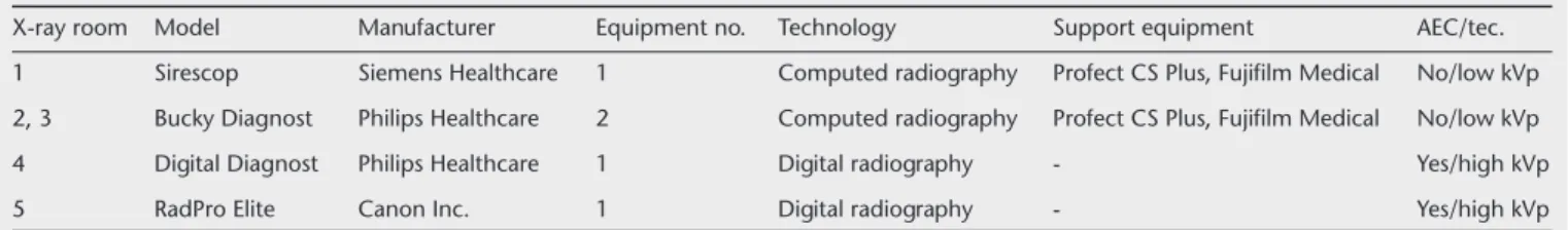
Measurements were made in five X-ray rooms at four local hospitals in the Ankara region; in one hospi-tal, doses were measured in two X-ray rooms. Patients were observed for each examination with each system, result-ing in dose data for 200 X-ray exam-inations: 100 from PA chest and 100 from AP KUB examinations. Three of the systems were CR systems, and the other two were DR systems (Table 1). The survey was conducted after local ethics comitte approval.
One X-ray room had a Sireskop sys-tem (Siemens Healthcare, Erlangen, Germany), two rooms had Bucky Di-agnost systems (Philips Healthcare, Eindhoven, The Netherlands) with FCR Profect CS Plus CR systems (Fuji-film Medical, Tokyo, Japan), one room had a Digital Diagnost system (Philips Healthcare), and one room had a Rad-PRO Elite system (Canon Inc., Lake Success, New York, USA). Although all of the systems have been subjected to established quality control protocols since the date of installation, all of the X-ray tubes and generators were tested before measuring patient doses
accord-ing to Institute of Physicists and Engi-neers in Medicine procedures (8). Each patient record was stored in a digital data file to facilitate the calculation of entrance surface doses (ESDs). A form, containing information on the patient (gender, age, weight) and technical ex-posure parameters used (applied tube voltage, tube current, and exposure time, and X-ray field size in the film plane) was filled out for each examina-tion.
Doses were measured without chang-ing the exposure parameters that the local technologists use and prefer in their daily practice. The preferences for exposure parameters and positioning techniques were also observed in every department.
Two different tools were used to measure the doses. First, the X-ray tube outputs were measured at a distance of one meters the X-ray tube voltages were increased from 50 to 150 kVp in 10 kVp steps using a Model 90X6-6 cal-ibrated 6 cc ion chamber, connected to a Radiation monitor controller model 9010 (Radcal Corporation, Monrovia, California, USA). All ESD values were calculated retrospectively from the tube output measurements (9).
In addition, a kerma area product (KAP) meter (M4DK, PTW, Freiburg, Germany) KAP ion chamber was used to calculate the tube outputs to cor-relate the previous measurement tech-nique and entrance surface air kerma (ESAK) estimations.
The KAP meter had two ion cham-bers: one transparent area ion chamber for the KAP and one at the center for air kerma measurements. The central ion chamber of the KAP meter was used for crosschecking of the ESAK calculations obtained from the tube output measurements (10). The KAP values were not included in the
pa-tient dose database. The ion chambers of the KAP meter were calibrated in situ against an ion chamber (Radcal Cor-poration, Monrovia, California, USA) before the measurements began using the procedure proposed by the manu-facturer and other researchers (11, 12). The ESD was calculated from the tube output measurements using the following formula:
where Y(d) is the X-ray tube output measured at distance d from the tube focus; a is the focus-to-bucky-surface distance; b is the patient thickness;
BSF is the backscatter factor; (μen/ρ)tiss and (μen/ρ)air are the mass energy
ab-sorption coefficients for tissue and air, respectively; P is the mAs value that was used for the patient exposure; and
T is the mass energy absorption
coef-ficient ratio and equaled 1.06 for the kVp range used in this study (13). Y(d) was measured for all possible kV set-tings and was also divided by the mAs value with which the measurement was made (9). To obtain the ESD from the air kerma, the BSF used for adult radiography was 1.35, as suggested in the European Guidelines (4). An expe-rienced radiologist reviewed all of the radiographs using the image criteria in the European Guidelines.
ESD values that were measured with two methods were compared via linear correlation analysis, and an interclass correlation analysis was also carried out.
Results
The mean patient weight was 74.1±8.62 kg (range, 55–115 kg), and the mean height was 1.75±0.05 m (range, 1.60–1.94 m). Most of the
pa-Table 1. Specifications of the radiography equipments included in this survey
X-ray room Model Manufacturer Equipment no. Technology Support equipment AEC/tec. 1 Sirescop Siemens Healthcare 1 Computed radiography Profect CS Plus, Fujifilm Medical No/low kVp 2, 3 Bucky Diagnost Philips Healthcare 2 Computed radiography Profect CS Plus, Fujifilm Medical No/low kVp 4 Digital Diagnost Philips Healthcare 1 Digital radiography - Yes/high kVp
5 RadPro Elite Canon Inc. 1 Digital radiography - Yes/high kVp
AEC, automatic exposure control; tec., exposure technique.
tients were young males, between 18 and 23 years old, and were close in height and weight to the standard human body size defined by ICRP (3). The mean patient body mass index was 24.44±3.3 kg/m2. ESAKs were
calculat-ed from tube output measurements and from the KAP meter central point ion chamber measurements in order to compare and cross-check all ESAK calculations. The correlation coeffi-cient was R2=0.90 for the PA chest and
R2=0.94 for the AP KUB ESAK
calcula-tions, as seen from Figs. 1 and 2, re-spectively. The high correlation coeffi-cients proved that the measured ESAK values were reliable in a methodolog-ical sense. In addition, an interclass correlation coefficient analysis was car-ried out for both dose datasets, giving correlation coefficients of 0.92 for the PA chest and 0.87 for the AP KUB dose measurements. These high interclass correlation coefficients indicate that
either of the dose datasets obtained from the different dose measurement methods can be used. Table 2 gives the ESAK values with their range for both examinations.
The maximum patient dose mea-sured for chest examinations was 45-fold greater than the minimum patient dose, while for the AP KUB ex-aminations this difference was 24-fold (Table 2). These big differences are due to the very low dose results obtained for one of the X-ray rooms. The room equipped with Philips Digital Diagnost (room 4) had a direct current X-ray generator and all exposures were much shorter than for the mean exposure times for the other four room. In ad-dition, this department used 150 kVp for all chest examinations. Due to the very short exposure times and high kVp used, all of the measured ESAK values from this room were very low. The ESAK measurements for the
oth-er four rooms woth-ere close to each othoth-er in both types of examination. Table 3 gives the exposure parameters for both examinations.
The differences in the kVp values used in the PA chest examinations were larger than the differences in the AP KUB examinations (Table 2). This was due to the technologists’ pref-erence for low kVp settings with the CR equipped X-ray systems, since CR plates have lower spectral sensitivity, while the technologists working with DR preferred high kVp settings for chest examinations. The parameter settings of the CR and DR equipment used for abdominal X-rays were closer to each other compared to the chest images. The European Guidelines sug-gest using a high kVp for PA chest ex-aminations, e.g., 125kV values (i.e., 120 and 150 kVp) were used in the two X-ray rooms (rooms 4 and 5) equipped with DR systems (4).
All radiographs from both the CR and DR systems were adequate and had good image quality for diagnosis according to image criteria recom-mended by the European Guidelines (4). Using a high kV with DR systems results in shorter exposure times, which lowers the mAs values and pa-tient dose.
In the clinics with CR systems, the technologists prefer manual settings with a low kV and high mAs. It has been observed that positioning the X-ray tube close to the patient also causes a higher patient dose.
Discussion
The differences in the kV values for the AP KUB examinations were less significant due to the low kVp used in both CR and DR systems. The AP KUB examination is a low kV application, al-though sometimes this technique pro-duces results with high mAs numbers in large patients. However, the maximum calculated ESD was still lower than in the DRL for this examination in the European Guidelines (Table 1). All of the radiographs obtained with both the CR and DR systems had good image quality and were adequate for diagnosis according to the European Guidelines criteria (4).
The chest examination is probably the most predominant use of a
low-Table 2. ESD values in comparison with European Union diagnostic reference levels for posteroanterior chest and anteroposterior kidney-ureter-bladder radiography examinations
Entrance surface dose value (mGy)
Diagnostic reference
Examination level Mean±SD Minimum Maximum
Posteroanterior chest 0.3 0.346±0.2 0.024 1.087 Anteroposterior kidney-ureter-bladder 10 1.948±1.1 0.264 6.288 SD, standard deviation.
Figure 1. The entrance surface doses (ESDs) calculated from tube output vs. the value calculated from reading for the central ion chamber of the kerma area product (KAP) meter for the postero anterior chest examinations. The correlation coefficient (R2) between the datasets was 0.9038.
ESDs calculated from KAP meter central ion chamber
0.00 0.20 0.40 0.60 0.80 1.00 1.20 1.40 1.20 1.00 0.80 0.60 0.40 0.20 0.00 y=1.0299x+0.0469 R2=0.9038 ESDs calcula
kVp technique with typical tube po-tentials of 64.5–87 kVp. This contrasts the European recommendation to use a high-kVp technique (i.e.,125 kVp). As mentioned, only the two rooms equipped with DR systems used a high kVp. The CR system users preferred low-kVp techniques for chest exam-inations based on service engineer rec-ommendations and the recommended target S values in the manufacturer’s manuals (i.e., 150–300). The Euro-pean Guidelines recommend using a high kVp with a 400-speed film-screen combinations using automatic expo-sure control (AEC). This is not the case with CR systems used clinically. In this study group, the CR system technolo-gists did not use AEC, but set the expo-sure parameters manually.
To implement the high-kVp chest technique in CR-equipped X-ray rooms,
the AEC systems must be recalibrated for CR plates. In Turkey, most AEC sys-tems are calibrated for 400-speed film-screen usage; when compared with film-screen combinations, CR plates refer to 200-speed film-screens. We are conducting an ongoing study to recal-ibrate AEC systems for CR plate usage in order to implement high-kVp usage for PA chest examinations. The kVp settings for the AP KUB examinations were in the range of kVp settings (75– 90 kV) recommended in the European Guidelines. All measured ESDs were be-low the DRL of 10 milligrays (mGy) in the European Guidelines.
A further survey of patient doses after establishing local DRLs will be a good tool for patient dose management and an example of the effective adjustment of patient doses. Even if patient age is not the main concern in
determin-ing DRLs, it is easier to collect data on patients of average weight and size by measuring young patients (5). Since most of the measured ESD values for the PA chest examinations exceeded the European Union DRL of 0.3 mGy and the International Atomic Energy Agency DRL of 0.4 mGy, further dose measurements and recalibration of the AEC systems are needed to optimize them and establish a national DRL for this examination. In our ongoing study to recalibrate AEC ion chambers for CR plates, the preliminary results show that the goal can be achieved in some systems. All of the measured ESD values were below the European Union DRL of 10 mGy for KUB AP examina-tions, and there does not seem to be a need to optimize this examination; however, extending the measured data will also help to achieve an accurate national DRL for this examination. A practical procedure for patient dose monitoring with respect to the DRLs is used only rarely in many hospitals in Turkey. The main reason for the values being two times higher than the Euro-pean reference level for chest radiogra-phy (PA) is likely the predominant use of a low-kVp technique with typical tube potentials of 60–80 kV and anti-scatter grid use in CR installations. The possible reasons for the large dose dis-crepancies seen are the use of non-opti-mized digital systems, different viewing preferences of clinicians, and different postprocessing parameters. Moreover, older and newer plates were used during the same time period for every room and both examination type.
This study had some limitations. First, measuring different technol-ogies, such as CR and DR, increases the differences between dose values, which introduces difficulty in compar-ing measured patient doses. Second, a group of five X-ray machines is smaller than needed to make a strong decision regarding DRLs, even for local settings. Third, instead of using an ion chamber for ESAK measurements, using thermo-luminesence dosimeters on the skin of the patients might produce more reli-able data, since it measures the energy delivered to the body directly. Despite these drawbacks, we have obtained the preliminary results to present to the lo-cal radiology community.
Table 3. Exposure factors and focal-skin distance values for posteroanterior chest and anteroposterior kidney-ureter-bladder radiography examinations
kVp mAs Focal-skin distance (cm) Examination Mean Range Mean Range Mean Range Posteroanterior chest 98.14 64.5–150 12.06 0.25–31.7 148.41 135.1–162.5 Anteroposterior 74.32 64.5–87.5 26.45 0.98–66.1 89.14 76.6–103.4 kidney-ureter-bladder
kV, kilovolt; mAs, milliamper per second.
Figure 2. The entrance surface doses (ESD) calculated from tube output vs. the value calculated from reading for the central ion chamber of the kerma area product (KAP) meter for the anteroposterior kidney-ureter-bladder radiography examinations. The correlation coefficient (R2) between the data sets was 0.9479.
ESDs calculated from KAP meter central ion chamber
0.00 1.00 2.00 3.00 4.00 5.00 6.00 7.00 9.00 8.00 7.00 6.00 5.00 4.00 3.00 2.00 1.00 0.00 y=1.4021x-0.3457 R2=0.9479 ESDs calcula
In conclusion, as patient dose values for general radiography can increase during the transition from conven-tional screen-film radiography to CR, dose management programs for digi-tal techniques, specific training of ra-diographers, and frequent patient dose audits can improve practice, while maintaining or reducing patient doses (14–16). In this study, the patient dos-es were generally acceptable, except for chest X-rays at two CR installations, compared to the published dose levels. Problematic areas that need further in-vestigation and improvement included improper adjustment of the exposure parameters, uncalibrated AEC systems, and hesitation of the technologists to use AEC systems correctly, especially for CR systems.
Acknowledgements
Authors would like to thank Assoc. Prof. Turan Olgar and Prof. Dr. Do¤an Bor from the Department of Physics Engineering, Ankara University Faculty of Engineering for their in-valuable assistance in collecting the data and also critics about the manuscript. We are also very grateful to engineer Ekrem Malkoç, PhD, for his kind efforts to review the manuscript for language corrections.
Conflict of interest disclosure
The authors declared no conflicts of interest.
References
1. Aroua A, Bize R, Buchiler-Decka I, Vad-er JP, Valley JF, SchnydVad-er F. X-ray imag-ing of the chest in Switzerland in 1998: a nationwide survey. Eur Radiol 2003; 13:1250–1259.
2. The Institute of Physics and Engineering in Medicine (IPEM). Guidance on the establishment and use of diagnostic ref-erence levels for medical x-ray examina-tions. IPEM report no. 88. IPEM, 2004. 3. International Commission on Radiation
Protection (ICRP). 1990 Recommenda-tions of the international commission on radiological protection. Ann. ICRP Publi-cation 60. ICRP 1991; 21:1–3.
4. European guidelines on quality criteria for diagnostic radiographic images. European commission report EUR 16260EN. Avali-able at: ftp://ftp.cordis.lu/pub/fp5-eura-tom/docs/eur16260.pdf. Accessed April 9, 2013.
5. Guidance on diagnostic reference levels (DRLs) for medical exposure European commission radiation protection report 109. Avaliable at: http://ec.europa.eu/en-ergy/nuclear/radiation_protection/doc/ publication/109_en.pdf. Accessed April 9, 2013.
6. Johnston DA, Brennan PC. Reference dose levels for patients undergoing common diagnostic x-ray examinations in Irish hospitals. Br J Radiol 2000; 73:396–402. 7. Torres CR, Espana Lopez ML, Ruiz
Man-zano P, et al. A comparative study of sev-eral digital flat panel x-ray units: patients doses and image quality in chest radiog-raphy. Paper presented at: 11th Interna-tional Congress of the InternaInterna-tional Radi-ation Protection AssociRadi-ation; May 23–28, 2004; Madrid, Spain.
8. The Institute of Physics and Engineering in Medicine (IPEM).Recommended stan-dards for the routine performance test-ing of diagnostic x-ray imagtest-ing systems. IPEM report no. 91. IPEM, 2005.
9. Faulkner K, Broadhead DA, Harrison RM. Patient dosimetry measurements meth-ods. Appl Radiat Isot 1999; 50:113–123.
10. McParland BJ. Entrance skin dose esti-mates derived from dose-area product measurements in interventional radio-logical procedures. Brit J Radiol 1998; 71:1288–1295.
11. Griftner H, Stieve FE, Wild J. A new dia-mentor for measuring kerma-area product and air-kerma simultaneously. Med Phys 1997; 24:1954–1959.
12. Bor D, Sancak T, Olgar T. Comparison of effective doses obtained from dose-ar-ea product and air kerma mdose-ar-easurements in interventional radiology. Br J Radiol 2004; 77:315–322.
13. International Atomic Energy Agency standards. International basic safety standards for protection against ionizing radiation and for the safety of radiation sources. Avaliable at: http://www.ilo.org/ wcmsp5/groups/public/@ed_protect/@ protrav/@safework/documents/publica-tion/wcms_152685.pdf. Accessed April 9, 2013.
14. Aldrich JE, Duran E, Dunlop P, Mayo JR. Optimization of dose and image quality for computed radiography and digital ra-diography. J Digit Imaging 2006; 19:126– 131.
15. Vano E, Fernandez Soto JM. Patient dose management in digital radiography. Biomed Imaging Interv J 2007; 3:1–5. 16. Vano E, Fernandez JM, Ten JI, et al.
Tran-sition from screen-film to digital radiog-raphy: evolution of patient radiation dos-es at projection radiography. Radiology 2007; 243:461–466.