INVESTIGATION OF THE EFFECTS OF BIOACTIVE PEPTIDE NANOFIBERS ON ACUTE MUSCLE INJURY REGENERATION
A THESIS SUBMITTED TO
MATERIALS SCIENCE AND NANOTECHNOLOGY PROGRAM OF THE GRADUATE SCHOOL OF ENGINEERING AND SCIENCE
OF BILKENT UNIVERSITY
IN PARTIAL FULFILLMENT OF THE REQUIREMENTS FOR THE DEGREE OF
MASTER OF SCIENCE
By
ÇAĞLA EREN ÇİMENCİ
II
INVESTIGATION OF THE EFFECTS OF BIOACTIVE PEPTIDE NANOFIBERS ON ACUTE MUSCLE INJURY REGENERATION
By Çağla Eren Çimenci, October, 2016
We certify that we have read this thesis and that in our opinion it is fully adequate, in scope and in quality, as a thesis for the degree of Master of Science.
Ayşe Begüm Tekinay (Advisor)
Aykutlu Dana
Reyhan Neslihan Gürsoy
Approved for the Graduate School of Engineering and Science:
Ezhan Karaşan
III
ABSTRACT
INVESTIGATION OF THE EFFECTS OF BIOACTIVE PEPTIDE NANOFIBERS ON ACUTE MUSCLE INJURY REGENERATION
Çağla Eren Çimenci
MSc in Materials Science and Nanotechnology Supervisor: Assist. Prof. Dr. Ayşe Begüm Tekinay, PhD
October, 2016
Skeletal muscle constitutes a large part of the human body. It is a hierarchically organized heterogeneous tissue and is composed of muscle fiber bundles, muscle fibers, myofibrils and myofilaments. Since muscle cells are terminally differentiated, they have limited capacity to renew themselves. Only new cells can fuse with muscle fibers and increase the size and volume of skeletal muscle. Myosatellite cells or satellite cells are small, mononuclear progenitor cells with virtually no cytoplasm. They are located in between the sarcolemma and basement membrane of terminally-differentiated muscle fibers. Satellite cells are precursors to skeletal muscle cells, and are able to give rise to satellite cells or differentiated skeletal muscle cells. They are normally found in silent state in adult muscle, but act as a reserve cell population that is able to proliferate in response to injury and give rise to regenerated muscle and to more satellite cells. Formation of the new muscular tissue is called myogenesis. During this event, satellite cells differentiate into myoblasts, and then myoblasts fuse with each other in order to form myofibers. There are many genes that regulate the myogenesis process and each of them is
IV
activated in a different step of myogenesis. Increased or decreased levels of gene expression determine the differentiation capacity.
Peptide nanofibers are supramolecular structures formed via self-assembly and they are promising molecules in regenerative medicine and tissue engineering. Peptide-based molecules for tissue regeneration is a widely studied area and currently used in the treatment-investigation of many different tissues such as bone, cartilage, skin and nerve.
Since laminin is one of the most abundant proteins found in the basal membrane of the skeletal muscle; in this thesis, we designed and synthesized a laminin-mimetic bioactive (LM/E-PA) molecule functionalized with bioactive groups for mimicking laminin activities and capable of accelerating satellite cell activation. Our research group had previously shown that LM/E-PA containing nanofibers can support muscle differentiation in vitro. In this thesis, the clinical relevance was demonstrated further by assessing laminin-mimetic bioactive scaffold in acute muscle injury in an in vivo rat model. Our findings revealed that this scaffold system significantly promotes satellite cell activation in skeletal muscle and accelerates regeneration following acute muscle injury. In addition, our findings show that the regeneration capacity of the skeletal muscle was increased and consequently regeneration time was reduced. This study is one of the first examples of molecular level and tissue level regeneration of skeletal muscle by using bioactive peptide nanofibers following acute muscle injury, and shows that laminin mimetic nanofiber system is a promising material for development of new therapeutic curatives for acute skeletal muscle injuries.
Keywords: Skeletal muscle, acute muscle injury, peptide nanofibers, functional self-assembly, biomaterials, in vivo, rat model, laminin
V
ÖZET
BİYOAKTİF PEPTİT NANOFİBERLERİN AKUT KAS HASARI ÜZERİNDEKİ
ETKİLERİNİN İNCELENMESİ
Çağla Eren Çimenci
Malzeme Bilimi ve Nanoteknoloji, Yüksek Lisans Tez Danışmanı: Asist. Prof. Dr. Ayşe Begüm Tekinay
Ekim, 2016
İskelet kası insan vücudunun büyük bir kısmını kaplamaktadır. Hiyerarşik bir şekilde organize olmuş yapılardan meydana gelen heterojen bir dokudur. Bu hiyerarşik yapı, kas demetleri, kas fiberleri, miyofibriller ve miyofilamentlerden meydana gelmektedir. Kas hücreleri farklılaşmalarını tamamladıklarından dolayı kendilerini yenileme kapasiteleri oldukça kısıtlıdır. Kas yenilenmesi yalnızca yeni hücrelerin kas fiberleri ile birleşerek iskelet kasının boyutunu ve hacmini artırması ile gerçekleşmektedir. Kas uydu (satellit) hücreleri küçük ve tek çekirdek içeren kas öncül hücreleridir ve neredeyse hiç sitoplazmaları yoktur. Sarkolemma ve kas fiberlerinin bazal membranı arasında yer alırlar. Satellit hücreler iskelet kası öncül hücreleridir ve gerektiğinde kasa farklılaşabildikleri gibi satellit hücre havuzunu korumak için asimetrik bölünme ile satellit hücre olarak da kalabilirler. Normalde sessiz durumda beklerler fakat bir hasar durumu söz konusu olduğunda aktifleşip farklılaşmaya başlarlar. Yeni kas dokusu oluşturulması işlemine miyogenez adı verilir. Bu olay sırasında, satellit hücreler miyoblastlara farklılaşır ve miyoblastlar biraraya gelerek miyofiberleri meydana getirir. Miyogenezi
VI
düzenleyen birçok gen vardır ve bu genlerden her biri miyogenezin farklı bir aşamasında aktifleşmektedir. Genlerin artması ve azalması ile hücrelerin farklılaşma kapasiteleri belirlenmektedir.
Peptit nanofiberler kendiliğinden biraraya gelerek oluşan çok moleküllü yapılardır ve rejeneratif tıp/doku mühendisliği alanında gelecek vaat eden moleküllerdir. Doku mühendisliğinde peptit tabanlı moleküllerin kullanılması geniş bir çalışma alanıdır ve kemik, kıkırdak, cilt ve sinir gibi birçok dokunun tedavi araştırmalarında kullanılmaktadır.
İskelet kasında en fazla bulunan moleküllerden biri olması sebebi ile bu tez çalışmasında laminini taklit eden peptit molekülleri (LM/E-PA) tasarlanıp sentezlenmiştir. Biyoaktif grubu laminin molekülünü taklit edecek şekilde işlevsel hale getirilmiştir ve bu grup satellit hücre farklılaşmasını hızlandırmaktadır. Daha önce grubumuzda yapılmış olan bir çalışma da LM/E-PA molekülünün hücresel düzeyde kas farklılaşmasına destek olduğu gösterilmiştir. Bu tez çalışmasında ise araştırmalar bir adım ileriye taşınarak malzemenin klinik önemi in vivo sıçan modeli üzerinde araştırılmıştır. Bulgularımız iskelet kasının yenilenme kapasitesinin arttığını ve buna bağlı olarak yenilenme süresinin kısaldığını göstermektedir. Bu çalışma, biyoaktif peptit nanofiberlerin akut kas hasarı üzerindeki doku ve molekül seviyesindeki iyileşmeye etkilerinin araştırıldığı ilk çalışmalardan biridir. Özet olarak, laminini taklit eden biyoaktif iskele sistemleri kas hasarına karşı yeni tedavi yöntemlerinin geliştirilmesinde gelecek vaat eden malzemelerdir.
Anahtar kelimeler: İskelet kası, akut kas hasarı, peptit nanofiberler, işlevsel supramoleküler nanoyapılar, biyomalzemeler, in vivo, sıçan modeli, laminin
VII
ACKNOWLEDGEMENTS
It would not have been possible to write this thesis without to help and support of the kind people around me. First, I would like to express my sincere appreciation and gratitude to my advisors, Prof. Ayşe Begüm Tekinay and Prof. Mustafa Özgür Güler for their guidance and support to my research. I'm grateful for their patience, motivation and immense knowledge helped me develop a trans-disciplinary research skills, which I can carry with me throughout my research career.
I would like to acknowledge the graduate scholarship from TÜBİTAK (The Scientific and Research Council of Turkey) BIDEB 2210-C for financial support.
I also would like to thank Gözde UZUNALLI for her helpful suggestions and comments during my first year study, Elif ARSLAN for her patience, encouragement and precious answers for my endless questions. I would like to express my special thanks to Özge UYSAL for her diligence and assistance while I was working in the lab.
I also would like to thank other “cartilage subgroup” members; Özüm Şehnaz ÇALIŞKAN, Nurcan HAŞTAR and İbrahim ÇELİK for creating such a warm working environment with their team spirit and friendship.
Special thanks to Melike SEVER (one of the reason why I’m here) for teaching me the survival methods in graduate life and her excellent friendship. If I had a chance, I would like to carry Seren HAMSİCİ everywhere with me, I appreciated for her true friendship.
Thanks to the current and former members of Nanobiotechnology and Biomimetic Materials laboratory group at the UNAM. I would like to wish the best for their graduate journey to Zeynep ORHAN, Gökhan GÜNAY, İdil UYAN, Canelif YILMAZ, Gülistan TANSIK, Mustafa
VIII
BETER, Merve ŞEN, Fatih YERGÖZ, Nuray GÜNDÜZ, Ahmet Emin TOPAL, Oğuz TUNCAY and Burak DEMİRCAN who became my family and they have made my graduate life unforgettable.
I would like to acknowledge Dr. Ebru UMAY and Dr. Ece ÜNLÜ who believe in my project and helped me for electromyographical analysis in animal studies. I also would like to thank to Alper Devrim ÖZKAN for his fruitful comments and proofread.
Moreover, I would like to express my most sincere thanks to my roommate Behide SALTEPE for making my home life spirited and peaceful. She was always with me when I wanted to talk and we shared so many things that two people can share. A very special thank you to my friends Recep Erden AHAN and Onur APAYDIN for the cheer and support you have lent me over all my graduate life. I had chance to know better Tuğçe ÖNÜR in Ankara, she was always ready for every kind of outdoor activities with me and deserves all compliments with her friendship.
Moreover, I would like to thank to my beloved sister Başak EREN who is not just a sister but also my best friend. She is the best architect I’ve ever known, helped me a lot with her advanced photoshop skills. My little brother Can EREN who was always cheering me up with his sense of humor. Also, my father Cahit EREN and my mother Şükran EREN who always encouraged me and helped me became who I am. I am grateful for their patience and sacrifice that will remain my inspiration throughout my life. I am also very much grateful to all my family members for their constant inspiration, unconditional confidence and love. Their positive outlook on life has given me the strength to do it all.
IX
Finally, I want to thank to my husband Kadir ÇİMENCİ who makes me feel motivated, who was always supporting and encouraging me when I encounter difficulties. His continued and unfailing love helped me to keep dreaming and deserves all compliments.
Last, but not least, I send big thanks to all my friends from İstanbul, who always support me, give me good advices and make my life colorful!
I love you with all my heart.
X
CONTENTS
LIST OF FIGURES ... XIV
ABBREVIATIONS ... XVII
1. Objective ... 1
2. Introduction: Skeletal Muscle Tissue Engineering ... 3
2.1. Muscle Tissue ... 3
2.2. Problems Related with Muscle Tissue... 4
2.3. Muscle Tissue Repair ... 6
2.4. Skeletal Muscle Proteins ... 13
2.4.1. Structural and Functional Proteins ... 14
2.4.1.1. Actin ... 14 2.4.1.2. Myosin ... 14 2.4.1.3. Troponin ... 15 2.4.1.4. Tropomyosin ... 15 2.4.1.5. Titin ... 15 2.4.2. ECM Proteins ... 15
XI
2.4.2.1. Collagen ... 16
2.4.2.2. Laminin ... 17
2.5. Artificial Muscle ... 17
2.6. Other Treatment Methods ... 18
2.7. Clinical Significance of Biomaterials ... 18
2.8. Peptide Nanofiber Gels for Muscle Regeneration ... 19
3. Experimental Section ... 21
3.1. Materials ... 21
3.1.1. Animal Model ... 21
3.2. Methods ... 22
3.2.1 Peptide Amphiphile Synthesis, Purification and Characterization ... 22
3.2.2. Self-Assembled Nanofiber Network Formation ... 23
3.2.3. Structural and Mechanical Characteristics of Peptide Nanofibers ... 24
3.2.4. Acute Muscle Injury Creation via Toxin Injection ... 25
3.2.5. Treatment with ECM mimetic PA Network ... 25
3.2.6. Behavioral Analysis ... 26
XII
3.2.6.2 Walking Track Step Test Analysis ... 28
3.2.7. Histological Analysis ... 28
3.2.7.1. Morphological Analysis (H&E) ... 28
3.2.7.2. Muscle Cross Section Area Calculation ... 29
3.2.7.3. Centrally Located Nuclei Calculation ... 29
3.2.8. Gene Expression Analysis by RT-qPCR ... 30
3.2.8.1. RNA Isolation ... 30
3.2.8.2. RNA Quantification ... 31
3.2.8.3. Quantitative reverse transcription PCR (RT-qPCR) ... 31
3.2.9. Protein Detection by Western Blotting ... 33
3.2.9.1. BCA Assay ... 34
3.2.9.2. SDS PAGE ... 35
3.2.9.3. Protein Transfer onto a PVDF Membrane... 37
3.2.9.4. Coomassie Staining ... 39
3.2.9.5. Antibody Incubation ... 40
3.2.9.6. Visualizing Results ... 43
XIII
3.2.9.8. Quantification and Statistical Analysis ... 44
4. Results and Discussion ... 45
4.1. Synthesis of PA Molecules and Characterization of Their Self-assembly into Nanofibers ... 45
4.2. Structural and Mechanical Characteristics of Peptide Nanofibers ... 47
4.3. Animal Behavior Analyses ... 51
4.3.1. Step Test Results ... 51
4.3.2. EMG Results ... 53
4.4. Histological Analyses ... 56
4.5. Gene Expression Analyses ... 62
4.6. Protein Expression Analyses ... 71
5. Conclusion ... 77
6. Future Perspectives ... 79
LIST OF FIGURES
Figure 1 Schematic illustration of the hierarchical organization of skeletal muscle ... 3
Figure 2 Localization of satellite cells (muscle progenitors) into the skeletal muscle fibers ... 6
Figure 3 Schematic representation of the steps involved in skeletal muscle regeneration ... 9
Figure 4 Schematic representation of muscle myogenesis and myogenesis markers ... 11
Figure 5 Schematic illustration of the asymmetric division of satellite cells. ... Error! Bookmark not defined. Figure 6 Schematic illustration of the interaction between laminin and type IV collagen in the ECM of the skeletal muscle ... 16
Figure 7 Schematic Illustration of Electromyography Setup ... 27
Figure 8 Gel and membrane setup for semi-dry electrophoretic transfer system. ... 39
Figure 9 Chemical structures of peptide amphiphile molecules ... 46
Figure 10 Structural characterizations of PAs by using Scanning Electron Microscopy and Transmission Electron Microscopy ... 47
Figure 11 Liquid chromatograms and mass spectra of PAs ... 48
Figure 12 Structural characterization of PAs by using circular dichroism ... 49
Figure 13 Mechanical characterization of PAs by using oscillatory rheology ... 50
Figure 14 Schematic illustration of a step test cabin and experimental setup ... 51
Figure 15 Quantitative step length analyses of both PA and negative control ... 52
XV
Figure 17 Representative light microscopy images of skeletal muscle tissue sections after
Hematoxylin & Eosin (H&E) staining ... 58
Figure 18 Quantitative analysis of average skeletal muscle cross section area calculation ... 59
Figure 19 Histology images of centrally located nuclei and quantitative analysis of percentage of myofibers with central nuclei ... 61
Figure 20 Pax7 gene expression analysis ... 63
Figure 21 MyoD1 gene expression analysis ... 65
Figure 22 Myf5 gene expression analysis ... 67
Figure 23 Myogenin gene expression analysis ... 69
Figure 24 Western blots showing the expression and quantification of Pax7 ... 72
Figure 25 Western blots showing the expression and quantification of MyoD1 ... 73
Figure 26 Western blots showing the expression and quantification of Myf5 ... 74
LIST OF TABLES
Table 1 Skeletal Muscle Specific Primer Sequences for RT-qPCR... 32
Table 2 Content of RIPA Lysis Buffer... 33
Table 3 Content of Stacking and Separating Acrylamide Gel ... 35
Table 4 Content of 5X Running Buffer for SDS-PAGE (1L) ... 37
Table 5 Content of 10X Towbin Buffer (Anode and Cathode Buffers are prepared from this buffer) ... 38
Table 6 Content of Anode Buffer ... 38
Table 7 Content of Cathode Buffer ... 38
Table 8 Coomassie Brilliant Blue Staining Solution Content ... 40
Table 9 Coomassie Destaining Solution Content ... 40
Table 10 Content of 1X TBS Buffer for Electrophoresis... 41
Table 11 Content of 1X TBS-T Buffer for Western Blot Applications ... 41
Table 12 Working Concentrations and Catalog Numbers of Primary Antibodies ... 42
Table 13 Working Concentrations and Catalog Numbers of Secondary Antibodies ... 42
XVII
ABBREVIATIONS
BCA : Bicinchoninic acid
BSA : Bovine serum albumin
β-gal : β-galactosidase CD : Circular dichroism
CD34 : Hematopoietic progenitor cell antigen CD34
Col-I : Collagen type I
Col-III : Collagen type III
DCM : Dichloromethane
DMF : N, N-Dimethylformamide
DMEM : Dulbecco’s modified Eagle’s medium
DMSO : Dimethylsulfoxide
ECL : Enhanced chemiluminescence
ECM : Extracellular matrix
EDTA : Ethylenediaminetetraacetic acid
XVIII EMG : Electromyography
FBS : Fetal bovine serum
FDA : U.S. Food and Drug Administration
bFGF : Basic fibroblast growth factor
GAG : Glycosaminoglycan
GAPDH : Glyceraldehyde 3-phosphate dehydrogenase
GF : Growth factor
H&E : Hematoxyline & eosin staining
HGF : Hepatocyte growth factor
HUVEC : Human umbilical vein endothelial cell
HPLC : High performance liquid chromatography
HRP : Horseradish peroxidase
IGF : Insulin like growth factor
IgG : Immunoglobulin G
kDa : Kilo dalton
LC-MS : Liquid chromatography-mass spectrometry
XIX
MLC3F-tg : Myosin light chain 3F transgenes
MRFs : Myogenic regulatory factors
Myf5 : Myogenic factor 5
MyoD1 : Myogenic differentiation 1
NSAIDs : Non-steroidal anti-inflammatory drugs
IHC : Immunohistochemistry
PA : Peptide amphiphiles
Pax7 : Paired box 7
PBS : Phosphate-buffered saline
PCR : Polymerase chain reaction
Q-TOF : Quadrupole time of flight
RICE : Rest, ice, compression, elevation
RIPA Buffer : Radioimmunoprecipitation assay buffer
RT-qPCR : Reverse transcriptase quantitative polymerase chain reaction
SC : Satellite cell
SDS : Sodium dodecyl sulfate
XX SEM (M) : Scanning electron microscope
SEM (S) : Standard error of the mean
STEM : Scanning transmission electron microscope
TA : Tibialis anterior
TBS : Tris-buffered saline
TBS-T : Tris-buffered saline-Tween 20
TIS : Triisopropylsilane
TCP : Tissue culture plate
TEM : Transmission electron microscope
TEMED : Tetramethylethylenediamine
TFA : Trifluoroacetic acid
TGF-β1 : Transforming growth factor beta 1
VEGF : Vascular endothelial growth factor
1
Investigation of the Effects of Bioactive Peptide Nanofibers on Acute Muscle Injury Regeneration
This work is partially described in the following publication:
Cagla Eren Cimenci *, †, Gozde Uzunalli *, †, Ozge Uysal *, †, Fatih Yergoz *, †, Ebru Karaca Umay ‡, Mustafa O. Guler *, †, §, Ayse B. Tekinay *, †, §
(Manuscript under Submission)
1. Objective
While the causes of muscle injury are numerous and diverse, acute injuries caused by excess pressure, shocks, falls and crushing damage are among the most widespread. Sports, heavy lifting and other strength-intensive tasks are both ubiquitous in modern life and likely to cause acute skeletal muscle injury. Even though skeletal muscle exhibits the capacity for self-renewal, this regeneration may take months. As such, speeding up the regeneration of skeletal muscle injuries would not only shorten the duration of recovery for the patient, but also support the general health and functionality of the repaired muscle tissue. In this work, we designed and synthesized a laminin-mimetic PA molecule that is functionalized with bioactive groups for mimicking laminin activities. We then created an acute tibialis anterior muscle injury in a rat model and injected the PA hydrogel to the injury site. Peptide amphiphile hydrogels are ideal scaffold systems for regenerative medicine studies due to their ease of injection. Following the local administration of PA scaffolds, we evaluated the regeneration and differentiation capacity of skeletal muscle through behavioral, histological/physiological
2
and molecular analyses. Our results suggest that the laminin-mimetic biomaterial system assists skeletal muscle regeneration and that peptide nanofibers are highly promising for the regeneration of acute muscle injuries. This work demonstrates for the first time that laminin-mimetic self-assembled peptide nanofiber networks facilitate myogenic differentiation in vivo without the need for additional treatment.
3
2. Introduction: Skeletal Muscle Tissue Engineering
2.1. Muscle Tissue
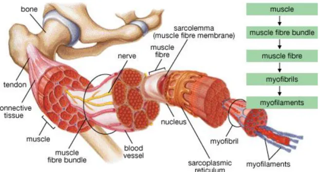
Skeletal muscle constitutes a great proportion of the vertebrate body. It provides postural support and facilitates the movements of the body through its contractions. Skeletal muscle is the most common muscle type among the three varieties of muscle. It has a hierarchical fiber organization (Figure 1) that encompasses multinucleated and parallel-aligned muscle fibers in each muscle.
Figure 1 Schematic illustration of the hierarchical organization of skeletal muscle. Adapted from Helene Colbourne et al, Inquiry into Biology (Toronto: McGraw-Hill Ryerson, 2007), p. 335.
4
In contrast to smooth muscle and cardiac muscle, skeletal muscle provides and controls intentional body movements. It is similar in shape to the myocardium; however, skeletal muscle has multinucleated, thin and long fibers. Nerves and blood vessels assist the skeletal muscle and provide communication between individual fibers, while connective tissue facilitates the attachment of these fibers to each other1. Muscle fibers are affected by several types of physical activity and status; including exercise, physical injury, disease and aging. Skeletal muscle has remarkably high adaptive capacity and readily alters its size and shape in response to external stimuli2. Correct measurement of muscle mass is an important parameter because it is strongly and directly related with the skeletal muscle strength3.
2.2. Problems Related with Muscle Tissue
Skeletal muscle tissue is susceptible to damage following exposure to excess pressure, shocks, drops, crushing damage, exercise, and sports injuries. Consequently; sports, heavy lifting and high performance-tasks are likely to cause acute skeletal muscle injury. Acute muscle injuries arise suddenly and cause severe and painful strains. Skeletal muscle damage is among the most frequent sports- and exercise-related injuries4. Its treatment is very limited and mostly ameliorative, often focusing on the mitigation of local pain and swelling. Long-term muscle problems may occur in 10% to 55% of all muscle injuries5.
Besides being adapted to environmental changes, skeletal muscle presents a spectacular regeneration capacity in response to small injuries6. In such cases, restoration of the slightly damaged muscle is possible without any therapeutic intervention7. However; if
5
damage is severe and left untreated, it may lead to loss of muscle mass and function. In these cases, skeletal muscle regeneration cannot be completely facilitated by the help of medical treatment or surgical reconstruction8. Accordingly, improving effective treatment methods against major skeletal muscle injuries is a growing area of interest9. Novel methods have been developed to support the recovery of heavy muscle injuries, often with spectacular results.
Skeletal muscle injury diagnosis is primarily performed by checking the history of the injury and using this as a guideline to determine the requisite set of clinical investigations. Non-invasive imaging is another helpful technique for the detection of muscle mass loss, and allows one to make accurate predictions about the prognosis of the injury10. The optimal therapeutic method can be determined for each individual case through the use of these approaches.
Muscular abnormalities can also arise as a result of problems with the nervous system, genetic mutations, malnutrition, and abuse or straining of muscles. Muscular dystrophy is caused by abnormal neurological signals and presents through the degeneration or abnormal development of muscular fibers11. Muscular hypertrophy involves the aberrant increase of muscle mass and may result from repeated, minor skeletal muscle damage, often as a consequence of physically demanding work. In this case, muscle cells grow continuously to compensate for the muscle damage, creating an increase in fiber diameters and overall muscle mass12. Muscular atrophy describes misused or unused muscles that shrink because of the lack of neurological connections, resulting in a degenerate morphology and decreased muscle mass13. Muscle injuries related to the
6
indirect causes. However, our main focus in this project is acute muscle injuries (which are caused by traumas) and their treatments. These type of injuries occur by the effect of direct causes such as crushing pressure, excess work, shocks, fall injuries etc.
2.3. Muscle Tissue Repair
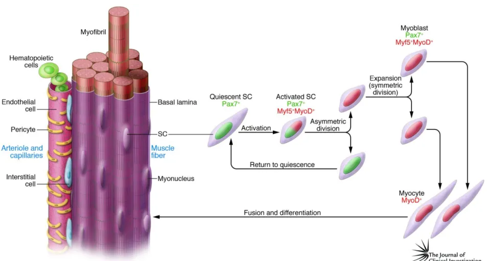
Satellite cells are considered to be skeletal muscle progenitor cells and are found in between the basal lamina and the plasma membrane (Figure 2). They promote the growth, repair and regeneration of skeletal muscle fibers14. They are unipotent cells and
have self-renewal capacity.
Figure 2 Localization of satellite cells (muscle progenitors) into the skeletal muscle fibers16. In normal myofibers, quiescent satellite cells are found in between the plasma membrane and the basal lamina. However in regenerating myofibers, activated satellite cells merge with the fiber and are localized at the center of the muscle fibers.
7
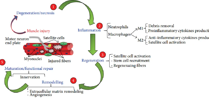
Through these characteristics, satellite cells maintain their existence within the muscle while protecting the self-repair ability of muscle after damage. When skeletal muscle is damaged, satellite cell activation takes place and muscle regeneration occurs through four main stages; degeneration, inflammation, regeneration and remodeling15. Following injury, activated satellite cells initially merge with muscle fibers and are localized at their centers. As such, centrally located nuclei are often observed within newly regenerated fibers and this situation is a strong indicator of active muscle regeneration.
Myogenesis is the process of muscle development. New muscle fibers are formed through the fusion of satellite cells with the existing fibers; which are multinucleated, post-mitotic and highly differentiated17. Sequentially ordered events constitute the skeletal muscle regeneration process (Figure 3). The first step of myogenesis is the degeneration step, where the integrity of skeletal muscle is disrupted because of trauma. Basal lamina and sarcolemma are demolished and the fibers are exposed to extracellular calcium, which results in their rupture and destruction18.
Degeneration is followed by the inflammation step, which is an early response to degeneration and provides a connection between the site of injury and the immune system. Activated macrophages and neutrophils are then recruited and tissue debris is removed.
Pro-inflammatory and anti-inflammatory cytokines are produced by these cells, resulting in the activation of satellite cells and leading to the regeneration phase19. In muscle tissue, regeneration begins at the first week (approximately 4th and 5th day) after injury20. After satellite cells are completely activated, stem cells are recruited and muscle fibers
8
start to regenerate. Myoblasts also proliferate and differentiate at this step, which culminates in their fusion into myofibers and the formation of mature myofibers21.
Growth factors like IGF-1, bFGF, EGF, HGF and TGF-β1 are known to play vital roles in the regeneration step22.
Overlapping with regeneration is the remodeling phase, which is the fourth step of skeletal muscle regeneration and entails the remodeling of the extracellular matrix, which is very crucial for muscle integrity. This phase starts at day 7-10 and lasts up to 14-28 days, depending on the injury. VEGF is also secreted at this phase and triggers local angiogenesis in the regenerating skeletal muscle. This leads to the formation of an extensive capillary network and allows the effective delivery of O2, nutrients and growth
factors to newly formed muscle fibers23.
The last and the longest phase of the skeletal muscle regeneration is maturation. Skeletal muscle gains its functional ability in this phase, but this phase can last up to 3 or 6 months depending on the injury24. Neurological connections are established completely and intramuscular nerve branches reach to the muscle fibers in the maturation step.
9
Figure 3 Schematic representation of the steps involved in skeletal muscle regeneration: Degeneration, Inflammation, regeneration, remodelling and maturation (Musarò et al., 2014).
10
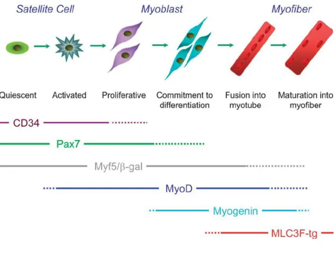
Each regeneration step of the skeletal muscle actually overlaps with both the previous step and the following step (Figure 4). Such fusion is triggered by the temporally ordered changing of gene expression profiles of the myoblast cells, which allows the formation, maturation and differentiation of myotubes25.
Pax7 is a transcription factor and expressed from quiescent satellite cells. Activation of satellite cells leads to Pax7 expression, which in turn mediates the maintenance of satellite cells. As seen in Figure 5, satellite cells undergo asymmetric division. One cell differentiates into a myocyte, while the other remains as a satellite cell in order to conserve the satellite cell pool26.
11
Figure 4 Schematic representation of muscle myogenesis and myogenesis markers. Specific markers for each step are illustrated in the order of their expression.
12
13
Myf5 is another transcription factor that is responsible for the activation of satellite cells during myogenesis. Myf5 is one of the earliest markers for myogenesis and it is a member of the myogenic regulatory factor family (MRFs). About 10% of Pax7+ satellite cells do not express Myf5; these cells are attached to the basal lamina at all times (even during SC proliferation) and considered to be stem cells for muscle tissue. Cells that express both Myf5 and Pax7, in contrast, are considered to be muscle progenitor cells and are more prone to differentiation27.
MyoD1 is also a member of the myogenic regulatory factor family (MRFs). MyoD is activated as a result of exercise or damage, and triggers satellite cell activation. It is known that the combination of MyoD and Myf5 is extremely necessary for skeletal muscle myogenesis28. Another study shows that satellite cells have different fates according to the expression balance between Pax7 and MyoD. Pax7+ / MyoD– cells return to quiescent satellite state while Pax7+ / MyoD+ cells are committed to myogenic differentiation29.
Myogenin is a transcription factor and also known as myogenic factor 4. It is one of the latest markers of skeletal muscle regeneration and essential for functional muscle formation. Myogenin controls the formation of myocytes from myoblasts and regulates terminal differentiation; consequently, this marker is upregulated in myogenesis30.
2.4. Skeletal Muscle Proteins
Skeletal muscle is characterized by well-defined bands that are composed of thick and thin filaments. Such filaments constitute a series of arranged structures called sarcomeres. Sarcomeres are protein-based architectures and those proteins are divided
14
into two main subgroups; structural & functional and extracellular proteins. Accordingly, some sarcomeric proteins are intracellular, while others are secreted to the extracellular matrix to perform their functions.
2.4.1. Structural and Functional Proteins
They are mainly responsible for controlling the shape and function of the skeletal muscle. Muscle fibers are the smallest functional units of muscle, and their main function is to contract. Actin, myosin, troponin, tropomyosin and titin are the main proteins that are vital for muscular shape and function31.
2.4.1.1. Actin
Actin is a major element of the thin filaments, which work in conjunction with myosin and provide contractile movements through the “sliding” of each protein over its neighboring component. This facilitates the contraction or shrinking of the actin filaments, which is translated to muscle movement32.
2.4.1.2. Myosin
Myosin is another structural protein and, together with the actin, is the most abundant protein found in the structure of skeletal muscle. Myosin is a major element of the thick filaments, where one myosin molecule is bound from its end to other independent myosins and creates cross bridge33.
15 2.4.1.3. Troponin
Troponin is a Ca+2-binding protein that plays an important role in muscle contraction.
Tropomyosin and actin both interact with troponin, and their relative affinity to this protein determines the behavior of the muscle fiber. Troponin ordinarily prevents the attachment of cross bridges and stops contraction in the relaxed muscle. If calcium channels open by the effect of stimulation, the incoming calcium ions bind to troponin. This causes a change in the conformation of troponin, resulting in a corresponding change in actin and myosin filaments and the contraction of the muscle fiber34.
2.4.1.4. Tropomyosin
Tropomyosin movement is led by Ca+2-bound troponin, which changes its conformation to facilitate the contraction process. This exposes the actin-affine site of tropomyosin, allowing it to interfere with actin-myosin interactions and mediate muscle contraction35.
2.4.1.5. Titin
Titin is a structural protein that is responsible for stabilizing myosin filaments, so that they remain in the middle of the z-lines in the skeletal muscle. When contraction ends, it allows the muscle fibers to return to the resting state. In short, mechanical characteristics of the muscle are determined by titin36.
2.4.2. ECM Proteins
Skeletal muscle extracellular matrix (ECM) is composed of a variety of proteoglycans and proteins that support muscle structure, provide cellular aggregation and create a suitable environment for cell migration37. Apart from these direct effects, the ECM can
16
also affect muscle cells indirectly by mediating binding interactions between growth factors and their receptors. As such, cellular communication occurs largely through muscle ECM38. Glycoproteins and polysaccharides are the main constituents of the ECM of the skeletal muscle and their assembly creates a macro-structured fiber architecture. Laminin, collagen, fibronectin and tenascins are protein-based components of the skeletal muscle, while glycosaminoglycans and proteoglycans are the main polysaccharides of the muscle ECM37. For this thesis project, I focused on the laminin
protein, but information on collagen is also provided on account of its profound importance for the muscle extracellular matrix.
2.4.2.1. Collagen
Collagen IV is one of the main extracellular matrix components of skeletal muscle, and interacts with laminin (Figure 6) to form sheet-shaped networks that provide skeletal muscle with most of its elastic properties39. Collagen IV and laminin can therefore be regarded as the main building blocks of the muscle ECM. In addition to collagen IV, collagen I also covers the outer layer of the muscle fibers40.
Figure 6 Schematic illustration of the interaction between laminin and type IV collagen in the ECM of the skeletal muscle.
17 2.4.2.2. Laminin
Laminin is one of the best-studied ECM proteins and can occur in several different forms. Laminin has α, β and γ chains that are arranged in a distinctive cross shape. As muscle basal membranes are rich in laminin and formed very early in embryonic development41, this protein can be tracked to follow the formation of skeletal muscle.
Laminin is also one of the major ECM elements in skeletal muscle tissue and binds directly to the collagen IV42. Laminins constitute a group of glycoproteins that are
present in all basement membranes and play a role in the protection of muscle fibers from damage, as well as in the regeneration of the skeletal muscle43.
The natural environment of satellite cells contains inducers of myogenic differentiation and muscle fiber development, and its emulation is a vital aspect of successful muscle injury treatment methods. It was previously proven that laminin has a crucial role in myogenesis and triggers the fusion of satellite cells, resulting in higher proliferation and skeletal muscle repair4445. The IKVAV peptide sequence is derived from the β1 and α1 cell-binding domains of laminin and has been utilized to mimic the natural extracellular environment of skeletal muscle tissue46.
2.5. Artificial Muscle
Although the concept of the artificial muscle is exciting, its current implementation leaves much to be desired. Both polymer and electronic artificial muscles are still substantially slower compared to biological muscles. Intentional movements have not been elicited in these muscles, and contractions over 60% are not possible without permanent damage. Furthermore, regeneration is impossible in current artificial muscle
18
systems: While synthetic materials capable of replicating the functions of muscle are promising for the treatment of muscle injuries47, no self-repairing artificial muscle
system has been described to date.
2.6. Other Treatment Methods
RICE (rest, ice, compression and elevation) is the traditional treatment method for muscle damage, and focuses on the alleviation of pain and secondary symptoms. Physical therapy and NSAIDs (non-steroidal anti-inflammatory drugs) are also recommended for the treatment of skeletal muscle injures48. No consensus exists on whether these treatments are truly effective.
The administration of growth factors (GFs) is a more promising approach for supporting the total functional regeneration of muscle tissue, as GFs have been suggested to control the progenitor cell differentiation and myotube formation from satellite cells. TGF-β1, for example, has been shown to be inhibited by the effect of TGF-β1 antagonists, which prevent skeletal muscle fibrosis by blocking the accumulation of collagen49.
Antioxidant treatment was also shown to improve the regeneration of skeletal muscle50. One disadvantage of this method is that treatment has to be continued for 6 months, which is a considerably lengthy recuperation period for muscle injuries.
2.7. Clinical Significance of Biomaterials
Recent developments in biomaterial fabrication methods have allowed the design and production of novel materials for the regeneration of skeletal muscle14. Tissue engineering scaffolds are designed to bear physiochemical and biological characteristics
19
similar to the tissues they are intended to supplement. As such, both natural and synthetic biomaterials have been used to mimic the architectural and biomechanical features of various tissue types51. Peptides are promising materials for this purpose, as their amino acid sequences can be engineered to emulate the functional motifs found in proteins. In this thesis project, a laminin-mimetic peptide-based hydrogel was used for the treatment of acute muscle injury.
2.8. Peptide Nanofiber Gels for Muscle Regeneration
Natural ECM contains a complex network of signals that interact with each other to organize cellular behavior and responses. Considering their natural biocompatibility and adaptable characteristics, peptide amphiphiles are ideal as functional tools for designing artificial extracellular matrix-like scaffolds52. It is possible to design a complex nano-network system through programmed self-assembly, which is driven by several non-covalent interactions53. Under physiological conditions, higher-order complex nanostructures are formed from primary amino acid sequences and replicate the biochemical characteristics and architecture of native tissue. Owing to their special characteristics, peptide amphiphile molecules are excellent bioactive scaffold candidates for tissue engineering and regenerative medicine applications54,55.
A peptide sequence derived from the active site of laminin was previously shown to provide an environment similar to that found in the basal lamina of skeletal muscle. The laminin-mimetic PA (LM/E-PA) was also shown to be biocompatible and support the adhesion, growth and differentiation of myoblasts56. The LM/E-PA scaffold system has
20
further been shown to not inhibit the differentiation of the satellite cells, making it an ideal candidate for in vivo experiments for the scaffold-mediated repair of muscle injury.
21 3. Experimental Section
3.1. Materials
9-Fluorenylmethoxycarbonyl (Fmoc) and tert-butoxycarbonyl (Boc) protected amino acids, lauric acid, [4-[α-(2’4’-dimethoxyphenyl) Fmoc-amino methyl] phenoxyl] acetomidonorleucyl-MBHA resin (Rink amide MBHA resin), Fmoc-Asp (OtBu)-Wang resin and 2-(1H-benzotriazol-1-yl)-1,1,3,3-tetramethyluronium hexafluorophosphate (HBTU) and diisopropylethylamine (DIEA) were purchased from Merck, NovaBiochem, Sigma-Aldrich and ABCR. Cover glasses and tissue culture plates were purchased from NEST Biotechnology and Corning. Antibodies were obtained from Abcam. Chondrogenesis Differentiation Kit was purchased from Thermo Fisher Scientific. All other materials and chemicals used in the study were analytical grade and purchased from Invitrogen, Fisher, Merck and/or Sigma-Aldrich. All the chemicals were used as provided.
3.1.1. Animal Model
Animal studies were performed in accordance with the Diskapi Yildirim Beyazit Research and Training Hospital (ADACELL facility) for animal care and handling, according to protocols approved by local and governmental Ethics Committees. 50 male, 2 months old healthy Sprague-Dawley rats were used in this study (n=10), weighing 200-250 g. Male rats were chosen to eliminate the potential contradictory effect of hormonal fluctuations on body in this age range. Rats were housed in standard plastic cages in a temperature and light controlled environment (24 ºC; 12h/12h light/dark cycle) and they were allowed to access food and water throughout the experiment.
22 3.2. Methods
3.2.1 Peptide Amphiphile Synthesis, Purification and Characterization
Peptide amphiphile molecules were synthesized manually by the standard solid phase peptide synthesis chemistry57. Laminin-mimetic PA (LM-PA, Lauryl-VVAGKKIKVAV-Am) was constructed on ring amide MBHA resin and E-PA (Lauryl-VVAGE) was constructed on Fmoc-Asp (OtBu)-Wang resin. Couplings of amino acids were performed with 2 equivalents of amino acids activated with 1.95 equivalents of HBTU, and 3 equivalents of N,N-diisopropylethylamine (DIEA) for one unit of starting resin. Each amino acid coupling proceeded 2 h and cleavage of Fmoc protecting group was performed by treating the solid phase with 20% (v/v) piperidine in dimethylformamide (DMF) for 20 min. After coupling all Fmoc protected amino acids, alkyl tail was attached by using lauric acid addition similarly to amino acid coupling except that coupling time was 4 h. In order to block remaining free amine groups by acetylating the unreacted amine groups after each amino acid coupling, 10% (v/v) acetic anhydride solution in DMF was added and treated for 30 min. After each step, resin was washed with DMF, Dichloromethane (DCM) and DMF respectively (three times each). Cleavage of protecting groups and peptide molecules from the resin was carried out by 95% trifluoroacetic acid (TFA) containing cleavage cocktail (95% TFA, 2.5% triisopropylsilane (TIS), 2.5% water) for 3 h. Excess TFA was removed by rotary evaporation. As a following step, ice-cold diethyl ether was used to precipitate the remaining viscous PA solution overnight at -20 ºC. Diethyl ether was expelled after centrifugation at 8000 rpm for 15 min and the resulting precipitate was dissolved in ddH2O and freeze-dried for two days.
23
Lyophilized PA molecules were characterized by liquid chromatography-mass spectrometry (LC-MS) and mass spectrum was obtained with Agilent 6530 quadrupole time of flight (Q-TOF) mass spectrometry with electrospray ionization (ESI) source equipped with reverse-phase HPLC system with Zorbax Extend-C18n 21.2 x 150 mm column for basic conditions and Zorbax SB-C8 21.2 x 150 mm column for acidic conditions. To purify the PA molecules and remove the residual TFA, preparative-HPLC system (Agilent 1200 series) was used with a mobile phase of optimized gradient 0.1% TFA/water and 0.1% TFA/acetonitrile for acidic conditions or 0.1% ammonium hydroxide/water and 0.1% ammonium hydroxide/acetonitrile for basic conditions. 0.1% HCl treatment was processed for positively charged peptide amphiphiles. After purification step, PAs were lyophilized and stored as a powder form at -20 ºC for further use.
3.2.2 Self-Assembled Nanofiber Network Formation
PA solutions were prepared by dissolving in sterile ddH2O and sonicating for 15 min. In
order to form nanofibers, oppositely charged peptide amphiphiles were mixed. Positively charged bioactive LM-PA and negatively charged E-PA (at pH 7.4) were mixed to form nanofiber at 2:3 molar ratio, respectively to have final neutral charge. Mixing process was carried out within the syringe right before injection. To see the difference of gelation ability of the peptides, mixing process was also carried out outside the syringe and viscoelastic properties of the gel was analyzed via oscillatory rheology once more.
24
3.2.3 Structural and Mechanical Characteristics of Peptide Nanofibers
PA nanofiber network was observed by imaging with a scanning electron microscope (SEM). To prepare the sample, oppositely charged PA solutions (1 wt%) were mixed on the silicon wafer surface to produce gel with neutral charge as explained above. After waiting for 10 min of gelation, hydrogel was dehydrated in graded ethanol solutions (20%, 40%, 60%, 80% and 100% v/v) for 10 min at each step. After dehydration, gel was critical point dried by using Autosamdri®-815B from Tousimis. Dried gel was coated with 6 nm Au/Pd and SEM (FEI Quanta 200 FEG) images were taken by using an Everhart-Thornley Detector (ETD) at high vacuum mode.
For transmission electron microscopy (TEM) analysis, sample preparation was done with the same annealing procedure with 1 mM PA concentration. Nanofiber was imaged with TEM (FEI Tecnai G2 F30 TEM). For sample analysis, 1 mM LM-PA and 1 mM E-PA were mixed at 2:3 ratio. Sample was put on a 200-mesh carbon TEM grid for 1 min followed by 2 wt% uranyl acetate staining for 40 s and drying under flow hood.
In order to probe the viscoelastic properties of the PA network, oscillatory rheology measurement was performed with an Anton Paar Physica RM301 rheometer operating with a 25 mm parallel plate configuration at 25 ºC. Samples of LM-PA and E-PA were mixed within the syringe at 2:3 molar ratio and placed on the lower stage of the rheometer. Measurement was done with 0.5 mm gap distance, 100-0.1 rad/s angular frequency and 0.5% shear strain. Storage moduli (Gˈ) and loss moduli (Gˈˈ) values were scanned and recorded at each strain.
25
Circular Dichroism (CD) (JASCO J815 CD spectrapolarimetry) was used to analyze the secondary structures of PA molecules. To perform the analysis, 0.2 mM aqueous solutions of peptide amphiphiles were diluted from 1 mM stock solution. For each measurement, 300 µL of the sample was put into a 1 mm quartz cuvette. Scanning was done between 190 nm to 300 nm; data interval and data pitch were 0.1 nm and all measurements were performed with three accumulations. Molar ellipticity was calculated with the data obtained from measurements using the equation: [θ] = 100 x θ / (C x l), where C is the concentration in molar, and l is the cell path length in centimeters.
3.2.4 Acute Muscle Injury Creation via Toxin Injection
Following the anesthesia with ketamine (100 mg/kg) and xylazine (10 mg/kg), the anterolateral aspect of the hindlimbs were shaved and treated with the aseptic solution. A longitudinal incision was made to expose the tibialis anterior (TA) muscle. TA muscle was injured by the injection of 150 µL (10 µM) cardiotoxin (from Naja Mosambica Mosambica). After acute muscle injury formation, the skin was closed with a 4-0 suture. Rats were then randomly divided into 5 groups: day 1, 3, 5, 7 and 14.
3.2.5 Treatment with ECM mimetic PA Network
1 day after the operation, right legs of each animal were treated with 150 µL laminin mimetic PA hydrogel application. Gel was made by mixing LM-PA and E-PA into the syringe. Likewise, left legs of the animals were used as a negative control group and 150 µL physiological saline was applied. In order to inject the hydrogel and physiological saline to the correct location, a longitudinal incision was made so as to be identical to the damaged area. After treatment, the skin was closed with a 4-0 suture. All animals were
26
carefully evaluated during the first 24 h after operation. No animals were observed to be infected throughout the experimental period.
3.2.6 Behavioral Analysis
In order to see the physical outputs of the experiment, behavioral tests were processed. Animal behavior is directly affected from trauma, that is why we need to check some physical parameters. First thing that we measured is motor action potential of the TA muscle via EMG. Second physical analysis was catwalk step test which measures the stride step length of rats. Step length is related with the TA muscle function because of the dorsiflexion movement. Since behavioral analyses are directly affected by acute injury, by doing these two analyses we can observe the functional recovery of the skeletal muscle.
3.2.6.1 Electromyography
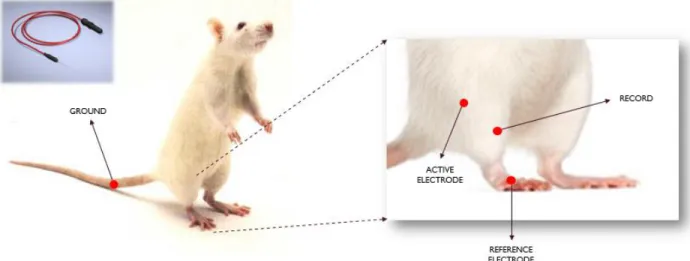
Electrophysiological studies were performed to measure the amplitude of the compound motor action potentials (CMAP). Measurements were taken by using Neuro-MEP-Micro two channels EMG (Neurosoft, Russia) device. Recordings were collected from pre-operation (healthy), right after the pre-operation, day 7 and day 14 post-pre-operation. After anesthesia with ketamine (100 mg/kg) and xylazine (10 mg/kg), evaluation place was cleaned with alcohol and allowed to dry before procedure. Schematic representation of the experimental setup can be seen from Figure 7.
27
Figure 7 Schematic Illustration of Electromyography Setup. Ground was placed to the tail, reference electrode was placed to the feet, active electrode was placed to the beginning of the leg, and measurements were recorded by using needle electrode from the tibialis anterior muscle.
For this evaluation, recording monopolar needle electrode was placed in TA muscle, reference electrode was placed in dorsum of the foot and common reference (ground electrode) was placed on the tail. In order to observe the amplitude of the motor action potentials sweep speed/sensitivity were set to 4 mV and 4 msec/division. The common peroneal nerve found in the knee at fibula’s head was stimulated with bipolar concentric needle electrode. The intensity was gradually increased until dorsiflexion movement was observed and up to supramaximal intensity (current intensity 30% above the value to evoke the maximal CMAP). CMAP amplitude (negative peak’s amplitude) was recorded from the TA muscle. Stimulation was repeated 3 times for all measurements and average values were used for statistical analysis.
28
3.2.6.2 Walking Track Step Test Analysis
At 1, 3, 5, 7 and 14 days after treatment, animals underwent a postoperative behavioral walking-track analysis. The rats were walked over a white sheet of paper (8.2 cm wide, 58 cm long) covering the bottom of a wooden alley ending in a dark destination box. Hind paws of the animals were painted with black ink, and then they were placed into the track to walk. Stride lengths were measured manually as the distance between two pawprints. By considering three longest steps from each walk, stride lengths were calculated58 and first & last paw prints were excluded because of velocity changes. Obtained data were analyzed and processed in Graphpad software Prism 5, as the mean with SD for each limb. Statistical comparisons were made by paired t-test for paired values and unpaired t-test for unpaired values.
3.2.7 Histological Analysis
3.2.7.1 Morphological Analysis (H&E)
After 1, 3, 5, 7 and 14 days of recovery, 10 rats for each day point were sacrificed and their TA muscles were extracted. TA muscles were divided into 3 pieces in order to proceed with histological, gene expression and protein detection analysis. Tissue allocated for histological examination was put into 4% paraformaldehyde solution for fixation. After 24 h, samples were dehydrated with a graded series of ethanol from 70% to 100% and prepared for paraffin embedding with two changes of xylene. Cleared samples were embedded in paraffin blocks and sectioned at 5 µm thickness by microtome. To assess regeneration, hematoxylin and eosin staining was performed,
29
slides were mounted by Histomount® mounting medium. Cross-sections were prepared for histological examination on Axio Scope (Zeiss) light microscope.
3.2.7.2 Muscle Cross Section Area Calculation
We next determined the loss of skeletal muscle mass in response to acute injury. Since degeneration causes the depletion of skeletal muscle, cross section thickness of skeletal muscle is associated with the skeletal muscle mass loss. Measurements were conducted at 20X magnification. Per each cross section, five random fields were chosen and area of fibers were calculated by using Image J software. Changes in fiber cross section area between skeletal muscle fibers were determined using parametric (2-way ANOVA) test.
3.2.7.3 Centrally Located Nuclei Calculation
Centrally located myonuclei observation indicates the degree of skeletal muscle regeneration because satellite cells merge with the existing fibers when they are stimulated for regeneration. By the time, they merged with the fibers and located the center of the skeletal fibers. Thus, I calculated the centrally located myonuclei for each group. Measurements were conducted in day 7 at 40X magnification per each cross section by bright-field microscopy. The number of central nuclei per muscle fiber was determined by counting a minimum of five random muscle fiber images per animal and five animals from each group were analyzed. Percentage of myofibers with central nuclei were calculated and statistical analysis was done by using parametric t-test.
30
3.2.8 Gene Expression Analysis by RT-qPCR
For analyzing molecular level myogenic differentiation, gene expression profiles of skeletal muscle tissue samples were assessed by quantitative real time PCR (qRT-PCR) method. Myogenesis markers such as Pax7, MyoD1, Myf5 and Myogenin were used for this experiment.
3.2.8.1 RNA Isolation
Tissue parts allocated for gene expression analysis were put into test tubes right after animals were sacrificed and immediately frozen with liquid nitrogen. RNA from each sample was isolated with TRIzol reagent (Invitrogen) according to the manufacturer’s instruction. Tissues were smashed with a tissue grinder and homogenized with TRIzol reagent by using homogenizer. After homogenization, centrifugation was performed at 4ºC with 2000 G for 5 minutes in order to get rid of tissue debris. 1 mL of supernatant from each sample was taken and 200 µL of chloroform was added into the test tube. Tubes were incubated for 3 min at room temperature and then centrifugation was done for 17 min with 15000 rpm at 4°C. After centrifugation, upper clear phase from each tube was taken and put into a new test tube for ½ v/v isopropanol would be added. 15 times gentle shaking (upside-down) was done and tubes were incubated for 10 min inside the ice container. Next, tubes were centrifuged for 12 min with 15000 rpm at 4 °C and then supernatant was discarded for further steps. Pellets were washed via slowly addition of 70% cold ethanol and tubes were centrifuged at 8000 rpm at 4 °C. Second washing step was applied via slowly addition of 100% cold ethanol and tubes were centrifuged at 8000 rpm at 4 °C again. Tubes were allowed to dry in order to get rid of
31
ethanol from the samples and 20 μL of DNAse & RNAse free ddH2O was added to each
test tube.
3.2.8.2 RNA Quantification
Purity and yield of isolated RNA were determined with Nanodrop 2000 spectrometer (Thermo Scientific). This system allows us to determine the concentration of RNA by using only 1 µL RNA sample. After quantifying the concentration, purity of RNAs was checked by considering the absorption ratio between 260/280 and 260/230. 260/280 ratio gives an information about protein contamination and should be between 1.8 - 2.0. However, 260/230 ratio gives an information about EDTA, TRIzol and phenol contamination and it should be in between 1.8 - 2.2. In this thesis project, we used only pure RNA for qRT-PCR.
3.2.8.3 Quantitative reverse transcription PCR (RT-qPCR)
Both cDNA synthesis and qRT-PCR were performed with one-step qRT-PCR kit (SuperScript III Platinum SYBR Green) according to the manufacturer’s instructions. Primer sets that were used in this project are represented in Table 1 below.
32
Table 1 Skeletal Muscle Specific Primer Sequences for RT-qPCR
Primer Sequence (Forward) Sequence (Reverse) Annealing Temp. Efficiency
Pax7 GTGCCCTCAGTGAGTTCGATT GGGAGGTCGGGTTCTGATTC 59 °C 101.6 %
MyoD1 CATAGACTTGACAGGCCCCG GCAGGTCTGGTGAGTCGAAA 59.2 °C 102 %
Myf5 AACCAGAGACTCCCCAAGGT AGCACATGCATTTGATACATCAGG 58.6 °C 97 %
Myogenin TGGTCCCAACCCAGGAGATC AGAAGTGGTGGCGTCTGAC 60 °C 97 %
33
Reaction conditions were briefly as follows: 55 ºC for 5 min and 95 ºC for 5 min were applied as a first step. Polymerase chain reaction continued with 40 cycles of 95 ºC for 15 s, 60 ºC for 30 s and 40 ºC for 1 min. Product specificity was confirmed by following a melting curve and the reaction efficiencies for each primer set were evaluated with standard curve using 5-fold serial dilutions of total RNA. For the analysis of expression, primary gene expression data were normalized by the expression level of healthy group and the expression level of GAPDH. A comparative Ct method was used to analyze the results.
3.2.9 Protein Detection by Western Blotting
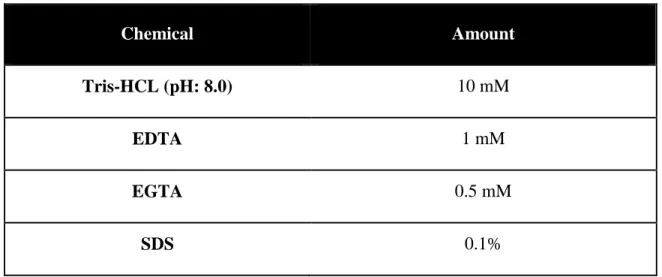
For detecting myogenic differentiation related proteins, protein profiles were assessed by western blot. Tissue part allocated for protein detection was put into a test tube right after animals were sacrificed and immediately frozen with liquid nitrogen. Total protein from each sample was homogenized with RIPA buffer. Ingredients of the RIPA buffer are presented in Table 2 below.
Table 2 Content of RIPA Lysis Buffer
Chemical Amount
Tris-HCL (pH: 8.0) 10 mM
EDTA 1 mM
EGTA 0.5 mM
34
NaCl 140 mM
Triton X-100 1%
Sodium deoxycholate 0.1%
Protease Inhibitor Cocktail According to the manufacturer’s instructions
Proteins were denaturated, resolved on 12% SDS-PAGE and transferred to polyvinylidene difluoride membranes (Thermo Scientific). Membranes were blocked with 5% freeze-dried nonfat milk in TBS-T for 1 h at room temperature and then incubated with primary antibody at 4 ºC overnight. Afterwards, membranes were washed extensively with TBS-T and incubated with HRP conjugated secondary antibody for 1 h and visualized by enhanced chemiluminescence (Bio-Rad) according to the manufacturer’s protocol on ChemiDoc™ Imaging System with Image Lab™ Software - Bio-Rad.
3.2.9.1 BCA Assay
Protein concentrations were determined with BCA Assay (Thermo Scientific). BCA is a bicinchoninic acid based colorimetric detection for proteins. Standards for BCA assay was prepared via serial dilution from bovine serum albumin (BSA) and known concentration gradient was obtained in the 2000-0 µg/µL range were prepared. 25 µL of each sample was put into a microplate and 200 µL developing solution (which was prepared from BCA protein assay kit, for 200 μL; 196 μL BCA reagent A + 4 μL BCA reagent B were mixed) was added onto samples. Microplate was incubated at 37 °C for
35
30 min and absorbance was measured at 560 nm. By using BSA standard curve, unknown concentrations of samples were determined. Protein stock samples were diluted so as to reach 50 µg/mL final concentration for SDS PAGE.
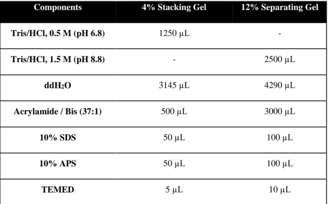
3.2.9.2 SDS PAGE
Molecules are separated according to their molecular weight with SDS Polyacrylamide Gel Electrophoresis technique. Two different acrylamide gel systems were used in these experiments. Stacking gel helps proteins to stack prior to separation. Separating gel is responsible for separation of proteins by their size. Contents of stacking and separating gels are represented in Table 3 below. Protein separation by SDS-PAGE gives an information about the molecular mass of the proteins, and also purity of proteins can be determined by this technique.
Table 3 Contents of Stacking and Separating Acrylamide Gels
Components 4% Stacking Gel 12% Separating Gel
Tris/HCl, 0.5 M (pH 6.8) 1250 µL - Tris/HCl, 1.5 M (pH 8.8) - 2500 µL ddH2O 3145 µL 4290 µL Acrylamide / Bis (37:1) 500 µL 3000 µL 10% SDS 50 µL 100 µL 10% APS 50 µL 100 µL TEMED 5 µL 10 µL
36
For preparation of 10 mL of 12% separating gel, 4290 µL of ddH2O, 3000 µL of
Acrylamide / Bis (37:1), 2500 µL of 1.5 M Tris/HCl (pH 8.8), 100 µL of 10% SDS, 100 µL of 10%APS and 10 µL TEMED were mixed. TEMED and APS were added at the end, because they cause faster polymerization of gel.
Separating gel was poured between the PAGE glasses and approximately 2 cm empty space was allowed. Isopropanol was added into such space to remove bubbles and obtain flat gel surface. After 15 min of polymerization, 4% stacking gel was prepared by mixing 3145 µL of ddH2O, 500 µL of Acrylamide / Bis (37:1), 1250 µL of 0.5 M Tris /
HCl (pH 8.8), 50 µL of 10% SDS, 50 µL of 10%APS and 5 µL TEMED.
Isopropanol was removed from separating gel and stacking gel was poured. Next, comb was placed into stacking gel and allowed for polymerization for 15 min. 35 μL of protein samples were prepared to obtain 50 µg/mL final protein concentration. For this purpose, 6X Laemmli loading dye and ddH2O were used. Protein solutions were incubated at 95
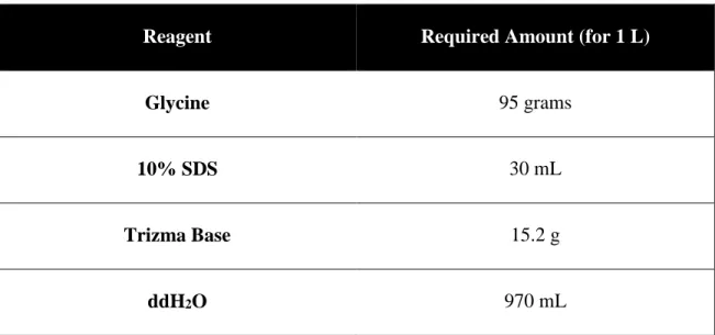
ºC for 5 min and gels were loaded by putting 4 μL of protein ladder and 30 μL of protein solutions. Separation was processed at 70 V for 20 min and 110 V for 2 h at room temperature. The content of stock running buffer is shown in Table 4 below. While running, ladder was always checked to observe whether its bands separate uniformly or not. In this thesis work, broad ranged (11-190 kDa) Blue Prestained Protein Standard was used (Invitrogen).
37
Table 4 Content of 5X Running Buffer for SDS-PAGE (1L)
Reagent Required Amount (for 1 L)
Glycine 95 grams
10% SDS 30 mL
Trizma Base 15.2 g
ddH2O 970 mL
3.2.9.3 Protein Transfer onto a PVDF Membrane
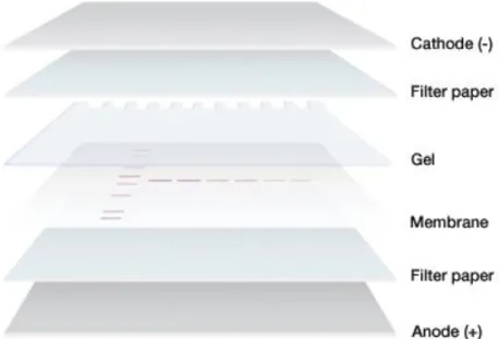
Separated proteins were transferred onto polyvinylidene difluoride (PVDF) membrane after electrophoresis. First, glass plate was opened and stacking gel was removed from the separating gel. In order to transfer proteins from gel to PVDF membrane Semi-Dry Electrophoretic Transfer Cell electroblot system was used. Filter papers were equilibrated in anode and cathode buffers and experimental setup was created as represented in Figure 8. Anode and cathode buffers were prepared from Towbin stock buffer. The content of the buffers are represented in Table 5, 6 and 7 below. To avoid air bubbles, glass tube was rolled onto papers and electroblot was performed at 12 V for 50 min for each gel.
38
Table 5 Content of 10X Towbin Buffer (Anode and Cathode Buffers are prepared from this buffer)
Reagent Stock Concentration Required Amount (for 500 mL)
Tris HCl 2 M 62.5 mL
Glycine - 72.0672 g
ddH2O - 437.5 mL
Table 6 Content of Anode Buffer
Reagent Stock Concentration Required Volume (for 100 mL)
Towbin Buffer 10 X 20 mL
Methanol 100% 10 mL
ddH2O - 70 mL
Table 7 Content of Cathode Buffer
Reagent Stock Concentration Required Volume (for 100 mL)
Towbin Buffer 10 X 10 mL
SDS 10% 1 mL