http://journals.tubitak.gov.tr/medical/ © TÜBİTAK
doi:10.3906/sag-1706-158
Isolation and culture of adult mouse vestibular nucleus neurons
Aydın HİM1,*, Serap ALTUNTAŞ2, Gürkan ÖZTÜRK3, Ender ERDOĞAN4, Nurettin CENGİZ5 1Department of Biophysics, Ondokuz Mayıs University, Samsun, Turkey
2Antalya Education and Research Hospital, Antalya, Turkey 3Department of Physiology, İstanbul Medipol University, İstanbul, Turkey 4Department of Histology and Embryology, Selçuk University, Konya, Turkey 5Department of Histology and Embryology, Sakarya University, Sakarya, Turkey
1. Introduction
Culturing isolated central nervous system (CNS) neurons provides a powerful tool for neuropharmacological studies and the study of mechanisms of neurodegeneration and neuroregeneration. Here we present a method for culturing isolated vestibular neurons from adult mice. Although it is relatively easy to isolate neurons from embryonic (1) or early postnatal animals (2) before they form synapses, the isolation of adult CNS neurons is often difficult due to tight adhesion of cell bodies, thousands of synapses, presence of few glial cells, requirement of trophic factors, and damage to axons and dendrites during the process (1,3,4). Although embryonic neurons represent an easy and effective source of primary neurons, their developmental stage is not always appropriate for the topic of interest. Many neurodegenerative diseases often occur later in life. In many cases, it is clear that embryonic neurons behave differently than adult neurons in terms of pharmacology,
electrophysiology, development, and regenerative and pathological characteristics. In such cases as calcium dynamics (5), glutamate sensitivity, and mitochondrial functions (6), the difference between the embryonic and adult neurons could be significant.
The use of cell cultures in the study of the vestibular system has been increasing. The vestibular system is the sensory system that detects the position and motion of the head in space. This information is used to stabilize the eyes when the head is moving and adjust the neck and body muscle tone to control posture. The receptive structures of the vestibular system are located in the labyrinth, which contains the three semicircular canals that sense the angular acceleration of the head in space, and the otolith organs (utricle and saccule) that sense linear acceleration. The vestibular sensory information is transmitted mainly to the superior, medial, inferior, and lateral vestibular nuclei and the cerebellum. These structures send ascending Background/aim: Isolated cell cultures are widely used to study neuronal properties due to their advantages. Although embryonic
animals are preferred for culturing, their morphological or electrophysiological properties may not reflect adult neurons, which may be important in neurodegenerative diseases. This paper aims to develop a method for preparing isolated cell cultures of medial vestibular nucleus (MVN) from adult mice and describe its morphological and electrophysiological properties.
Materials and methods: Vestibular nucleus neurons were mechanically and enzymatically isolated and cultured using a defined medium
with known growth factors. Cell survival was measured with propidium iodide, and electrophysiological properties were investigated with current-clamp recording.
Results: Vestibular neurons grew neurites in cultures, gaining adult-like morphological properties, and stayed viable for 3 days in
culture. Adding bovine calf serum, nerve growth factor, or insulin-like growth factor into the culture medium enhanced neuronal viability. Current-clamp recording of the cultured neurons revealed tonic and phasic-type neurons with similar input resistance, resting membrane potential, action potential amplitude, and duration.
Conclusion: Vestibular neurons from adult mice can be cultured, and regenerate axons in a medium containing appropriate growth
factors. Culturing adult vestibular neurons provides a new method to study age-related pathologies of the vestibular system.
Key words: Vestibular nucleus, primary culture, regeneration, mouse, whole-cell recording
Received: 22.06.2017 Accepted/Published Online: 11.10.2017 Final Version: 19.12.2017 Research Article
and descending projections to stabilize gaze and posture via reflexes, such as the vestibulo-ocular reflex and the vestibulospinal reflex (7).
Cell cultures have been used to investigate the electrophysiological properties of vestibular ganglion cells (8), the effects of growth factors on their development (9), and the glutamate receptor function on vestibular ganglion neurons (10). Electrophysiological properties of medial vestibular nucleus (MVN) neurons were studied in microexplant cultures (11). These culture systems used embryonic or early postnatal animals. The use of cell cultures from adult animals is especially important for vestibular research, since many problems of the vestibular system, such as dizziness, falls, balance disorders, and vertigo, are commonly seen in advanced age. Isolated cell cultures have not been used for physiological studies of MVN neurons, which have been implicated in the mechanisms of vestibular compensation, an experimental model for lesion-induced plasticity, and in age-related vestibular disorders (12–19).
The present study develops a method for preparing isolated cell cultures of MVN neurons from adult animals. Cell survival in serum-free media was investigated using common growth factors. Whole-cell patch clamp studies were performed to describe the electrophysiological properties of cultured vestibular neurons.
2. Materials and methods 2.1. Animals
Male young adult (6–8 weeks) mice (Balb/C) were obtained from the Animal House of the Faculty of Medicine, Yüzüncü Yıl University. The animals were housed in an environment with natural day–night cycles and kept in standard cages with food and water ad libitum. Approval was obtained from the Animal Ethics Committee and all experiments were conducted in accordance with the NIH Guide for the Use and Care of Laboratory Animals. All efforts were made to minimize pain and discomfort.
2.2. Culture preparation
The method described by Brewer (3) was used for culture preparation with some modification. The animals were anesthetized with intraperitoneal injection of ketamine (100 mg/kg, Pfizer), and their brains were removed by orbital approach and placed into a 2-mL ice-cold culture medium containing B27, Neurobasal A (NBA), antibiotics, and glutamine (a complete description is provided below). Bilateral MVN was dissected from the brain on a cold plate under an operation microscope and incubated for 30 min at 4 °C in an enzyme solution containing papain (6 U/mL, prepared in NBA) in an Eppendorf tube. The cells were dissociated by mechanical trituration using
blue and yellow pipette tips and an insulin needle. The cell suspension was kept in an agitator (50 Hz) for 15 min in DNase (100 mg/mL). Then it was placed on top of a gradient, prepared in NBA. The gradient was made in four 1-mL steps of 60%, 30%, 20%, and 10% Percoll. The cell suspension was centrifuged for 25 min at 4 °C at 5000 rpm. Then 1 mL of cell suspension was collected from the 30% band and resuspended in 5 mL of culture medium. The cell suspension was centrifuged for 5 min at 4 °C at 2000 rpm, and the top 5 mL supernatant was removed giving a 200-µL cell suspension. Cells were plated in four-well dishes previously coated with 10% poly-L-lysine (Sigma). Fifty microliter cell suspension was placed in each well and topped up to 400 µL with culture medium. Poly-L-lysine was applied for 2 h, aspirated and rinsed once with distilled water, and allowed to dry for about 1 h before plating. One hour after plating and incubation (5% CO2, 37 °C), the medium was aspirated to remove unattached cells and debris and was rinsed once with 400 µL of culture medium. For electrophysiological recordings, the cells were plated in 35-mm plastic dishes, previously coated with poly-L-lysine and topped up to 1500 µL with culture medium. The basic culture medium consisted of 2 mM GlutaMaxI (Gibco), 100 units/mL penicillin, 100 µg/ mL streptomycin, and 2% B27 (NBA-B27)(Gibco). To test their effects on cell survival, fetal calf serum (FCS (10% and 20%), nerve growth factor (NGF)(50 ng/mL), brain-derived neurotrophic factor (BDNF)(50 ng/mL), neurotrophin-3 (NT-3)(50 ng/mL), leukemia inhibitory factor (LIF)(10 ng/mL), basic fibroblast growth factor (bFGF)(5 ng/mL), epidermal growth factor (EGF)(10 ng/ mL), or insulin like growth factor-1 (IGF-1)(10 ng/mL) were added to the culture medium. A six-well plate was used to test the effects of each serum or growth factors. Plating density was 1163 cells/cm2. FCS and growth factors were obtained from Sigma-Aldrich (UK).
2.3. Immunohistochemistry and viability essay
For the immunohistochemistry experiments, the cells were cultured on 10-mm coverslips coated with 10% poly-L-lysine. After 2 days in vitro (DIV), the cells were stained for glial fibrillary acidic protein (GFAP), neuroflament 200 (NF200), βIII tubulin, and neurotrophin receptors TrkA, TrkB, and TrkC, using standard immunohistochemical techniques. Briefly, the cells were fixed in 4% paraformaldehyde, freshly prepared in phosphate buffer saline (PBS) for 30 min. The cultures were washed in PBS and moved into a blocking solution (BSA 3%, Triton X 0.3%, Na-azide 0.01%, prepared in PBS) for 30 min. After incubation in 1:100 dilution of the primary antibody overnight at 4 °C, the cultures were washed 3 times for 5 min. The fluorescent-conjugated secondary antibodies were applied for 3–4 h and the cultures were rinsed 3 times
for 5 min. After mounting (Slow Fade Light Antifade Kit, Mol. Probes), the cells were visualized using a fluorescent microscope (Carl Zeiss, USA). Propidium iodide (10 µM), which stains the nuclei of dead cells, was used to test the viability of the cells.
2.4. Electrophysiological recordings
Whole cell patch-clamp recordings were obtained 48 h after
plating. Prior to the recordings, the culture medium was exchanged with 1500 µL of recording medium, consisting of (in mM) 124 NaCl; 5 KCl; 1.2 KH2PO4; 1.3 MgSO4; 2.4 CaCl2; 26 NaHCO3; and 10 D-(+) glucose, adjusted to pH 7.4 and osmolarity 300 mOsm. The petri dishes were placed on the stage of an inverted microscope (TE300, Nikon, Japan) and kept at room temperature during recordings. The glass microelectrodes (5–6 MW, WPI) were back-filled with (in mM) 145 potassium gluconate, 2 MgCl2, 5 K2ATP, 0.1 EGTA, and 5 HEPES. The osmolarity and acidity of the filling solution were adjusted to 290 mOsm and pH 7.2, respectively. Electrophysiological recordings were performed with continuous current-clamp in bridge mode using an AxoClamp-2B amplifier, stored digitally via Digidata 1322A interface and analyzed offline with pClamp 10 (Molecular Devices, USA). The signal from the amplifier was low-pass filtered at 3 kHz and digitally sampled at 25 kHz. The bridge balance and capacity compensation were adjusted to minimize the transient voltage change at the beginning of the current injection. A neuron was accepted for study only when it exhibited a resting membrane potential (RMP) more negative than –40 mV, and had a spontaneous activity or fired action potential in response to depolarizing stimuli. From each neuron, a continuous recording was obtained for 1 min without the delivery of any external stimulus, to see if the cell was spontaneously active. The reading on the digital voltmeter on the front panel of the AxoClamp 2B amplifier was taken as the resting membrane potential, which was later corrected for an estimated liquid junction potential of –10 mV. The input resistance (Rin, MΩ) was calculated from membrane potential change, evoked in response to a 100-ms hyperpolarizing current step of –0.2 nA. To find the duration and amplitude, the neurons were held at –70 mV membrane potential, and action potential was evoked by injecting current steps of 5 ms duration from 0.05 nA in increments of 0.05 nA. Action potentials evoked by the smallest current injection (rheobase) were used for assessing action potential shape. To study the firing characteristics of the cells, a series of constant depolarizing current steps of increasing amplitude (0.05 nA increments from 0.05 nA) was injected for a duration of 500 ms. Depolarizing and hyperpolarizing current steps of 500-ms duration (0.05 nA decrements starting at +0.05 nA) were injected to study the current/voltage responses of the cells.
3. Results
At the time of plating, most cells were spherical with no processes. Within a few hours of plating, some cells began to extend neurites. At 3 DIV, the somata and length of the neurites were measured in neuron cultures with 20% FCS using AxioVision 3 (Carl Zeiss Vision, Germany). The cells were multipolar and the average number of primary processes emanating from the soma was 6.8 ± 2.3 per cell (n = 53). Cells extend neurites as long as 107 µm (mean length 37.4 ± 12.1 µm). The somata of the cells were typically round or triangular, and had 192.4 ± 36.6 µm2 soma area (n = 53) and 16.6 ± 1.9 µm diameter (n = 53) (Figure 1). Due to low plating density (1163 cells/ cm2), the neurons were usually found in isolation and were not in contact with glial cells. Glial cells were very scarce throughout the culture period. Most cells were positive for neuronal markers NF200 and βIII tubulin and negative for the glial cell marker GFAP (90%), which confirmed the neuronal identity of the cultured cells. There was no TrkB-positive cell, although many cells were TrkB-positive for TrkA and TrkC (Figure 2).
The percentage of live cells incubated in culture medium was 52%, 9%, and 1% at 1, 2, and 3 DIV, respectively. When 10% FCS was added to the culture medium, the cells survived longer (92%, 29%, and 11%), and 20% FCS further increased their viability (92%, 68%, and 33%) (P < 0.05, χ2 test). Furthermore, cell survival was prolonged with the addition of NGF (68%, 34%, and 13% at 1, 2, and 3 DIV, respectively) or IGF-1 (66%, 29%, and 5% at 1, 2, and 3 DIV, respectively) into the culture medium, whereas BDNF, NT3, LIF, bFGF, and EGF did not affect cell survival (P < 0.05, χ2 test)(Figure 3).
Among 28 neurons cultured with 20% FCS and recorded at 2 DIV with current-clamp, only one cell (4%) showed spontaneous action potentials, whereas the rest responded by producing action potentials on depolarizing stimuli. According to their responses to depolarizing current pulses, two types of neurons were identified: tonic (93%) and phasic (7%) neurons (Figure 4). Tonic neurons produced multiple action potentials with a single component after hyperpolarization throughout the current injection period. Phasic neurons responded to the current pulse with a single action potential appearing at the beginning of the depolarization. Input resistance, resting membrane potential, action potential amplitude, and duration were not different in tonic and phasic neurons. When two types of neurons combined, they had 91.07 ± 48.6 MΩ input resistance, –53.57 ± 6.4 mV resting membrane potential, 76.02 ± 16.4 mV action potential amplitude, and 1.02 ± 0.2 ms action potential duration.
4. Discussion
This study cultured MVN neurons from adult mice for up to 3 days. Although microexplant cultures of MVN neurons were prepared beforehand, isolated cell culture of MVN from adult mice has been described here for the first time. Culturing mammalian cells in serum-free media has been a challenge (20), but is considered indispensable, especially in studies regarding actions of biologically active molecules. As we observed in this study, supplementation of fetal serum to the culture enhances the survival of neurons and neurite regeneration alike, although it renders the medium undefined. In this study, we replaced serum with a defined supplement, B27, which was shown to specifically support neurons in vitro (1). Additionally,
it managed to obtain a survival rate of 52% for the first 24 h of incubation, which might permit the design of short experimental paradigms such as electrophysiology. While using BDNF, NT-3, LIF, bFGF, or EGF in culture medium did not prove useful in extending the culture period, adding NGF and IGF-1 to the medium increased the survival rate of cultured vestibular neurons, which expressed TrkA and TrkC. Although cultured vestibular neurons expressed TrkC, they did not benefit from the neurotrophic effect of NT-3, which can be explained by its low level of expression. For longer periods of culturing, the addition of either NGF or IGF-1 will still allow us to exercise a defined medium. Use of these two factors has been implicated in other neuronal culture protocols, Figure 1. Microscopic appearance of isolated medial vestibular neurons up to 4 days in culture with 20% fetal calf serum. Although
neurons did not have any processes when first cultured, soon after plating they started growing neurites as long as 107 µm within a few days. Scale bar = 10 µm.
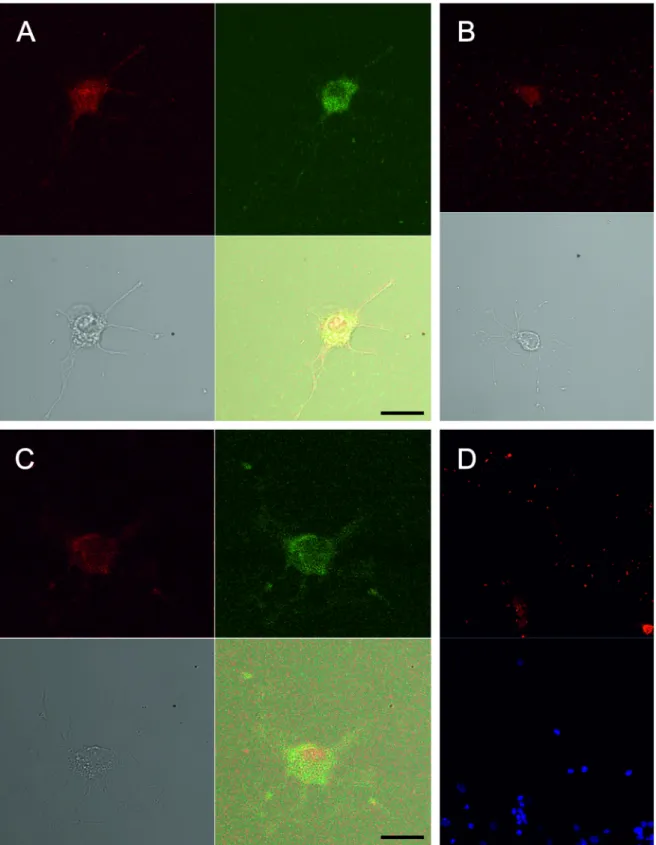
Figure 2. Immunostaining for neurotrophin receptors TrkA (A), TrkB (B), and TrkC (C). Florescent antibody stains
neuroflament 200, a neuron marker, as red, and florescent antibodies stain TrkA and TrkB receptors as green. The neuronal identity of the cells was confirmed by staining with florescent antibodies against βIII tubulin (green) and GFAP (red) (D). DAPI staining shows cell nuclei as blue. Scale bar = 10 µm.
along with those proved ineffective in this culture method (3,21–25). Since the culture additionally contained glial cells, though in low density, it is possible that the effect of growth factors is not direct. They may influence the survival of glial cells and stimulate them to secrete factors that indirectly support neuronal survival (26).
Papain is a widely used enzyme in CNS tissue dissociation and is praised for its compatibility with neurons (3,27). However, when we used this enzyme at 37 °C, as suggested, we obtained a very low neuronal viability (data not shown). By lowering the temperature to 4 °C, we aimed to slow down the metabolism and reduce stress-induced cell death (28). Although we do not have data to explain the mechanism of action, this has worked for MVN neurons. It is noteworthy that at such a low temperature, papain was just as effective in the digestion of tissue as at physiological temperature, without being required to increase its concentration.
Most MVN neurons were not spontaneously active in cultured conditions. At 2 DIV, only one neuron (4%) produced spontaneous action potentials. As in many cell culture systems, neurons start producing spontaneous action potentials a few days after cell plating, and this may increase and later decrease (29). Fitzakerley et al. found only 2 spontaneously active neurons out of 22 cochlear neurons in culture (30), whereas no neuron was
spontaneously active in microexplant cultures of MVN (11). Low plating density may be responsible for the lack of resting activity, since it may depend on cell-to-cell contacts in some cells (31). According to action potential characteristics, cultured MVN neurons were similar to Type A neurons that were described in brain slices. MVN neurons recorded in slices are classified either as Type A, which show a single deep after-hyperpolarization (AHP), or Type B, which show an early fast AHP followed by a delayed slow AHP (32,33). It is possible that only Type A cells are thriving in culture, or expression of additional channels needed for Type B cells may require a longer culture period. Genlain et al. (11) found only Type A cells, even after 4 weeks in microexplant cultures of MVN. The electrophysiological characteristics of the medial vestibular neurons 2 days after culturing are not entirely similar to those of the adult vestibular neurons in brain slices. For example, very low percentage of spontaneously firing neurons in culture reflects the missing pacemaker conductances, which generates resting discharge of action potentials. Performing electrophysiological recordings at a later culture period, such as at the 3rd and 4th days, when about 10%–30% cell viability is achieved, may reveal neurons with electrical membrane properties more similar to those of the mature neurons. Since vestibular neurons in culture extended neuritis more than 100 µm, Figure 3. Effects of serum and growth factors on cell survival in cultured medial vestibular nucleus neurons.
While adding fetal calf serum (FCS) (10% and 20%), nerve growth factor (NGF) and insulin like growth factor-1 (IGF-1) increased cell survival, adding brain-derived neurotrophic factor (BDNF), neurotrophin-3 (NT3), leukemia inhibitory factor (LIF), fibroblast growth factor (FGF), and epidermal growth factor (EGF) did not affect cell survival. *P < 0.05, χ2 test.
it is possible that the mechanisms of axon degeneration, induced by laser microdissection, can be studied in central nervous system neurons as in the peripheral nervous system (34). Molecular changes, as well as changes in the current components of the vestibular neurons, such as hyperpolarization-activated cationic current, low threshold calcium current, and pacemaker current, can be investigated in cultured vestibular neurons during axonal degeneration and regeneration. In vitro studies of neurodegenerative diseases, such as Parkinson and Alzheimer diseases, often include cell lines such as HEK293 and SH-SY5Y, which do not express neuronal phenotype of the related neupathology or primary cell cultures obtained from embryonic tissues, which are easily prepared and maintained in vitro (35). However, the developmental stage of embryonic primary cell cultures is not always suitable for studies of neurodegeneration, which often occurs later in life (36). Hence, it would be desirable to use primary neuronal cultures for exploring
neurodegenerative mechanisms, since they reproduce more adult-like phenotypic properties.
This study described culturing of isolated MVN neurons from adult animals, and demonstrated that they retain some electrophysiological properties in culture. Adult CNS neurons are inherently much more difficult to culture than those from fetal or neonatal animals. However, in vitro observations are more relevant to physiological or pathological conditions in adult organisms. In this study, we developed a protocol to culture adult MVN neurons for up to 3 days, which may be useful for neuropharmacological studies of vestibular neurons from adult or even aged animals. The culture period and neuronal survival rate can be increased by manipulating growth factors such as adding EPO, CNTF, and GDNF, or using more than one growth factor in culture. Depending on the particular experiment, desired vestibular neurons could be cultured with glial cells, which play a role in supporting neuronal development (26). Although not performed in the current Figure 4. Current-clamp recording of medial vestibular nucleus neurons cultured with
20% fetal calf serum for 2 days revealed two types of neurons: tonic (A) and phasic (B). Tonic neurons responded to depolarizing current injections with multiple action potentials throughout the stimulus period, whereas phasic neurons responded to the current pulse with a single action potential appearing at the beginning of the stimulus.
study, dissociating current components in tonic and phasic neurons in young and old animals may clarify mechanisms of age-related changes in electrical properties of vestibular neurons.
Acknowledgment
This work was supported by a grant from the Scientific and Technological Research Council of Turkey (TÜBİTAK, Grant No. SBAG2971-104S506).
References
1. Brewer GJ, Torricelli JR, Evege EK, Price PJ. Optimized survival of hippocampal neurons in B27-supplemented Neurobasal, a new serum-free medium combination. J Neurosci Res 1993; 35: 567-576.
2. Ahlemeyer B, Baumgart-Vogt E. Optimized protocols for the simultaneous preparation of primary neuronal cultures of the neocortex, hippocampus and cerebellum from individual newborn (P0.5) C57Bl/6J mice. J Neurosci Meth 2005; 149: 110-120.
3. Brewer GJ. Isolation and culture of adult rat hippocampal neurons. J Neurosci Meth 1997; 71: 143-155.
4. Goslin K, Banker G. Experimental observations on the development of polarity by hippocampal neurons in culture. J Cell Biol 1989; 108: 1507-1516.
5. Brewer GJ, Reichensperger JD, Brinton RD. Prevention of age-related dysregulation of calcium dynamics by estrogen in neurons. Neurobiol Aging 2006; 27: 306-317.
6. Parihar MS, Brewer GJ. Simultaneous age-related depolarization of mitochondrial membrane potential and increased mitochondrial reactive oxygen species production correlate with age-related glutamate excitotoxicity in rat hippocampal neurons. J Neurosci Res 2007; 85: 1018-1032. 7. Khan S, Chang R. Anatomy of the vestibular system: a review.
Neurorehabilitation 2013; 32: 437-443.
8. Chabbert C, Chambard JM, Valmier J, Sans A, Desmadryl G. Voltage-activated sodium currents in acutely isolated mouse vestibular ganglion neurones. Neuroreport 1997; 8: 1253-1256. 9. Lefebvre PP, Van de Water TR, Represa J, Liu W, Bernd P,
Modlin S, Moonen G, Mayer MB. Temporal pattern of nerve growth factor (NGF) binding in vivo and the in vitro effects of NGF on cultures of developing auditory and vestibular neurons. Acta Oto-Laryngol 1991; 111: 304-311.
10. Rabejac D, Devau G, Raymond J. AMPA receptors in cultured vestibular ganglion neurons: detection and activation. Eur J Neurosci 1997; 9: 221-228.
11. Genlain M, Nonclercq D, Laurent G, Toubeau G, Godaux E, Ris L. Properties of neurons from the rat medial vestibular nucleus in microexplant culture. Neurosci Lett 2003; 338: 45-48.
12. Paterson JM, Short D, Flatman PW, Seckl JR, Aitken A, Dutia MB. Changes in protein expression in the medial vestibular nuclei during vestibular compensation. J Physiol 2006; 575: 777-788.
13. Beraneck M, Idoux E, Uno A, Vidal PP, Moore LE, Vibert N. Unilateral labyrinthectomy modifies the membrane properties of contralesional vestibular neurons. J Neurophysiol 2004; 92: 1668-1684.
14. Lacour M, Helmchen C, Vidal PP. Vestibular compensation: the neuro-otologist’s best friend. J Neurol 2016; 263: 54-64. 15. Smith PF. Age-related neurochemical changes in the vestibular
nuclei. Front Neurol 2016; 7: 20.
16. Arshad Q, Seemungal BM. Age-related vestibular loss: current understanding and future research directions. Front Neurol 2016; 7: 231.
17. Him A, Johnston AR, Yau JL, Seckl J, Dutia MB. Tonic activity and GABA responsiveness of medial vestibular nucleus neurons in aged rats. Neuroreport 2001; 12: 3965-3968. 18. Chabbert C. New insights into vestibular neuropharmacology:
from bench to bedside. J Vestib Res 2013; 23: 107-111. 19. Uffer DS, Hegemann SC. About the pathophysiology of acute
unilateral vestibular deficit – vestibular neuritis (VN) or peripheral vestibulopathy (PVP)? J Vestib Res 2016; 26: 311-317.
20. Maurer HR. Towards chemically-defined, serum-free media for mammalian cell culture. In: Freshney RI, editor. Animal Cell Culture: A Practical Approach. Oxford, UK: IRL Press, 1986. pp. 13-31.
21. Kelly CM, Tyers P, Borg MT, Svendsen CN, Dunnett SB, Rosser AE. EGF and FGF-2 responsiveness of rat and mouse neural precursors derived from the embryonic CNS. Brain Res Bull 2005; 68: 83-94.
22. Chihara Y, Iwasaki S, Kondo K, Yamasoba T. Responsiveness of rat vestibular ganglion neurons to exogenous neurotrophic factors during postnatal development in dissociated cultures. Brain Res 2011; 1408: 1-7.
23. Inoue A, Iwasaki S, Fujimoto C, Nakajima T, Yamasoba T. Developmental changes in the protective effect of exogenous brain-derived neurotrophic factor and neurotrophin-3 against ototoxic drugs in cultured rat vestibular ganglion neurons. Cell Tissue Res 2014; 356: 299-308.
24. Yang Q, Ke Y, Luo J, Tang Y. Protocol for culturing low density pure rat hippocampal neurons supported by mature mixed neuron cultures. J Neurosci Meth 2017; 277: 38-45.
25. Klausmeyer A, Stern D, Wiese S. Isolation and culture of spinal cord motor neurons. Curr Protoc Cell Biol 2015; 66: 191-210. 26. Roppongi RT, Champagne-Jorgensen KP, Siddiqui TJ.
Low-density primary hippocampal neuron culture. J Vis Exp 2017; 122: doi: 10.3791/55000.
27. Tabata T, Sawada S, Araki K, Bono Y, Furuya S, Kano M. A reliable method for culture of dissociated mouse cerebellar cells enriched for Purkinje neurons. J Neurosci Meth 2000; 104: 45-53.
28. Varathan S, Shibuta S, Varathan V, Takemura M, Yonehara N, Mashimo T. Effects of deep hypothermia on nitric oxide-induced cytotoxicity in primary cultures of cortical neurons. J Neurosci Res 2003; 72: 613-621.
29. Yang J, Thio LL, Clifford DB, Zorumski CF. Electrophysiological properties of identified postnatal rat hippocampal pyramidal neurons in primary culture. Brain Res Dev Brain Res 1993; 71: 19-26.
30. Fitzakerley JL, Schaefer KL, Kitko RA, Manis PB. Properties of cochlear nucleus neurons in primary culture. Hear Res 1997; 114: 148-168.
31. Siebler M, Koller H, Stichel CC, Muller HW, Freund HJ. Spontaneous activity and recurrent inhibition in cultured hippocampal networks. Synapse 1993; 14: 206-213.
32. Darlington CL, Gallagher JP, Smith PF. In vitro electrophysiological studies of the vestibular nucleus complex. Prog Neurobiol 1995; 45: 335-346.
33. Mathews MA, Murray A, Wijesinghe R, Cullen K, Tung VW, Camp AJ. Efferent vestibular neurons show homogenous discharge output but heterogeneous synaptic input profile in vitro. PLoS One 2015; 10: e0139548. doi:10.1371/journal. pone.0139548.
34. Cengiz N, Ozturk G, Erdogan E, Him A, Oguz EK. Consequences of neurite transection in vitro. J Neurotraum 2012; 29: 2465-2474.
35. Schlachetzki JCM, Saliba SW, de Oliyeira ACP. Studying neurodegenerative diseases in culture models. Rev Bras Psiquiatr 2013; 35: 92-100.
36. Eide L, McMurray CT. Culture of adult mouse neurons. Biotechniques 2005; 38: 99-104.