312
|
© 2020 The American Ceramic Society wileyonlinelibrary.com/journal/ijac Int J Appl Ceram Technol. 2021;18:312–318.1
|
INTRODUCTION
One of the primary goals of dentistry is to restore defected tooth structures using restorative materials with mechanical properties similar to those of the natural tooth structures.1 In the conventional approach, indirect restorations are made with ceramic powders or composite resin pastes mostly in laboratories after taking an impression, which increases the treatment time and cost. Nowadays, computer-assisted de-sign and computer-assisted manufacturing (CAD/CAM) technology is getting more popular and dentists can perform aesthetic ceramic restorations in a short time with lesser appointments compared with the conventional approach.
Ceramics have long been preferred in dental practice because of their color stability, superior aesthetics, and biocompati-bility.1,2 However, ceramics are microstructurally brittle ma-terials with low fracture toughness, and they cause wear to opposing tooth during the mastication.3-5 Another material that can be used for CAD/CAM technology is composite res-ins. Composite resins also have some disadvantages such as lower wear resistance and poor color stability compared with ceramics.6-8 In order to improve these unfavorable properties, polymer-based CAD/CAM blocks have been produced.
The polymer-based CAD/CAM blocks aim to combine the advantages of ceramics and polymers.9,10 These materials can be classified according to their microstructural geometry into
O R I G I N A L A R T I C L E
Mechanical properties and repair bond strength of
polymer-based CAD/CAM restorative materials
Soner Sismanoglu
1|
Aliye Tugce Gurcan
2|
Zuhal Yildirim-Bilmez
3|
Burak Gumustas
41Department of Restorative Dentistry,
Faculty of Dentistry, Altinbas University, Istanbul, Turkey
2Department of Pediatric Dentistry,
Faculty of Dentistry, Altinbas University, Istanbul, Turkey
3Department of Restorative Dentistry,
Faculty of Dentistry, Hatay Mustafa Kemal University, Hatay, Turkey
4Department of Restorative Dentistry,
Faculty of Dentistry, Istanbul Medipol University, Istanbul, Turkey
Correspondence
Soner Sismanoglu, Department of Restorative Dentistry, Faculty of Dentistry, Altinbas University, Bakirkoy, TR-34147 Istanbul, Turkey.
Email: [email protected]
Abstract
Information on the mechanical properties and repairability of computer-aided de-sign/computer-aided manufacturing (CAD/CAM) blocks is scarce. Five CAD/CAM blocks with similar indications were evaluated, including four resin nanoceramics, one polymer-infiltrated ceramic network (PICN), and one feldspathic ceramic. CAD/ CAM blocks were sectioned into 4 mm × 1.2 mm × 13 mm bars for flexural strength (FS), and 4 mm thick blocks were prepared for elastic modulus (EM), nanohardness (NH), and microshear bond strength (µSBS) testing. FS of the CAD/CAM blocks was determined using a three-point bending test, whereas EM and NH values were measured using a nanoindenter. The reparability of the tested block materials was determined by the µSBS test. One-way ANOVA was conducted for FS, EM, NH, and µSBS followed by Tukey's pairwise comparison (α < 0.05). FS ranged from 115 to 207 MPa, EM from 8.21 to 44 GPa, NH from 0.76 to 7.24, and µSBS from 24.9 to 30.6 MPa. The findings of the present study revealed that resin nanoceramic blocks exhibited higher FS values than PICN and feldspathic ceramic, and they have accept-able mechanical properties for the fabrication of single-unit restorations according to the ISO 6872:2008. Furthermore, all CAD/CAM blocks tested can be successfully repaired regarding their recommended repair protocol.
K E Y W O R D S
two types: nanoceramics (resins with dispersed fillers) and polymer-infiltrated ceramic network (PICN) materials or du-al-network ceramics.11 In general, polymer-based CAD/CAM materials undergo more elastic deformation prior to failure, so that these materials tend to get less brittle, and are more flex-ible.3 Furthermore, studies are revealing that polymer-based CAD/CAM blocks have better flexural strength (FS) perfor-mance, lower elastic modulus (EM), and hardness compared with ceramic ones.12-14 Studies have reported that these materi-als are suitable for single-unit crown restorations, occlusal ve-neer restorations, and inlay/onlay restorations.7,10,15
Despite the sufficient physical properties reported, resto-rations may fail due to various reasons, especially in the pos-terior region.16 These failures may present themselves in the form of bulk fracture or chipping. The previously published literature indicates that the mechanical properties, such as frac-ture strength,12 fracture toughness,12 and microhardness,12,17 of polymer-based CAD/CAM restorative materials are affected by aging. Furthermore, it was stated that the thickness of CAD/ CAM restorations was also effective on the fracture against chewing forces. In cases of CAD/CAM restoration failures, such as chipping and fracture, intraoral repair should be consid-ered as a minimally invasive approach, conserving the existing restoration and sound dental structures.18 Studies suggest that intraoral repair applications are both cost-effective and prolong the life of the fractured restoration.18,19 Various techniques, such as hydrofluoric acid etching20,21 and aluminum-oxide air abrasion20-22, can be used in intraoral repair processes. There are a number of polymer-based CAD/CAM block materials available on the dental market. They vary in their compositions (such as microstructural geometry, filler type, and load) and properties (such as FS, EM, and hardness). These differences may influence their adhesion to the repair substrate.
Although the mechanical properties of restorative ma-terials provide information on longevity, the repairability of these materials is also of great importance. To date, mechanical behavior characteristics and repairability of polymer-based CAD/CAM blocks have not been exten-sively investigated. Therefore, the aim of this in vitro study was to investigate the mechanical properties, including FS, EM, and nanohardness (NH), of CAD/CAM blocks. Furthermore, the repairability of CAD/CAM blocks was evaluated by performing microshear bond strength (µSBS) test. The tested null hypotheses were that (1) the FS, EM, and NH values of CAD/CAM blocks tested would be sim-ilar, and (2) there would be no difference among the µSBS values of the repaired CAD/CAM blocks.
2
|
EXPERIMENTAL PROCEDURE
Five CAD/CAM blocks with similar clinical indications, including CeraSmart (CS; resin nanoceramic; GC Corp.),
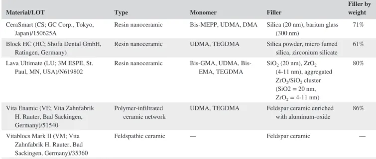
Block HC (HC; resin nanoceramic; Shofu Dental GmbH), Lava Ultimate (LU; resin nanoceramic; 3M ESPE), Vita Enamic (VE; dual-network ceramic; Vita Zahnfabrik, H. Rauter), and Vitablocs Mark II (VM; feldspathic ceramic; Vita Zahnfabrik, H. Rauter) were tested. The detailed infor-mation about compositions of the tested CAD/CAM blocks is given in Table 1.
2.1
|
Flexural strength test
Flexural properties were measured using a three-point bend-ing test accordbend-ing to ISO 6872:2008. Sixteen specimens were fabricated for each of five CAD/CAM blocks. CAD/CAM blocks were cut into rectangular-shaped bars (4 mm wide, 1.2 mm thick, and 13 mm length)12,15 using a high precision cutter (IsoMet High Speed Pro, Buehler) under water-cooling (n = 16 for each material). The specimen surfaces were then wet grounded by using silicon carbide (SiC) papers with a grit of 600, 800, and 1200 in ascending order. The specimen dimensions were checked with a micrometer, and any mens out of the range (±50 µm) were discarded. The speci-mens were then ultrasonically cleaned in deionized water for 10 min. After allowing specimens to air-dry for 24 h at room temperature, the three-point bending test was con-ducted. Each specimen was loaded to failure on a 10 mm long support span by a universal testing machine (MOD Dental, Esetron Smart Robotechnologies) at a crosshead speed of 0.5 mm/min (Figure 1). The three-point FS was calculated under the following formula: FS = 3Nl/2bd2, where N is the fracture load (N), l is the support span in mm, b is the width of the specimen in mm, and d is the thickness of the specimen in mm.12,15
2.2
|
Elastic modulus and nanohardness
measurement
Forty specimens of 3 mm thickness were obtained using the same precision cutter (n = 8 for each material). They were wet grounded by using SiC papers with a grit of 600, 800, and 1200. The specimens were then ultrasonically cleaned in deionized water for 10 minutes. After cleaning, the specimens were air-dried at room temperature for 24 h. Nanoindentation was used to measure the EM and NH of the tested CAD/CAM blocks. Five indentations were per-formed on each specimen using a nanomechanical tester (TI 950 TriboIndenter, TI 950, Hysitron Inc.) equipped with a Berkovich diamond indenter tip of a nominal radius of 100 nm. An objective lens with ×20 magnification was utilized for accuracy. The loading/unloading rates used were 0.5 mN/s, with a dwelling time of 10 s. The maximum load was set to 5.0 mN.23
2.3
|
Microshear bond strength test
The blocks were sliced into 3-mm thick slices using the same precision cutter. They were then wet grounded with SiC pa-pers (600-, 800-, and 1200-grit). For surface treatment, CS, HC, and LU were air-abraded with 50 µm aluminum-oxide particles using a mini sandblaster (Airsonic® mini sandblaster, Hager Werken) at 2.0 bar pressure and 10 mm above the specimen surface, whereas VE and VM were acid-etched with 9% hy-drofluoric acid (Ultradent) for 60 s, as recommended by their manufacturers. After the surface treatment, the specimens were ultrasonically cleaned for 10 min. After that, a silane cou-pling agent (Clearfil Ceramic Primer Plus, Kuraray Noritake Dental Inc.) was applied and followed by an universal adhe-sive (Clearfil Universal Bond, Kuraray Noritake Dental Inc.) application. The adhesive was light-cured using an LED curing unit (Elipar Deep Cure, 3M ESPE). The polyvinyl tube with an inner diameter of 1 mm was sliced into microtubules with 0.5 mm height. Each transparent microtubule was adjusted over
the specimen surface. The microtubules were carefully filled with composite resin (Filtek Ultimate Flowable, 3M ESPE). Composite resins were cured via light-activation following the manufacturers’ instructions using the LED curing unit for 20 s (n = 8 for each material), and then polymerized specimens were stored in distilled water for 24 h. After 24 h, the microtubes were removed using a surgical blade.
A microshear testing device (MOD Dental, Esetron Smart Robotechnologies) was performed a shear force to the ad-hesive interface at 0.5 mm/min crosshead speed. The load at failure was recorded, and the µSBS value was calculated by dividing the load at failure (N) to the area of adhesive interface (mm2). The failure modes were observed under a stereomicroscope at 30× magnification. Failure modes were classified as adhesive failure, cohesive failure, or mixed failure.
2.4
|
Statistical analysis
Differences in FS, EM, NH, and bond strength values were analyzed using one-way analysis of variance (ANOVA). Post hoc tests were performed using the Tukey HSD for pair-wise analysis among the materials. All statistical analyses were performed with the software Prism (GraphPad Software) with α = .05.
3
|
RESULTS AND DISCUSSION
The present in vitro study examined the mechanical prop-erties including FS, EM, and NH of CAD/CAM blocks. In
TABLE 1 Materials used in the study.
Material/LOT Type Monomer Filler Filler by weight
CeraSmart (CS; GC Corp., Tokyo,
Japan)/150625A Resin nanoceramic Bis-MEPP, UDMA, DMA Silica (20 nm), barium glass (300 nm) 71%
Block HC (HC; Shofu Dental GmbH,
Ratingen, Germany) Resin nanoceramic UDMA, TEGDMA Silica powder, micro fumed silica, zirconium silicate 61%
Lava Ultimate (LU; 3M ESPE, St.
Paul, MN, USA)/N619802 Resin nanoceramic Bis-GMA, UDMA, Bis-EMA, TEGDMA SiO(4-11 nm), aggregated 2 (20 nm), ZrO2
ZrO2/SiO2 cluster
(SiO2 = 20 nm,
ZrO2 = 4-11 nm)
80%
Vita Enamic (VE; Vita Zahnfabrik H. Rauter, Bad Sackingen, Germany)/51540
Polymer-infiltrated
ceramic network UDMA, TEGDMA Feldspar ceramic enriched with aluminum-oxide 86%
Vitablocs Mark II (VM; Vita Zahnfabrik H. Rauter, Bad Sackingen, Germany)/35360
Feldspathic ceramic — Feldspar ceramic —
Abbreviations: Bis-EMA, ethoxylated bisphenol-A dimethacrylate; Bis-GMA, bisphenol A diglycidyl methacrylate; Bis-MEPP, 2,2-Bis
(4-methacryloxypolyethoxyphenyl) propane; DMA, dimethacrylate; TEGDMA, triethylene glycol dimethacrylate; UDMA, urethane dimethacrylate.
addition, repair bond strength of CAD/CAM blocks was evaluated by performing µSBS test. The findings of the one-way ANOVA tests revealed that FS, EM, NH, and bond strength values varied significantly among the tested materi-als (p < 0.01). Therefore, the first hypothesis—that FS, EM, and NH values of CAD/CAM blocks tested would be simi-lar—was rejected. Moreover, the second hypothesis—that there would be no difference among the µSBS values of the repaired CAD/CAM blocks—was accepted as no significant difference was detected among the repair µSBS values of the different CAD/CAM blocks (p > 0.05).
Uniaxial flexure test (three-point bending test) is a fre-quently preferred and well-established method for testing the fracture strength of dental materials.7,12 In the present study, statistically significant differences were detected between the FS values of the tested CAD/CAM materials (p < 0.01), as shown in Figure 2. The highest FS values were obtained for the resin nanoceramic blocks (LU, CS, and HC, respec-tively), followed by the PICN (VE) and feldspathic ceramic (VM) materials. These findings are in accordance with those of previous studies.8,15,24 The lowest FS value was observed for the VM group (p < 0.01). VM is a feldspar-reinforced aluminosilicate glass material that contains a weak glass ma-trix with amorphous crystalline phases (silica and alumina). Therefore, VM is more fragile than zirconia-reinforced ce-ramic blocks.1,2 With regard to the findings, the fact that the polymer-based CAD/CAM blocks exhibit better FS than the VM can be explained by the resin component in their micro-structure increasing their fracture strength.
Simply put, a PICN material is manufactured by means of polymer infiltration into the microstructure of feldspathic ceramics. Coldea et al. investigated the correlation between the ceramic network densities and the FS, strain at failure, EM, and surface hardness of a range of PICN materials.25 The authors examined four ceramic network densities vary-ing between 59% and 72% as well as pure polymer and dense
ceramic cross-sections, and they reported that the ratio be-tween the porous feldspathic ceramic and the polymer con-tent influences the mechanical properties, especially the FS (ranging from 131.1 to 159.9 MPa), EM (16.4-28.1 GPa), and hardness (1.1-2.1 GPa), of the PICN materials. In the present study, resin nanoceramic blocks were found to ex-hibit greater FS than PICN material, which is in line with the results of prior studies.3,15 However, Albero et al. noted this difference to be insignificant.14 This contrast in findings may stem from differences in the study methodologies, especially in terms of the specimen thickness. The differences in the fracture strengths of the polymer-based CAD/CAM blocks can be explained by their differing microstructures, including the chemical composition of the resin matrix and the size and distribution of the filler particles.8 According to the manu-facturers’ specifications, VE has an 86/14% ceramic to poly-mer ratio by weight, whereas LU, CS, and HC have ratios of 80/20%, 71/29%, and 61/39%, respectively (Table 1). The evident differences between the ceramic to polymer ratios may explain the lower FS performance of the PICN material. Further, LU contains 31% ZrO2 filler particles within its in-organic structure. This factor might have contributed to the greater FS exhibited by LU when compared with CS and HC.
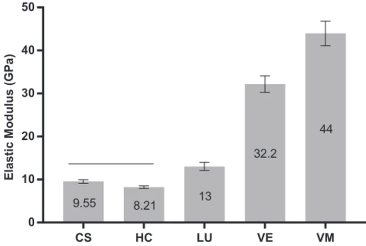
The EM of tested materials is given in Figure 3. The EM of CS (9.55 GPa), HC (8.21 GPa), and LU (13 GPa) are lower than VE (32.2 GPa), which is lower than VM (44 GPa) (p < 0.01). VE exhibited an EM that is approximately three-fold greater than the EM of the resin nanoceramic blocks, as shown in Figure 3. VE is a PICN material. Unlike conven-tional composites, a PICN material is produced via the inter-penetration of the resin component into the porous ceramic scaffold. Thus, it consists of two continuous interpenetrating networks: ceramic and polymer. A continuous interpenetrat-ing inorganic network provides stiffness to VE, and is also responsible for the higher EM. Resin nanoceramic blocks have one continuous resin matrix that is filled with inorganic
FIGURE 2 Mean flexural strength (MPa) of tested CAD/CAM blocks. Horizontal line above bars indicates that there is no statistical difference. CS HC LU VE VM 0 50 100 150 200 250 Fl exura lS tr en gt h( MPa ) 187 176 207 149 115
FIGURE 3 Mean elastic modulus (GPa) of tested CAD/CAM blocks. Horizontal line above bars indicates that there is no statistical difference. CS HC LU VE VM 0 10 20 30 40 50 El as ticM odulus (G Pa ) 9.55 8.21 13 32.2 44
particles, depending on the steric interaction between the in-organic particles. Moreover, such resins have a percolation threshold that restricts the reinforcement of the resin matrix. Therefore, resin nanoceramic blocks have lower elastic mod-uli than PICN material (p < 0.01).13,25,26
The aim is to develop restorative materials that have sim-ilar mechanical properties to those of the natural tooth struc-ture.27,28 According to previous studies, human dentin has an EM that varies from 16 to 20.3 GPa,13,29,30 whereas the EM of human enamel varies from 48 to 105.5 GPa.13,31,32 The EM of VE (32.2 GPa) is closer to that of a human tooth than the EM of VM (44 GPa). Furthermore, a recent study found that VE has an EM quite similar to that of enamel, which sup-ports the findings of the present study.33 However, the resin cements used for the luting of indirect restorations have an EM similar to that of polymer-based CAD/CAM blocks com-pared with ceramic ones due to their polymeric structure.31 Consequently, a system comprising a tooth, resin cement, and restorative material acts as a monoblock structure against the chewing forces and also shows a more uniform stress distri-bution.31,34 In the present study, the resin nanoceramic blocks showed relatively lower EM values (p < 0.01), which is in accordance with the findings of other studies.13,15 It has been stated that crown restorations fabricated using restoratives with low elastic moduli can cause microseparation from the dental tissues due to creating hoop stress, which is a form of mechanical stress associated with rotationally symmetric objects.7 This stress development restricts the areas of use of resin nanoceramic blocks.
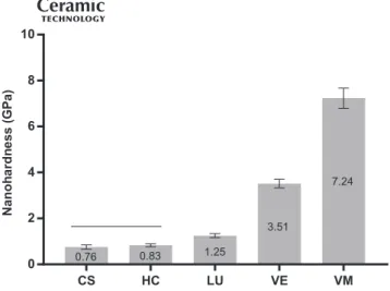
Nanoindentation is a confirmed method to precisely mea-sure the mechanical properties of dental materials.35 In the present study, it was used to measure the EM as well as the NH values of CAD/CAM blocks. Figure 4 shows NH values of the tested materials. The surface hardness can provide valu-able information about dental materials, such as the cross-linking density, wear resistance, abrasion potential against
antagonist teeth, and machinability.36,37 Previous studies reported that the surface hardness of dentin varies between 0.6 and 0.92 GPa,38,39 while the surface hardness of enamel varies between 3 and 5.3 GPa.32,40 In the present study, the highest NH value was obtained for VM (7.24 GPa), while the lowest NH values were obtained for the resin nanoceramics, namely CS (0.76 GPa), HC (0.83 GPa), and LU (1.25 GPa), respectively (p < 0.01). VE (3.51 GPa) has an in-between NH value. This finding is in agreement with the findings of previous studies.13,15 Alamoush et al. evaluated the me-chanical properties of various CAD/CAM composite blocks, including CS, HC, LU, VE, and VM, and found that the lowest NH was recorded for the specimens fabricated from resin nanoceramics, followed by VE and VM.13 The fact that polymer-based CAD/CAM blocks exhibit hardness values close to those of dental tissues indicates that these materi-als would not aggressively abrade the opposing dental tis-sues when functioning as feldspathic ceramics. In terms of the resin nanoceramic blocks, LU exhibits higher NH than CS and HC, which differ insignificantly. The greater NH of LU can be explained by its higher inorganic filler ratio (80% by weight), as it is reinforced with zirconium to a level of approximately 31%. According to the results of the present study, the first hypothesis was rejected as significantly differ-ent FS, EM, and NH values were observed for the differdiffer-ent CAD/CAM blocks.
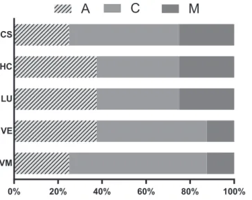
Although CAD/CAM blocks with physical properties that converge with those of the natural tooth structure have been produced, such materials, whether they are made of ceramic or composite, are known to fail due to various factors, in-cluding fatigue, occlusal thickness,16 and the EM of the lut-ing resin.31 Moreover, monolithic restoratives are associated with a high clinical failure tendency, especially in the poste-rior region,41,42 and so are frequently replaced. However, the replacement of a failed restoration may cause extra trauma and be time-consuming.43 Therefore, restorative materials should be repaired intraorally as much as possible within the minimally invasive approach. The repair protocols would in-evitably differ depending on the microstructural differences of materials. According to the manufacturer's instructions, air-borne particle abrasion is recommended for resin nano-ceramic blocks, whereas hydrofluoric acid etching is recom-mended for PICN and feldspathic ceramic blocks. Previous studies also confirm these recommendations.20,21 In the pres-ent study, no statistically significant difference was observed between the repair bond strengths of the tested CAD/CAM blocks (p > 0.05), as shown in Figure 5. All tested CAD/ CAM blocks were successfully repaired with their individual repair protocol, in terms of the acceptable repair µSBS range (15-25 MPa) suggested by Elsaka.44 Failure mode distribu-tion revealed that repaired specimens exhibit predominantly cohesive failure (Figure 6), which is supporting the µSBS results.
FIGURE 4 Mean nanohardness (GPa) of tested CAD/CAM blocks. Horizontal line above bars indicates that there is no statistical difference. CS HC LU VE VM 0 2 4 6 8 10 Nano ha rd ne ss (GPa ) 0.76 0.83 1.25 3.51 7.24
One of the main limitations of the study was that the spec-imen dspec-imensions recommended in ISO 6872:2008 could not be followed since CAD/CAM blocks are produced in certain sizes, it is impossible to provide recommended specimen dimensions. Considering that the physical properties of the materials may change and the bond strength can be affected after long-term use, the lack of aging process can be shown among the limitations of the study. Furthermore, clinical evi-dence is needed to support the findings of this in vitro study. Therefore, future studies should also include an appropriate aging protocol to simulate the oral environment.
4
|
CONCLUSIONS
Within the limitations of the present study, the following conclusions were drawn:
1. Resin nanoceramic blocks showed significantly higher flex-ural strengths than PICN and feldspathic ceramic. Besides, feldspathic ceramic exhibited significantly the highest elastic modulus and nanohardness, followed by PICN and others. 2. The mechanical properties of polymer-based CAD/CAM
blocks were within the acceptable range for the fabri-cation of single-unit restorations according to the ISO 6872:2008. Clinicians should consider material-based dif-ferences during treatment planning.
3. According to the bond strength testing, CAD/CAM blocks were successfully repaired with their recommended repair protocol and no significant differences were observed among them. Therefore, clinicians may apply a repair pro-tocol according to the manufacturer's recommendations when the repair is indicated.
ORCID
Soner Sismanoglu https://orcid.org/0000-0002-1272-5581
REFERENCES
1. Miyazaki T, Hotta Y, Kunii J, Kuriyama S, Tamaki Y. A review of dental CAD/CAM: Current status and future perspectives from 20 years of experience. Dent Mater. J. 2009;28:44–56.
2. Kelly JR, Benetti P. Ceramic materials in dentistry: historical evo-lution and current practice. Aust Dent J. 2011;56:84–96. 3. Ruse ND, Sadoun MJ. Resin-composite blocks for dental CAD/
CAM applications. J Dent Res. 2014;93:1232–4.
4. Vichi A, Carrabba M, Paravina R, Ferrari M. Translucency of ce-ramic materials for CEREC CAD/CAM system. J Esthet Restor Dent. 2014;26:224–31.
5. Della Bona A, Nogueira AD, Pecho OE. Optical properties of CAD–CAM ceramic systems. J Dent. 2014;42:1202–9.
6. Stawarczyk B, Liebermann A, Eichberger M, Güth JF. Evaluation of mechanical and optical behavior of current esthetic dental re-storative CAD/CAM composites. J Mech Behav Biomed Mater. 2016;55:1–11.
7. Lawson NC, Bansal R, Burgess JO. Wear, strength, modulus and hardness of CAD/CAM restorative materials. Dent Mater. 2016;32:275–83.
8. Sen N, Us YO. Mechanical and optical properties of monolithic CAD-CAM restorative materials. J Prosthet Dent. 2018;119:593–9. 9. Della Bona A, Corazza PH, Zhang YU. Characterization of
a polymer-infiltrated ceramic-network material. Dent Mater. 2014;30:564–9.
10. Johnson AC, Versluis A, Tantbirojn D, Ahuja S. Ahuja Sç fracture strength of CAD/CAM composite and composite-ceramic occlusal veneers. J Prosthodont Res. 2014;58:107–14.
11. Mainjot AK, Dupont NM, Oudkerk JC, Dewael TY, Sadoun MJ. From artisanal to CAD-CAM blocks: state of the art of indirect composites. J Dent Res. 2016;95:487–95.
12. Sonmez N, Gultekin P, Turp V, Akgungor G, Sen D, Mijiritsky E. Evaluation of five CAD/CAM materials by microstructural charac-terization and mechanical tests: a comparative in vitro study. BMC Oral Health. 2018;18:1–13.
13. Alamoush RA, Silikas N, Salim NA, Al-Nasrawi S, Satterthwaite JD. Effect of the composition of CAD/CAM composite blocks on mechanical properties. Biomed Res Int. 2018;2018:4893143.
FIGURE 5 Mean repair microshear bond strength (MPa) of tested CAD/CAM blocks. Horizontal line above bars indicates that there is no statistical difference.
FIGURE 6 Failure mode distribution.
0% 20% 40% 60% 80% 100% VM VE LU HC CS
A
C
M
14. Albero A, Pascual A, Camps I, Grau-Benitez M. Comparative characterization of a novel CAD-CAM polymer-infiltrated-ceram-ic-network. J Clin Exp Dent. 2015;7:495–550.
15. Lauvahutanon S, Takahashi H, Shiozawa M, Iwasaki N, Asakawa Y, Oki M, et al. Mechanical properties of composite resin blocks for CAD/CAM. Dent Mater J. 2014;33:705–10.
16. Chen C, Trindade FZ, de Jager N, Kleverlaan CJ, Feilzer AJ. The fracture resistance of a CAD/CAM resin nano ceramic (RNC) and a CAD ceramic at different thicknesses. Dent Mater. 2014;30:954–62. 17. Dayan SC, Mumcu E. Effect of different storage media on the
microhardness and wear resistance of resin-matrix ceramics. Int J Appl Ceramic Tech. 2019;16:2467–73.
18. Ozcan M, Barbosa SH, Melo RM, Galhano GA, Bottino MA. Effect of surface conditioning methods on the microtensile bond strength of resin composite to composite after aging conditions. Dent Mater. 2007;23:1276–82.
19. Attia A. Influence of surface treatment and cyclic loading on the durability of repaired all-ceramic crowns. J Appl Oral Sci. 2010;18:194–200.
20. Sismanoglu S, Yildirim-Bilmez Z, Erten-Taysi A, Ercal P. (2020) Influence of different surface treatments and universal adhesives on the repair of CAD-CAM composite resins: an in vitro study. J Prosthet Dent. 2020;124:238.e1–238.e9.
21. Sismanoglu S, Gurcan AT, Yildirim-Bilmez Z, Turunc-Oguzman R, Gumustas B. Effect of surface treatments and universal adhe-sive application on the microshear bond strength of CAD/CAM materials. J Adv Prosthodont. 2020;12:22–32.
22. Akay C, Cakirbay Tanis M, Sen M, Akkas KP. Strengthen adhe-sion between zirconia and resin cement using different surface modifications. Int J Appl Ceramic Tech. 2019;16:917–22. 23. Perea-Lowery L, Minja IK, Lassila L, Ramakrishnaiah R, Vallittu
PK. Assessment of CAD-CAM polymers for digitally fabricated complete dentures. J Prosthet Dent. 2020. https://doi.org/10.1016/j. prosd ent.2019.12.008
24. Coldea A, Swain MV, Thiel N. In-vitro strength degradation of dental ceramics and novel PICN material by sharp indentation. J Mech Behav Biomed Mater. 2013;26:34–42.
25. Coldea A, Swain MV, Thiel N. Mechanical properties of polymer-infiltrated-ceramic-network materials. Dent Mater. 2013;29:419–26.
26. Belli R, Wendler M, de Ligny D, Cicconi MR, Petschelt A, Peterlik H, et al. Chairside CAD/CAM materials. Part 1: measurement of elastic constants and microstructural characterization. Dent Mater. 2017;33:84–98.
27. Zhang Y, Kelly JR. Dental ceramics for restoration and metal ve-neering. Dent Clin North Am. 2017;61:797–819.
28. Petrini M, Ferrante M, Su B. Fabrication and characterization of biomimetic ceramic/polymer composite materials for dental resto-ration. Dent Mater. 2013;29:375–81.
29. Hairul Nizam BR, Lim CT, Chng HK, Yap AU. Nanoindentation study of human premolars subjected to bleaching agent. J Biomech. 2005;38:2204–11.
30. Lawn BR, Deng Y, Thompson VP. Use of contact testing in the characterization and design of all-ceramic crownlike layer struc-tures: a review. J Prosthet Dent. 2001;86:495–510.
31. Ausiello P, Rengo S, Davidson CL, Watts DC. Stress distributions in adhesively cemented ceramic and resin-composite Class II inlay restorations: a 3D-FEA study. Dent Mater. 2004;20:862–72. 32. He LH, Swain MV. Nanoindentation derived stress-strain
proper-ties of dental materials. Dent Mater. 2007;23:814–21.
33. Al-Shatti RA, Dashti GH, Philip S, Michael S, Swain MV. Size or hierarchical dependence of the elastic modulus of three ceram-ic-composite CAD/CAM materials. Dent Mater. 2019;35:953–62. 34. Ichim IP, Schmidlin PR, Li Q, Kieser JA, Swain MV. Restoration
of non-carious cervical lesions. Part II. Restorative material selec-tion to minimise fracture. Dent Mater. 2007;23:1562–9.
35. Doerner MF, Nix WD. A method for interpreting the data from depth-sensing indentation instruments. J Mater Res. 1986;1:601–9. 36. Murakami N, Wakabayashi N, Matsushima R, Kishida A, Igarashi
Y. Effect of high-pressure polymerization on mechanical proper-ties of PMMA denture base resin. J Mech Behav Biomed Mater. 2013;20:98–104.
37. Awada A, Nathanson D. Mechanical properties of resin-ce-ramic CAD/CAM restorative materials. J Prosthet Dent. 2015;114:587–93.
38. Lawn BR, Lee JJW. Analysis of fracture and deformation modes in teeth subjected to occlusal loading. Acta Biomater. 2009;5:2213–21.
39. Mahoney E, Holt A, Swain M, Kilpatrick N. The hardness and modulus of elasticity of primary molar teeth: an ultra-micro-inden-tation study. J Dent. 2000;28:589–94.
40. Park S, Quinn JB, Romberg E, Arola D. On the brittleness of enamel and selected dental materials. Dent Mater. 2008;24:1477–85. 41. Nandini S. Indirect resin composites. J Conserv Dent.
2010;13:184–94.
42. Alshehri SA. An investigation into the role of core porcelain thick-ness and lamination in determining the flexural strength of in-ce-ram dental materials. J Prosthodont. 2011;20:261–6.
43. Wady AF, Paleari AG, Queiroz TP, Margonar R. Repair technique for fractured implant-supported metal-ceramic restorations: a clin-ical report. J Oral Implantol. 2014;40:589–92.
44. Elsaka SE. Repair bond strength of resin composite to a novel CAD/CAM hybrid ceramic using different repair systems. Dent Mater J. 2015;34:161–7.
How to cite this article: Sismanoglu S, Tugce Gurcan A, Yildirim-Bilmez Z, Gumustas B. Mechanical properties and repair bond strength of polymer-based CAD/CAM restorative materials. Int J Appl Ceram
Technol. 2021;18:312–318. https://doi.org/10.1111/ ijac.13653