Clinical experience with coronary sinus catheterization in minimally invasive
aortic valve surgery under transesophageal echocardiography guidance
Ergun Demirsoy
1, Ugur Ozbek
2, Osman Bayindir
2& Bingur Sonmez
11
Department of Cardiovascular Surgery; 2Department of Anesthesiology, Kadir Has University, Florence Nightingale Hospital, Istanbul, Turkey
Received 9 January 2002;accepted in revised form 23 March 2002
Key words: aortic valve surgery, minimally invasive, retrograde coronary sinus catheter
Abstract
We aimed to show conventional coronary sinus (CS) catheter could be used with transesophageal echo-cardiography (TEE) guidance through the limited surgical field in aortic valve surgery with ‘J’ sternotomy. This method was performed in 14 patients and completed successfully in 12. We believe that in minimally invasive aortic valve surgery, the insertion of the conventional retrograde cardioplegia catheter to the CS with routine way may not be possible but application of TEE guidance is cost-effective and easily applicable method without significant complications.
Introduction
Cardioplegia delivery via coronary sinus (CS) with retrograde cannulation or combined with ante-grade is a preferable method in open heart surgery [1, 2]. Although delivery of cardioplegic solution through direct cannulation of both coronary ostia is possible but may cause intimal injury of the ostia, therefore retrograde cardioplegia has an important role in aortic valve surgery [2]. During minimally invasive aortic valve surgery, insertion of the con-ventional retrograde cardioplegia catheter to the CS is possible but may not be easy due to limited surgical field [3]. We have been inserting the retro-grade cardioplegia catheter with transesophageal echocardiography (TEE) guidance in minimally invasive aortic valve surgery for 9 months. Cathe-terization of CS becomes easy with the control of tip of the catheter position in two-dimensional TEE.
In this study, we aimed to share our experiences of CS catheterization with TEE guidance in 14 patients in minimally invasive aortic valve surgery with ‘J’ sternotomy.
Methods
Retrograde CS cannulation with TEE guidance in 14 patients in minimally invasive aortic valve sur-gery between October 2000 and June 2001 studied retrospectively.
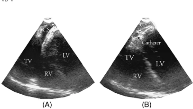
After anesthetic induction and central venous catheterization 5.0 MHz multiplane TEE probe was placed. Following the routine echocardio-graphic examination of the heart, the probe (GE Vingmed Ultrasound Horten, Norway) was ad-vanced in the transverse plan (0°) four chamber view until the CS was seen (Figure 1A).
Adequate surgical exposure was obtained to re-place the aortic valve with 6–8 cm ‘J’ sternotomy described by Gundry et al. [4]. Routine CS catheter (15 Fr., Medtronic DLP, Grand Rapids, MI, USA) was inserted into the right atrium through a small purse string. The tip of the catheter was guided with TEE to the CS ostium so that when the catheter turned towards the right ventricle or in-ferior vena cava, drawn back slightly and directed to the CS again. After the catheter entered to the
The International Journal of Cardiovascular Imaging 18: 453–455, 2002.
ostium, advanced 3–4 cm into the CS, subse-quently its position was confirmed by typical CS waveform after the balloon inflation (Figure 1B).
Results
In 12 of 14 patients who underwent minimally invasive aortic valve surgery with ‘J’ sternotomy, insertion of the retrograde cardioplegia catheter into the CS was completed successfully. Initially, we had longer cannulation time but got better. We were not able to cannulate in two patients despite many attempts and cardioplegic solution was given through the coronary ostia directly.
In one patient, we diagnosed catheter displace-ment during surgery with TEE and direct coronary ostial cannulation was used.
One patient produced atrial fibrillation during the insertion but since the catheter was in right position, cardioplegic solution delivered and maintained good arrest. Mean insertion time was 3.41 min (min–max: 0.45–11.30 min).
Discussion
Minimally invasive approaches for aortic valve replacement have been preferred because of re-duced postoperative pain and length of stay in hospital, and also cosmetic reasons [5–7]. But with this approach, surgeon may not feel safe if possi-bility of retrograde cardioplegic delivery is not possible due to difficult manipulations. Recently,
TEE monitoring is an available and important part of all cardiac surgical procedures as well as minimally invasive approaches and gives us very important informations about catheter positions, valve functions, wall motions and other patholo-gies of the heart all way through the surgery [8].
‘J’ sternotomy approaches can provide enough surgical exposure to establish cardio pulmonary bypass and perform the aortic valve replacement in a safe way [4, 9]. With this technique ascending aorta approached easily but manipulation of the heart and insertion of conventional retrograde cardioplegia catheter to the CS might be extremely difficult and sometimes impossible. On the other hand repeated blind attempts would increase the possibility of complications and false cannula-tions. After aortotomy, direct delivery of cardio-plegic solution through both coronary ostia is possible but may cause intimal injury which cause acute dissection or late ostial stenosis [2]. Using the percutaneous CS catheter insertion via internal jugular vein is possible but expensive, and also needs TEE guidance and experienced hands.
Plotkin et al. [3] reported their experiences about percutaneous CS cannulation via internal juguler vein guided by TEE. In this study they found that the mean CS cannulation time was 10.5 min after placement of the introducer (min–max: 1.5– 42 min). This period is longer than our mean cannulation time since the length of the catheter is shorter and needs less manipulation in our method. This is a improved technique which does not need a lot of experience and cheaper than percutaneous technique.
Since the insertion of the conventional retro-grade cardioplegia catheter to the CS with TEE guidance is cost-effective and easily applicable in short period of time and does not cause any morbidity. This method is expected to be popular in minimal invasive aortic valve surgery and with the advent of intracardiac echocardiography, it will be able to be applied much more easier.
References
1. Chitwood WR Jr. Retrograde cardioplegia: current methods. Ann Thorac Surg 1992;53(2): 352–355.
Figure 1. (A) TEE 0° image showing CS. (B) TEE 0° image showing the CS catheter with in the lumen of the CS. CS – coronary sinus;TV – tricuspid valve;LV – left ventricle;RV – right ventricle.
2. Menasche P, Kural S, Fauchet M, et al. Retrograde coronary sinus perfusion: a safe alternative for ensuring cardioplegic delivery in aortic valve surgery. Ann Thorac Surg 1982; 34(6): 647–658.
3. Plotkin IM, Collard CD, Aranki SF, Rizzo RJ, Shernan SK. Percutaneous coronary sinus cannulation guided by trans-esophageal echocardiography. Ann Thorac Surg 1998;66: 2085–2087.
4. Gundry SR, Shattuck OH, Razzaouk AJ, del Rio MJ, Sardari FF, Bailey LL. Facile minimally invasive cardiac surgery via ministernotomy. Ann Thorac Surg 1998;65(4): 1100–1104. 5. Luciani GB, Mazzuco A. Aortic valve replacement via
minimal access complete sternotomy. Tex Heart Inst J 2000; 27(3): 286–288.
6. Lee JW, Lee SK, Choo SJ, Song H, Song MG. Routine minimally invasive aortic valve procedures. Cardiovasc Surg 2000;8(6): 484–489.
7. Szerafin T, Jagamos E, Jaber O, et al. Mini-sternotomy for aortic valve surgery. Acta Chir Hung 1997;36(1–4): 352–355. 8. Gillam LD. Intraoperative transesophageal
echocardiogra-phy. Cardiol Rev 2000;8(5): 269–278.
9. Svensson Lg, D’Agostino RS. ‘J’ incision minimal-access valve operations. Ann Thorac Surg 1998;66(3): 1110–1112.
Address for correspondence: Dr Demirsoy, Necip Bey Sok., Melen Apt., 6/6 81010 Acibadem, Istanbul, Turkey
Tel./Fax: +90-212-2208910 E-mail: [email protected]