http://journals.tubitak.gov.tr/medical/ © TÜBİTAK
doi:10.3906/sag-1504-41
Evaluation of cutaneous anthrax cases during an outbreak in the east region of Turkey
Esra KURAL ÜNÜVAR1,*, Deniz Bahar AKGÜN KARAPINAR2, Nazlı DİZEN NAMDAR31Department of Dermatology and Venereology, Eren Hospital, İstanbul, Turkey
2Department of Medical Microbiology, İstanbul Faculty of Medicine, İstanbul University, İstanbul, Turkey 3Department of Dermatology and Venereology, Faculty of Medicine, Dumlupınar University, Kütahya, Turkey
1. Introduction
Anthrax is a zoonotic infection caused by Bacillus anthracis, which is a gram-positive, nonmotile, aerobic, spore-forming, encapsulated, large, rod-shaped bacterium (1). The major virulence factors of Bacillus anthracis are encoded on 2 virulence plasmids, pXO1 and pXO2. The tritoxin-bearing plasmid pXO1 codes for three toxins, which cause hemorrhage, edema, and necrosis. Bacillus anthracis has the lethal factor, edema factor, and protective antigen, which contains the host cell receptor component. The capsule-bearing plasmid pXO2 encodes three genes (cap A, cap B, cap C) involved in the synthesis of the polyglutamyl capsule that inhibits host phagocytosis of the vegetative form of Bacillus anthracis. Both plasmids are necessary for full virulence (2).
Anthrax can be found naturally in soil and commonly affects domestic and wild animals around the world. People can get sick with anthrax if they come in contact with infected animals or contaminated animal products. Anthrax could be used as a weapon of bioterrorism. Human anthrax is classified in two ways:
(i) Based on the occupation of the individual:
a) Nonindustrial anthrax, which occurs in farmers, butchers, veterinarians, etc.
b) Industrial anthrax, which appears in people who process animal products like bones, hides, and wool.
(ii) Based on how the disease was acquired:
a) Cutaneous anthrax is the most common clinical form. The spores get into the skin, usually through skin lacerations, abrasions, or biting flies when a person handles infected animals or contaminated animals products like wool, hides, or hair.
b) Pulmonary anthrax occurs via inhalation of the spores.
c) Gastrointestinal anthrax occurs via ingestion of spores.
Inside the infected host, the vegetative form of Bacillus anthracis multiplies in the lymphatics and the spleen. Simultaneously, a capsule and toxins are produced, which eventually lead to the death of the animal. Then Bacillus Background/aim: Anthrax is a zoonotic infection caused by Bacillus anthracis. We aimed to retrospectively evaluate cutaneous anthrax
cases that occurred during an outbreak in eastern Turkey (Hakkari-Yüksekova), where people mostly earn their living from animal husbandry.
Materials and methods: Forty-six cutaneous anthrax patients that were admitted to the hospital during a very short duration of 3
months (June–August 2011) were evaluated.
Results: Out of 46 patients, 27 (52%) were women and 19 (48%) were men. The mean age was 37 ± 13 years. The distribution of
occupations was 1 butcher, 1 cook, 5 farmers, 27 housewives, 11 shepherds, and 1 teacher. Multiple lesions were seen in 7 patients (15%) and the rest of the patients had only 1 lesion. We observed significant clinical differences among the cases and noted which particular symptoms were associated with the various skin lesions. We treated our patients with intramuscular procaine penicillin or oral ciprofloxacin/doxycycline.
Conclusion: Anthrax is an important health problem that can cause lethal outbreaks. Therefore, one should think about anthrax when
faced with a patient with history of animal contact that has a painless ulcer with edema and/or vesicles, especially in endemic countries like Turkey.
Key words: Bacillus anthracis, cutaneous anthrax, animal husbandry
Received: 21.04.2015 Accepted/Published Online: 20.01.2016 Final Version: 17.11.2016
anthracis is released into the environment. Sporulation requires the presence of free oxygen. Once outside the host with exposure to air, the sporulation occurs and is ready to be ingested by another animal. The ingestion by the next host may happen at any time, from less than one hour to many decades later. Humans can be infected with these spores via microinjury (cutaneous), via ingestion of infected meat or contaminated water (gastrointestinal), or via inhalation of spores (pulmonary) (3).
Bacillus anthracis toxins cause local skin lesions resulting in tissue necrosis. Organisms may also enter the lymphatic system, resulting in lymphangitis and lymphadenopathy. Occasionally skin lesions are accompanied by bacteremia and systemic symptoms (4).
2. Materials and methods
In this retrospective study, we aimed to evaluate cutaneous anthrax cases during an outbreak in the eastern region of Turkey, Hakkari-Yüksekova, where people mostly earn their living from animal husbandry. The diagnosis of patients was based upon a detailed history and physical examination and direct microscopic examination of Gram stain smears from the samples taken from the lesions. We evaluated 46 patients that were admitted to the hospital during a very short duration of 3 months (June–August 2011). Thirty-four patients were hospitalized and the other 12 patients were treated as outpatients. Age, sex, occupation, incubation period, what kind of animal the patients had contact with, the location, number, and type of lesion, the symptoms that accompanied the lesion, and the choice of treatment were recorded. All the characteristic lesions were photographed. Clinical samples were obtained with needle aspiration from the vesicle in the vesicular stage. During the eschar stage we lifted the edge of the eschar with tweezers and obtained the fluid. Gram stain was performed on the specimens and gram-positive bacilli were observed. We took pictures from the microscope, but imaging was unsuccessful. Culture tests of the samples could not be performed because of a lack of appropriate biosafety conditions of the laboratory in the small, local hospital. Moreover, the laboratories able to perform culture tests were far away and the transport of these dangerous samples was hazardous.
3. Results
In our study, 46 patients were assessed in 3 months. Out of 46 patients, 27 (59%) were female and 19 (41%) were male. The youngest patient was 12 years old and the oldest patient was 70 years old. The mean age was 37 ± 13.7 years (Table). The distribution of occupations was 1 butcher, 1 cook, 5 farmers, 27 housewives, 11 shepherds, and 1 teacher (Figure 1).
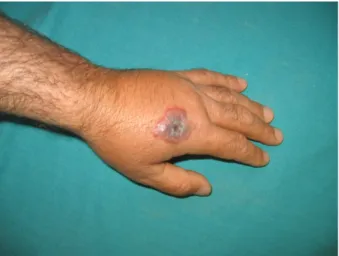
We could not note the incubation period because the patients could not precisely remember the day the lesions began to occur; however, they all remembered when they touched the contaminated animal products. Therefore, we noted the time between contamination and coming for examination was 4.67 ± 1.7 days. We recorded the parts of the body where the lesions were localized. In our cases, forearm, hand, or finger involvement was reported (Figure 2–4). Seven (15%) of the patients had multiple lesions and 39 (85%) of the patients had one lesion (Table).
We observed the clinical differences and noted what kinds of signs accompanied the skin lesion. Pain in the arm, fever, lymphadenopathy, and lymphangitis accompanied the lesions (Table). The treatment chosen and its success were also recorded. Only 4 of the patients had taken antibiotic treatments before coming to the hospital. After the spores of B. anthracis enter the skin through an abrasion or cut, or the vegetative form is transferred by insect bite, lesions usually appear and are followed by papules and vesicles around the papules; edema develops after the papule ulcerates and the characteristic eschar occurs. When the patients came to the hospital we observed papule, vesicle, and eschar forms of the disease. Sixteen of the patients had vesicles, 7 of the patients had papules, and 23 of the patients had eschars. Twenty-one patients had edema accompanied by lesions (Table). Out of the 46 patients, 14 patients were contaminated by cows and 32 patients were contaminated by sheep.
Clinical findings and Gram staining were positive in almost all (in 42 patients) of the cases. We were not able to see the agent in only 4 patients. These 4 patients had a history of contact with infected animals and typical clinical findings. They had taken antibiotics before coming for an examination and so we diagnosed anthrax without microbial confirmation for these 4.We treated our patients with intramuscular procaine penicillin or oral ciprofloxacin/doxycycline (Figure 5). All our cases were uncomplicated cutaneous anthrax. We treated all of our patients for 7 days. We used intramuscular procaine penicillin 2 × 1 800,000 units for 35 patients, oral ciprofloxacin 500 mg 2 × 1 for 10 patients who wanted outpatient treatment, and oral doxycycline 100 mg 2 × 1 for 1 patient who had a penicillin allergy. We had 1 pregnant patient and we treated her with intramuscular penicillin.
4. Discussion
History and clinical appearance of the lesions are important data for the diagnosis of cutaneous anthrax. The diagnosis can be confirmed by detecting the agent in staining (Giemsa, polychrome methylene blue, or gram-positive), the growth of the organism in a culture, or both. Nowadays serology, immunohistochemistry tests for anthrax-specific protective antigens, and PCR are available
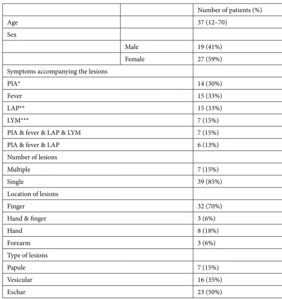
Table. Age and sex distribution of patients; symptoms accompanying the lesions; number, type, and location of lesions. Number of patients (%) Age 37 (12–70) Sex Male 19 (41%) Female 27 (59%)
Symptoms accompanying the lesions
PIA* 14 (30%)
Fever 15 (33%)
LAP** 15 (33%)
LYM*** 7 (15%)
PIA & fever & LAP & LYM 7 (15%)
PIA & fever & LAP 6 (13%)
Number of lesions
Multiple 7 (15%)
Single 39 (85%)
Location of lesions
Finger 32 (70%)
Hand & finger 3 (6%)
Hand 8 (18%) Forearm 3 (6%) Type of lesions Papule 7 (15%) Vesicular 16 (35%) Eschar 23 (50%)
*PIA: pain in arm,**LAP: lymphadenopathy,*** LYM: lymphangitis.
Figure 1. Occupational distribution of patients. Figure 2. A lesion on the forearm. Pain in arm, fever, and
in specialized laboratories for retrospective confirmation of diagnosis (3,5). In our cases, Gram staining was performed on the specimens and gram-positive bacilli were observed. However, B. anthracis will die quickly after appropriate antibiotic treatment. The characteristic lesion can survive many weeks longer, because of the toxin-induced damage. Clinicians need to be aware of the late resolution and not extend the treatment unnecessarily (3). The differential diagnosis of cutaneous anthrax includes impetigo, cellulites, orf (ecthyma contagiosum), milker’s nodules, cutaneous leishmaniasis, cutaneous tuberculosis, arachnoid bites, plague, ulceroglandular tularemia, and ecthyma gangrenosum (3,5,6). In our study, most of the patients were female, but there was no significant difference between sexes. In many studies it has also been shown that there were no differences between the sexes (7–10). The incubation period ranges from a few hours to 3 weeks, most often 2 to 6 days (2). We could not note the incubation period because the patients could not precisely remember the day the lesions began to occur; however, they all remembered when they touched the contaminated animal products. Therefore, we noted the time between
contamination and coming for examination, which was 4.67 ± 1.7 days. The duration was similar to the incubation period. In our cases, forearm, hand, or finger involvement was reported. Results were similar to the results of studies that were reported before in Turkey (7,8,11–13). Penicillin G is still the drug of choice, though doxycycline or ciprofloxacin are now accepted as the best alternatives in the treatment of naturally occurring cutaneous anthrax (3,11,12,14,15). Penicillin alone is not recommended for the life-threatening condition of anthrax (3,14). In severe life-threatening cases, intravenous penicillin or another chosen primary antibiotic, for example ciprofloxacin, may be combined with other antibiotics. For anthrax meningitis, the additional antibiotic is suggested to be vancomycin or rifampicin; for gastrointestinal anthrax, streptomycin or other aminoglycosides are suggested as well (3). Both the Center for Disease Control (CDC) and the European guidelines established that the first line of treatment should be ciprofloxacin or doxycycline, with an additional one or two antibiotics (either rifampicin, chloramphenicol, clindamycin, clarithromycin, erythromycin, gentamicin, streptomycin, or vancomycin) for inhalational anthrax. A 60-day treatment is recommended for just ciprofloxacin according to the CDC and the European guidelines (16,17). In studies in Turkey, penicillin resistance has not been shown yet (11,14). Penicillin G is still the drug of choice, though doxycycline or ciprofloxacin are now accepted as the best alternatives in the treatment of naturally occurring cutaneous anthrax (12,15). All of our cases were naturally occurring anthrax. Therefore, we treated our patients with intramuscular procaine penicillin or oral ciprofloxacin/ doxycycline. B. anthracis is generally sensitive to antibiotic therapy but the clinical effects of the toxin may continue for some time afterwards. Suggested durations are 3–7 days for uncomplicated cutaneous anthrax and 10–14 days in cases Figure 3. A lesion on the hand. Pain in arm, lymphadenopathy,
and fever accompanied the lesion. Figure 4. Multiple lesions on the fingers. Pain in arm, fever, and lymphadenopathy accompanied the lesions.
24% 2% 74% Ciprofloxaci Doxycyline Penicillin
of systemic anthrax (3). Penicillin (in combination with rifampicin or vancomycin in life-threatening infections) is suitable for pregnant women and nursing mothers, as well as with children (3). We treated our pregnant patient with intramuscular penicillin as well.
Studies have shown that contamination with cooked, infected animal meat is not a risk factor for cutaneous anthrax (18,19). Cooking (100–150 °C for 10 to 60 min) sterilizes raw animal materials, which may then be consumed safely (20,21). Education of the community to use personal protective equipment is important. They should be encouraged to wear gumboots, gloves, and aprons while slaughtering dead animals and handling meat and skins. We also should emphasize the importance of recognizing the disease and reporting it promptly. Early reports of animal cases would allow secondary prevention measures, including (a) decontamination of slaughtering sites, (b) surveillance of animals, (c) advice to avoid slaughtering ill and dead livestock, and (d) vaccination of herbivores (22,23).
District health authorities and animal husbandry personnel should be alerted promptly about the existence of anthrax in their area to start control measures as early as possible. In this way, mass vaccination of livestock, proper disposal of carcasses with lime, and soil decontamination in the affected areas and in nearby villages can be initiated early (24).
The outbreak took place during a very short period of time. A reason for the sudden outbreak is a lack of enforcement of health and safety standards in this region. As a result, there were a few animal clusters with sick animals that were taken care of by uneducated animal husbandry
personnel and were owned by people unfamiliar with the disease. Neither the owners nor the caretakers of the sick animals reported the health of the animals to government officials. This could be due to their ignorance or it could be a deliberate choice so as not to incur financial losses. As a consequence, preventative measures were not taken. The general population that lives in this district is uneducated about the disease as well and so they were contaminated by those sick animals. Therefore, it is very important to inform the general population and encourage animal husbandry personnel to take precautions in order to prevent an outbreak.
When we established the diagnosis of anthrax, we informed the district health authorities and the district governor; hence, we had the chance to contact the owners of animal clusters and veterinarians, as well as the population that lives in this district. Veterinarians educated people to promptly report the death and illness of animals. Informational flyers that include information about the disease and protection methods were prepared and information was given orally as well. Soil was decontaminated with bleaching powder, the dead animals were buried with lime, and healthy livestock was vaccinated. We also encouraged people, especially housewives, to use protective clothes like gloves while touching meat during cooking. This is due to the fact that we had 28 people who were infected by touching raw meat of ill animals during cooking (27 housewives and 1 teacher) (Figure 3).
In conclusion, one should think about anthrax when faced with a patient with a history of animal contact who has painless ulcers with edema, vesicles, or papules, especially in endemic countries like Turkey.
References
1. Turnbull PCB, Kramer JM. Bacillus. In: Balows A, Hausler WJ, Herrmann KL, Isenberg HD, Shadomy HJ, editors. 5th ed. Manual of Clinical Microbiology. Washington, DC, USA: ASM Press; 1991. pp. 296-303.
2. Little SF, Ivins BE. Molecular pathogenesis of Bacillus anthracis
infection. Microbes Infect 1999; 1: 131-139.
3. Turnbull P. Anthrax in Humans and Animals. 4th ed. Geneva, Switzerland: WHO Press; 2008.
4. Doğanay M, Almaç A, Hanağasi R. Primary throat anthrax. A
report of six cases. Scand J Infect Dis 1986; 18: 415-419.
5. Thappa DM, Karthikeyan K. Anthrax: an overview within the
Indian subcontinent. Int J Dermatol 2001; 40: 216-222.
6. Wenner KA, Kenner JR. Anthrax. Dermatol Clin 2004; 22:
247-256.
7. Kaya A, Tasyaran MA, Erol S, Ozkurt Z, Ozkan B. Anthrax in
adults and children: a review of 132 cases in Turkey. Eur J Clin Microbiol Infect Dis 2002; 21: 258-261.
8. Karahocagil MK, Akdeniz N, Akdeniz H, Calka O, Karsen H,
Bilici A, Bilgili SG, Evirgen O. Cutaneous anthrax in eastern Turkey: a review of 85 cases. Clin Exp Dermatol 2008; 33: 406-411.
9. Doğanay M, Kökkaya A, Hah MN. A review of 35 anthrax cases. Mikrobiyoloji Bülteni 1983; 17: 1-10 (article in Turkish with a summary in English).
10. Oncül O, Ozsoy MF, Gul HC, Koçak N, Cavuslu S, Pahsa A. Cutaneous anthrax in Turkey: a review of 32 cases. Scand J Infect Dis 2002; 34: 413-416.
11. Engin A, Elaldı N, Dökmetaş İ, Bakıcı MZ, Kaya Ş, Bakır M. Cutaneous anthrax in the central Anatolia region of Turkey: a review of 39 adults cases. Türkiye Klinikleri J Med Sci 2010; 30: 1032-1038.
12. Baykam N, Ergonul O, Ulu A, Eren S, Celikbas A, Eroglu M, Dokuzoguz B. Characteristics of cutaneous anthrax in Turkey. J Infect Dev Ctries 2009; 3: 599-603.
13. Demirdag K, Ozden M, Saral Y, Kalkan A, Kilic SS, Ozdarendeli A. Cutaneous anthrax in adults: a review of 25 cases in the eastern Anatolian region of Turkey. Infection 2003; 31: 327-330.
14. Metan G, Doğanay M. The antimicrobial susceptibility of
Bacillus anthracis isolated from human cases: a review of the
Turkish literature. Türkiye Klinikleri J Med Sci 2009; 29: 229-235.
15. Doganay M, Metan G, Alp E. A review of cutaneous anthrax and its outcome. J Infect Public Health 2010; 3: 98-105. 16. Bossi P, Tegnell A, Baka A, Van Loock F, Hendriks J, Werner
A, Maidhof H, Gouvras G. Bichat guidelines for the clinical management of anthrax and bioterrorism-related anthrax. Euro Surveill 2004; 9: 1-7.
17. Stern EJ, Uhde KB, Shadomy SV, Messonnier N. Conference report on public health and clinical guidelines for anthrax. Emerg Infect Dis 2008; 14: e1.
18. Ray TK, Hutin YJ, Murhekar MV. Cutaneous anthrax, West Bengal, India, 2007. Emerg Infect Dis 2009; 15: 497-499. 19. Woods CW, Ospanov K, Myrzabekov A, Favorov M, Plikaytis
B, Ashford DA. Risk factors for human anthrax among contacts of anthrax-infected livestock in Kazakhstan. Am J Trop Med Hyg 2004; 71: 48-52.
20. Turnbull PCB, Böhm R, Cosivi O, Doganay M, Hugh-Jones ME, Joshi DD, Lalitha MK, de Vos V. Guidelines for the Surveillance and Control of Anthrax in Human and Animals. 3rd ed. WHO/EMC/ZDI/98/6.
21. Rao GR, Padmaja J, Lalitha MK, Rao PV, Gopal KV, Kumar HK, Mohanraj P. An outbreak of cutaneous anthrax in a non-endemic district--Visakhapatnam in Andhra Pradesh. Indian J Dermatol Venereol Leprol 2005; 71: 102-105.
22. Chakraborty A, Khan SU, Hasnat MA, Parveen S, Islam MS, Mikolon A, Chakraborty RK, Ahmed BN, Ara K, Haider N et al. Anthrax outbreaks in Bangladesh, 2009-2010. Am J Trop Med Hyg 2012; 86: 703-710.
23. Reddy R, Parasadini G, Rao P, Uthappa CK, Murhekar MV. Outbreak of cutaneous anthrax in Musalimadugu village, Chittoor district, Andhra Pradesh, India, July-August 2011. J Infect Dev Ctries 2012; 6: 695-699.
24. Rao GRR, Padmaja J, Lalitha MK, Rao PVK, Kumar HK, Gopal KVT, Jaideep M, Mohanraj P. Cutaneous anthrax in a remote tribal area—Araku Valley, Visakhapatnam district, Andhra Pradesh, southern India. Int J Dermatol 2007; 46: 55-58.