0022-4766/16/5704-0731 © 2016 by Pleiades Publishing, Ltd. 731 Journal of Structural Chemistry. Vol. 57, No. 4, pp. 731-736, 2016.
Original Russian Text © 2016 A. Karahan, R. Kurtaran, Y. Yahsi, E. Gungor, H. Kara.
A DINUCLEAR OXYGEN-BRIDGED
SCHIFF BASE IRON(III) COMPLEX DERIVED
FROM N,N′-bis(4-METHOXY-2-HYDROXYBENZYLIDENE)-
2,2-DIMETHYLPROPANE-1,3-DIAMINE
A. Karahan1, R. Kurtaran2, Y. Yahsi3, E. Gungor3, and H. Kara3,4
UDC 548.73:541.49:546.72
The μ-oxo-bridged Fe(III) dimer complex [{Fe(4-MeOL1)}2(μ-O)]⋅HOCH3, (H2-4-MeOL1 =
N,N′-bis(4-methoxy-2-hydroxybenzylidene)-2,2-dimethylpropane-1,3-diamine), 1 is synthesized and characterized by single crystal X-ray diffraction. Complex 1 contains a [{Fe(4-MeOL1)}2(μ-O)] dimeric unit with a methanol solvent molecule of crystallization. Each Fe(III) ion has a distorted square-pyramidal coordination geometry. In the basal plane, the Fe(III) atom is coordinated by two N and two O atoms of the Schiff base ligand. The apical position is occupied by a bridging O2– ion, linking another Fe(III) ion in the complex. There are intermolecular C–H⋯O and C–H⋯π interactions among the dinuclear complexes. DOI: 10.1134/S0022476616040156
Keywords: Schiff-base ligand, iron(III) complex, X-ray crystal structure analysis, oxo-bridged structure.
INTRODUCTION
Schiff bases and their polynuclear metal complexes have attracted much attention because of their significant contribution to the field of molecular magnetism [1, 2], relevance to multielectron transfer centers in biological systems [3-5], and the production of new nanometric materials such as molecular magnets [6, 7]. Among them, the synthesis and characterization of μ-oxo-bridged diiron(III) complexes have been extensively studied [8-11]. These complexes have found utility in a wide range of applications in bioinorganic and organometallic syntheses [12, 13], catalysis [14, 15], proteins and enzymes [16-18]. The μ-oxo-bridged diiron(III) complexes have played an important role in the development of coordination chemistry due to the structural, electronic, magnetic, and spectroscopic properties [19-25].
Our research group has recently reported the structural and magnetic characterization of mononuclear and phenoxo-bridged binuclear iron(III) complexes containing tetradentate Schiff base ligands with an O, N, N, O, donor set [24-26]. In view of the importance of Fe(III) complexes and our interest in the characterization of transition metal complexes containing Schiff base ligands, we report here the synthesis and single crystal X-ray structure of a μ-oxo-bridged dinuclear iron(III)
1Suleyman Demirel University, Sutculer Prof. Dr. Hasan Gurbuz Vocat Sch., Dept. Property Protect & Safety,
Isparta, Turkey; [email protected]. 2Akdeniz University, Alanya Engineering Faculty, Materials Science and Engineering, Alanya, Antalya, Turkey. 3Department of Physics, Faculty of Science and Art, Balikesir University, Balıkesir, Turkey; [email protected]. 4Department of Physics, Faculty of Science, Mugla Sıtkı Koçman University, Mugla, Turkey. The text was submitted by the authors in English. Zhurnal Strukturnoi Khimii, Vol. 57, No. 4, pp. 770-775, May-June, 2016. Original article submitted November 30, 2015.
complex [{Fe(4-MeOL1)}2(μ-O)]⋅HOCH3 (H2-4-MeOL1 =
N,N′-bis(4-methoxy-2-hydroxybenzylidene)-2,2-dimethylpropane-1,3-diamine).
EXPERIMENTAL
Preparation of complex 1. The H2-4-MeOL1 ligand (N,N ′-bis(4-methoxy-2-hydroxybenzylidene)-2,2-dimethylpropane-1,3-diamine) was prepared by the reaction of 2,2-dimethyl-1,3-diaminopropane (1 mmol, 0.102 g) with 2-hydroxy-4-methoxybenzaldehyde (2 mmol, 0.304 g) in hot ethanol (100 ml). The yellow product of the ligand precipitated from the solution on cooling. Complex 1 was prepared by the addition of FeCl3 (1 mmol, 0.162 g) in 30 ml of hot methanol to
the ligand (1 mmol, 0.374 g) in 30 ml of hot methanol. This solution was warmed to 60°C and stirred for 2 h. The resulting solution was filtered rapidly and then allowed to stand at room temperature. Several weeks of standing led to the growth of red crystals of 1 suitable for the X-ray analysis.
Scheme. Synthetic route of the Schiff base ligand and complex 1.
X-ray structural determination. Diffraction measurements were carried out on a Bruker ApexII Kappa CCD diffractometer at 100 K for 1 using graphite monochromated MoKα radiation (λ = 0.71073 Å). The intensity data were
integrated using the APEXII program [27] and absorption corrections were applied based on equivalent reflections using SADABS [28]. The structures were solved by direct methods and refined by full-matrix least-squares against F2 using SHELXL [29]. All non-hydrogen atoms were assigned anisotropic displacement parameters and refined without positional constraints. Hydrogen atoms were included in idealized positions with isotropic displacement parameters constrained to 1.5 Ueq of their attached carbon atoms for methyl hydrogen atoms, and 1.2 Ueq of their attached carbon atoms for all the others. Disorder in the C42 atom and the methanol molecule for 1 was considered.
RESULTS AND DISCUSSION
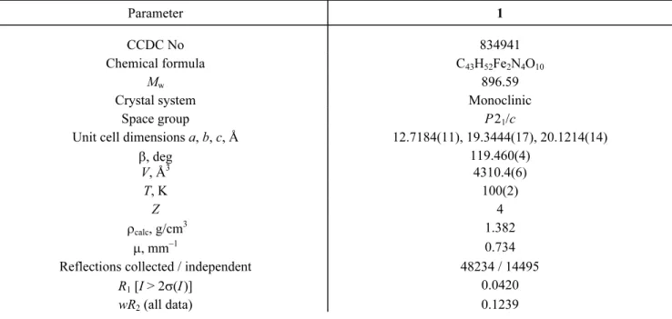
X-ray structural analysis of complex 1. The crystal data and structure refinement details for complex 1 are listed in Table 1. Selected bond lengths and angles are summarized in Table 2. Possible hydrogen bonds are also given in Table 3. Representative structural diagrams of complex 1 are shown in Fig. 1, while packing diagrams are given in Fig. 2.
Compound 1 is a μ-oxo-bridged dinuclear Fe(III) compound. The asymmetric unit of 1 consists of a [{Fe(4-MeOL1)}2(μ-O)] dimeric unit with a lattice methanol molecule. The iron(III) atoms (Fe1 and Fe2) surrounded by the four
TABLE 1. Crystal Data and Structure Refinement of Complex 1
Parameter 1
CCDC No 834941
Chemical formula C43H52Fe2N4O10
Mw 896.59
Crystal system Monoclinic
Space group P 21/c
Unit cell dimensions a, b, c, Å 12.7184(11), 19.3444(17), 20.1214(14)
β, deg 119.460(4) V, Å3 4310.4(6) T, K 100(2) Z 4 ρcalc, g/cm3 1.382 μ, mm–1 0.734
Reflections collected / independent 48234 / 14495
R1 [I > 2σ(I)] 0.0420
wR2 (all data) 0.1239
TABLE 2. Some Selected Bond Lengths (Å) and Angles (deg) for Compound 1
1 Fe–Nimine 2.1139(14); 2.1151(14); 2.1194(14); 2.1274(14) Fe–Ophenolic 1.9390(13); 1.9445(11); 1.9647(12); 1.9731(12) Fe–Ooxo 1.7752(13); 1.7818(12) Fe–O–Fe 161.58(8) Fe…Fe 3.511
TABLE 3. Hydrogen Bond Geometry (Å, deg) of Compound 1
D–H⋯A* D–H H⋯A D⋯A D–H⋯A Symmetry
O10A–H10A⋯O4 0.84 2.02 2.81 158 1+x, y, z C1–H1⋯O8 0.93 2.55 3.41 153 –x, –1/2+y, 1/2–z C–H⋯π C18–H18⋯R1 0.93 2.93 3.756 149 x, 1/2–y, 1/2+z C40–40B⋯R2 0.96 2.62 3.553 164 –x, –1/2+y, 1/2–z * D – donor, A – acceptor, R1 – C2–C3–C4–C5–C6–C7, R2 – C23–C24–C25–C26–C27–C28.
respectively. Above the best N2O2 least-squares plane the ligand exhibits the umbrella conformation (Fig. 1). The FeN2O2
coordination plane in each iron(III) atom is trans-oriented to the other relative to the oxo bridge in order to avoid interligand steric repulsions. For the coordination polyhedron of the metal atom, the distortion of the coordination environment from trigonal bipyramidal (TBP) to square pyramidal (SP) can be evaluated by the Addison distortion index τ defined as τ = (α – – β) / 60, where α and β are the two largest coordination angles. The coordination polyhedron of the metal atom is described as τ = 0 for perfect SP and 1 for ideal TBP [30]. In our case, the structural distortion indexes of 1 were found as τFe1 = 0.026
and τFe2 = 0.003 respectively, which indicates that Fe1 and Fe2 polyhedra are all close to a distorted square pyramid. The bond lengths between the metal and donor atoms in the base of the pyramid are as follows: Fe–Nimine 2.1139(14)-2.1274(14) Å; Fe–Ophenolic 1.9390(13)-1.9731(12) Å. The axial Fe–Ooxo bond length is 1.7752(13)-1.7818(12) Å. The Fe…Fe distance in 1 (3.511 Å) is in the same range as those already reported for complexes with the Fe–O–Fe core (3.35-3.55 Å)
Fig. 1. Molecular structure of 1.
Fig. 2. Molecular packing diagram in the bc plane of 1.
[18].The Fe1–O3–Fe2 angle is 161.58(8)°. These observed geometrical features of iron(III) centers in 1 are quite comparable to those of the similar dinuclear complexes reported in the literature [31, 52].
Complex 1 revealed the presence of intermolecular C–H⋯O interactions between the interconnected dinuclear complexes (Table 2). This hydrogen bonded networks lie in the bc plane and stack along to the a axis. (Fig. 2). However, these dimeric units are further linked by C–H⋯π interactions (H⋯R = 2.93 Å and 2.62 Å) to the other dimers present in the unit cell (Table 3).
Crystallographic data of the structural analysis have been deposited with the Cambridge Crystallographic Data Centre, CCDC No. 834941 (1). Copies of the data can be obtained free of charge from the Director, CCDC, 12 Union Road, Cambridge CB2 1EZ, UK (fax: +44-1223-336-033; e-mail: [email protected] or www: http://www.ccdc.cam.ac.uk).
The authors are grateful to the Research Funds of Balikesir University (BAP-2013/73) and TUBITAK (Grants No: TBAG-108T431) for the financial support. Dr. Kara would like to thank TUBITAK for NATO-B1 and the Royal Society short visit fellowship for financial support and Prof. Guy Orpen (School of Chemistry, University of Bristol, UK) for his hospitality.
REFERENCES
1. O. Kahn, Molecular Magnetism, VCH, Weinheim (1993).2. R. E. P. Winpenny, Adv. Inorg. Chem., 52, 1 (2001).
3. R. L. Lintvedt and J. K. Zehetmair, Inorg. Chem., 29, 2204 (1990).
4. W. Kaim and B. Schwederski, Bioinorganic Chemistry: Inorganic Elements in the Chemistry of Life, Wiley, Chichester, UK (1995).
5. E. Safaei, I. Saberikia, A. Wojtczak, Z. Jaglicic, and A. Kozakiewicz, Polyhedron, 30, 1143 (2011).
6. C. Boskovic, E. K. Brechin, W. E. Streib, K. Folting, J. C. Bollinger, D. N. Hendrickson, and G. Christou, J. Am. Chem. Soc., 124, 3725 (2002).
7. P. Artus, C. Boskovic, J. Yoo, W. E. Streib, L. C. Brunel, D. N. Hendrickson, and J. Christou, Inorg. Chem., 40, 4199 (2001).
8. D. M. Kurtz Jr., Chem. Rev., 90, 585 (1990). 9. L. Que and A. E. True, Prog. Inorg., 38, 97 (1990). 10. A. L. Feig and S. J. Lippard, Chem. Rev., 94, 759 (1994).
11. U. Bossek, H. Hummel, T. W. Muller, E. Bill, and K. Wieghardt, Angew. Chem., Int. Ed. Engl., 34, 2642 (1995). 12. K. S. Min, A. M. Arif, and J. S. Miller, Inorg. Chim. Acta, 360, 1854 (2007).
13. G. T. Karimpour, E. Safaei, A. Wojtczak, and Z. Jaglicic, Inorg. Chim. Acta, 405, 309 (2013). 14. Z. Chen, H. Morimoto, S. Matsunaga, and M. Shibasaki, J. Am. Chem. Soc., 130, 2170 (2008). 15. E. K. Noh, S. J. Na, S. Sujith, S. W. Kim, and B. Y. Lee, J. Am. Chem. Soc., 129, 8082 (2007). 16. S. J. Lippard, Angew. Chem., Int. Ed. Engl., 27, 344 (1988).
17. H. Fujii, T. Kurahashi, and T. Ogura, J. Inorg. Biochem., 96, 133 (2003). 18. E. Y. Tshuva and S. J. Lippard, Chem. Rev., 104, 987 (2004).
19. E. Q. Gao, L. H. Yin, J. K. Tang, P. Cheng, D. Z. Liao, Z. H. Jiang, and S. P. Yan, Polyhedron, 20, 669 (2001). 20. H. Fujii and Y. Funahashi, Angew. Chem., Int. Ed., 41, 3638 (2002).
21. M. A. Torzilli, S. Colquhoun, J. Kim, and R. H. Beer, Polyhedron, 21, 705 (2002). 22. A. Elmali, Y. Elerman, C. T. Zeyrek, and I. Svoboda, Z. Naturforsch., 58b, 433 (2003). 23. Z. L. You and H. L. Zhu, Acta Crystallogr., E60, m1046 (2004).
24. Y. Yahsi, H. Kara, L. Sorace, and O. Buyukgungor, Inorg. Chim. Acta, 366, 191 (2011). 25. A. Karakas, E. Donmez, H. Kara, and A. Elmalı, J. Nonlinear Opt. Phys. Mater., 16, 329 (2007).
26. Y. Yahsi, H. Kara, C. Kazak, A. Iakovenko, and L. Sorace, J. Optoelectron. Adv. Mater. – Symposia, 1, 566 (2009). 27. SAINT V760A, Bruker AXS (2008).
28. G. Sheldrick, SADABS V2008/1, University of Göttingen, Germany.
29. G. Sheldrick, Acta Crystallogr., Sect A: Found. Crystallogr., 64, 112 (2008).
30. A. W. Addison, T. Nageswara, J. Reedijk, J. van Rijn, and G. C. Verchoor, J. Chem. Soc., Dalton Trans., 1349 (1984). 31. R. Biswas, M. G. B. Drew, C. Estarellas, A. Frontera, and A. Ghosh, Eur. J. Inorg. Chem., 2558 (2011).
33. E. Wilkinson, Y. Dong, and L. Que Jr., J. Am. Chem. Soc., 116, 8394 (1994).
34. R. H. Fish, M. S. Konnings, K. J. Oberhausen, R. H. Fong, W. M. Yu, G. Christou, J. B. Vincent, D. K. Coggin, and R. M. Buchanan, Inorg. Chem., 30, 3002 (1991).
35. R. M. Buchanan, S. Chen, J. F. Richardson, M. Bressan, L. Forti, A. Morvillo, and R. H. Fish, Inorg. Chem., 33, 3208 (1994).
36. Y. Dong, H. Fujii, M. P. Hendrich, R. A. Leising, G. Pan, C. R. Randall, E. C. Wilkinson, Y. Zang, and L. Que Jr., J. Am. Chem. Soc., 117, 2778 (1995).
37. X. Wang, S. Wang, L. Li, E. B. Sundberg, and G. P. Gacho, Inorg. Chem., 42, 7799 (2003). 38. F. Corazza, C. Floriani, and M. Zehnder, J. Chem. Soc., Dalton Trans., 709 (1987).
39. G. Das, R. Shukla, S. Mandal, R. Singh, and P. K. Bharadwaj, Inorg. Chem., 36, 323 (1997). 40. A. R. Li, H. H. Wei, and L. L. Gang, Inorg. Chim. Acta, 290, 51 (1999).
41. K. Oyaizu, E. L. Dewi, and E. Tsuchida, Inorg. Chim. Acta, 321, 205 (2001). 42. S. Koner, S. Iijima, M. Watanabe, and M. Sato, J. Coord. Chem., 56, 103 (2003).
43. W. J. Ruan, G. H. Hu, S. J. Wang, J. H. Tian, Q. L. Wang, and Z. A. Zhu, Chin. J. Chem., 23, 709 (2005). 44. Z. L. You, L. L. Tang, and H. L. Zhu, Acta Crystallogr., E61, m36 (2005).
45. G. Ilyashenko, M. Motevalli, and M. Watkinson, Tetrahedron: Asymmetry, 17, 1625 (2006). 46. Q. Meng, L. Wang, Y. Liu, and Y. Pang, Acta Crystallogr., E64, m63 (2008).
47. L. N. Rusere, T. Shalumova, J. M. Tanski, and L. A. Tyler, Polyhedron, 28, 3804 (2009).
48. R. Mayilmurugan, H. Stoeckli-Evans, E. Suresh, and M. Palaniandavar, Dalton Trans., 5101 (2009). 49. J. H. Yan, X. P. Shen, and H. Zhou, Acta Crystallogr., E66, m1090 (2010).
50. J. B. H. Strautmann, C.-G. Freiherr von Richthofen, G. Heinze-Brückner, S. DeBeer, E. Bothe, E. Bill, T. Weyhermüller, A. Stammler, H. Bögge, and T. Glaser, Inorg. Chem., 50, 155 (2011).
51. M. S. Shongwe, U. A. Al-Zaabi, F. Al-Mjeni, C. S. Eribal, E. Sinn, I. A. Al-Omari, H. H. Hamdeh, D. Matoga, H. Adams, M. J. Morris, A. L. Rheingold, E. Bill, and D. J. Sellmyer, Inorg. Chem., 51, 8241 (2012).