Journal of Dentistry Indonesia
Journal of Dentistry Indonesia
Volume 27 Number 3 Article 6
12-30-2020
Effect of Craniofacial Growth Pattern on Head Posture
Effect of Craniofacial Growth Pattern on Head Posture
Burak KaleDepartment of Orthodontics, Faculty of Dentistry, Antalya Bilim University, Antalya, Turkey, [email protected]
Muhammed Hilmi Buyukcavus
Department of Orthodontics, Faculty of Dentistry, Suleyman Demirel University, Isparta, Turkey, [email protected]
Follow this and additional works at: https://scholarhub.ui.ac.id/jdi
Part of the Oral and Maxillofacial Surgery Commons, and the Orthodontics and Orthodontology Commons
Recommended Citation Recommended Citation
Kale, B., & Buyukcavus, M. Effect of Craniofacial Growth Pattern on Head Posture. J Dent Indones. 2020;27(3): 144-150
ORIGINAL ARTICLE
Effect of Craniofacial Growth Pattern on Head Posture
Burak Kale
1, Muhammed Hilmi Buyukcavus
21Department of Orthodontics, Faculty of Dentistry, Antalya Bilim University, Antalya, Turkey 2Department of Orthodontics, Faculty of Dentistry, Suleyman Demirel University, Isparta, Turkey
Correspondence e-mail to: [email protected]
ABSTRACT
Head posture refers to the upright position of the head of a standing or sitting subject. The literature reports that head posture is affected by many factors. Objective: To evaluate differences in head posture according to craniofacial growth pattern. Methods: A total of 163 individuals (83 females and 80 males) were included in this study. Patients were divided into three groups according to ANB angle as Class I, Class II, and Class III, and each group was divided further into three subgroups according to SN/GoGn angle as hyperdivergent, normodivergent, and hypodivergent. The patients were compared in terms of head posture measurements. Two-way ANOVA was used to evaluate the main and interactive effects of vertical growth pattern and malocclusion type on the head posture of the patients. Results were considered statistically significant at p < 0.05. Results: No statistically significant difference between malocclusion and face-type groups was observed in all head posture measurements (p > 0.05). Conclusion: Head posture is similar among subgroups of different malocclusion types separated by vertical growth pattern.
Key words: cephalometry, head posture, malocclusions, vertical growth pattern
How to cite this article: Kale B, Buyukcavus MH. Effect of craniofacial growth pattern on head posture. J Dent Indones. 27(3):144-150
INTRODUCTION
Head posture and facial morphology are affected by
respiratory functions during growth and development.1-6
Postural adaptation of the hyoid bone and tongue is not always sufficient to solve airway obstruction problems. Extension of the head and neck may occur as a
functional response to airway obstruction.3,7 Schwartz
first introduced the relationship between head posture and craniofacial morphology in 1928. According to the author, extension of the head, especially during sleep,
causes Class II malocclusions.8 In 1982, Rocarbado et
al. presented strong evidence of the relation between
malocclusion and head posture.9 Woodside et al. found
that the prevalence of mandibular anterior crowding is higher in impaired nose breathers than in normal
breathers.10 Liu et al. reported that distal occlusion and
increased overjet are caused by a flexed head posture.11
Basheer et al. observed postural changes resulting from total nasal obstruction among 25 adult individuals before and 1 hour after total nasal obstruction and found statistically significant differences in the spacing
of the lips, lower jaw, and hyoid bone position.12
Björk revealed the relationship between cranial base and posture by observing that individuals with retrognathia tend to raise their heads up whereas individuals with prognathies tend to bow their heads
forward.13 In his cephalometric study, Bench discussed
the relationship between facial form and cervical spine length and curvature. The scholar found that the vertical growth of the face after puberty is closely related to neck growth and that the cervical column is flat and long in dolichosphate individuals but inclined
forward in brachiocephalus individuals.14
Solow and Kreiborg explained the relationships between airway obstruction, postural changes caused by soft tissue tension, and changes in craniofacial morphology by using the “soft tissue tension” hypothesis. The researchers argued that postural changes due to airway obstruction disturb the balance of forces around the skeleton and, as a result, cause changes in craniofacial
morphology.15 Solow and Tallgren explained the
relationship between head posture and craniofacial
morphology as follows.16 During extension of the head
craniofacial size decreases, the height of the anterior surface decreases, the height of the anterior face increases, facial retrognatism occurs, the slope of the mandible relative to the anterior cranial base increases, the angle of the cranial base increases, and the nasopharyngeal airway decreases. During flexion of the head, the authors observed an increase in anteroposterior craniofacial size, an increase in the height of the posterior surface, a decrease in the height of the anterior face, facial prognatism, a decrease in the angle of the mandible compared with the anterior cranial base, an increase in the angle of the cranial base, and an increase in the nasopharyngeal airway. Previous studies associated head posture with height, ethnic difference, gender, age, facial morphology, dimensions, shape, nasorespiratory function,
temporomandibular dysfunction, and bruxism.17,18 The
relationships between head posture and skeletal–dental malocclusions, such as crowding in the maxillary and mandibular dental arches, spacing, overbite, crossbite, midline discrepancies, skeletal, and molar,
have been studied.18,19 Some authors have reported that
nasorespiratory dysfunction and malocclusions cause
changes in vertical growth patterns.18,20 Several studies
have also examined the effects of different variables on head posture. However, both the vertical growth pattern and the malocclusion type are considered, and it is not within the scope of our study information evaluated according to the craniofacial growth pattern. Therefore, in the present study, we aimed to compare head positions among subgroups of healthy patients with different malocclusions (i.e., Class I, Class II, and Class III) separated by different vertical growth patterns (i.e., hypodivergent, normodivergent, and hyperdivergent).
METHODS
A retrospective study of untreated patients with orthodontic malocclusion at the Oral and Dental Health Treatment Center, Antalya Bilim University, Turkey was conducted. Ethical approval for this retrospective study was obtained from Clinical Research Ethics Committee of University of Health Sciences Antalya Training and Research Hospital (12.03.2020-5/9), and informed consent was obtained from the parents of the patients included in this work. This study consisted of patients divided into three subgroups according to their vertical growth pattern (i.e., hypodivergent, normodivergent, and hyperdivergent) with skeletal Class I, Class II, and Class III malocclusions.
The sample size was calculated on the basis of a power analysis using G* Power (version 3.0.10, Kiel, Germany) for superior airway space at an α error probability of 0.05 and a power of 80%. The power analysis showed that a minimum of 17 individuals
was required for each subgroup. More individuals were included in the study to increase its power and compensate for possible losses.
The inclusion criteria are the absence of any craniofacial anomaly or systemic disorder, no airway pathologies, adequate imaging quality of cephalometric radiographs, and no previous orthodontic treatment. Patients whose head posture was erroneous during radiographic exposure, patients over 17 years of age, and patients with artifacts on their radiographs were excluded from the study. Patients who met the inclusion criteria in accordance with the necessary information derived from the anamnesis forms were determined without evaluation of their radiographic findings. The participants were then randomly divided into groups according to their analysis at the beginning of the treatment, regardless of head posture.
Out of the 243 patients included in the evaluation, 80 patients were excluded from the study. A total of 163 individuals (83 females and 80 males) were included in the study. The radiographs of the patients were exclusively obtained from the device in our clinic for orthodontic patients. During radiograph collection, the head was fixed with a cephalostat, and exposure was made in the natural head posture with the help
of a mirror located opposite the Orthophos SL 3D®
(Dentsply–Sirona, Bensheim, Germany) device (85 kV, 8 mA, 15 s exposure time). In addition, the patients’ heads were fixed with a cephalostat, and radiographs were obtained while the teeth were in centric occlusion. This standardization was routinely performed for each patient, and patients who kept their head under extension or flexion during radiography were excluded from the study.
During grouping according to craniofacial growth pattern, ANB angles were used to detect skeletal malocclusions in the sagittal direction and SN/GoGn angles were used to determine vertical growth patterns. Patients were divided into three groups according to ANB angle as Class I (0° < ANB < 4°), Class II (ANB > 4°), and Class III (ANB < 0°). Each group was further divided into three subgroups according to SN/GoGn angle as hyperdivergent (SN/GoGn > 38°), normodivergent (26° < SN/GoGn < 38°), and hypodivergent (SN/GoGn < 26°).
The patients were compared according to their skeletal malocclusion classification and vertical growth pattern, and the nine subgroups were compared in terms of head posture measurements.
Cephalometric analysis
Lateral cephalometric radiographies (LCR) were taken while in orthoposition, a postural recording method defined by Molhave (1958) and modified by Solow
methods, patients were asked not to swallow or move their head and tongue. Gender differences in LCR head posture dimensions were determined. Radiographs were traced and measured by the same investigator (BK). The cephalometric angular measurements, landmarks, and reference lines are shown in Figure 1. Linear measurements used in the study are in the Dolphin Imaging (Patterson Technology, Chatsworth, CA, USA) were performed on digital lateral cephalometric films after digital calibration. In this study, 35 cephalometric points, 8 cephalometric planes, and 5 head posture measurements were used.
SN-CVT: The angle between the anterior cranial base
(SN) and the cervical vertebrae tangent
SN-OPT: The angle between the anterior cranial base
(SN) and the odontoid process tangent (OPT)
FH-CVT: The Frankfurt horizontal (FH) and cervical
vertebrae tangent (CVT)
FH-OPT: The Frankfurt horizontal (FH) and odontoid
process tangent (OPT)
CVT-OPT: The cervical vertebrae tangent (CVT) and
the odontoid process tangent (OPT)
Statistical analysis
The Kolmogorov–Smirnov test was used to determine whether the data had a normal distribution. Because the parameters evaluated in this study were generally distributed homogenously among the subgroups, parametric tests were used. The gender distribution of the patients was compared using Pearson’s chi-squared test. Two-way ANOVA (generalized linear model [GLM]) was used to evaluate the main and interactive effects of two factors among patients subdivided according to vertical growth pattern and malocclusion type. Tukey’s post-hoc tests were used
for bilateral comparisons of significant parameters. The error margin of the measurements was determined from 45 randomly selected films obtained from 163 lateral cephalometric films by the same researcher after the first measurements. Cronbach α coefficients were determined for each measurement. Repeatability coefficients were found to be high for each measurement (α ≥ 863), and the results of a paired t-test showed that the data were free of systematic error (p > 0.05). The SPSS package for Windows (version 21.0; SPSS Inc., Chicago, IL, USA) was used for data analysis. Results were considered statistically significant at p < 0.05.
RESULTS
The distribution of patients grouped according to craniofacial growth pattern (i.e., malocclusion type and vertical growth pattern), chronological age, and sex is shown in Table 1. The mean age of 55 patients (29 females and 26 males) with Class I malocclusion was found to be 14.45± 2.35 years. In this group, 31% of the patients were hypodivergent (17 patients; 9 females, 8 males; mean age, 14.61± 2.01 years), 34% of the patients were normodivergent (19 patients; 10 females, 9 males; mean age, 14.36± 2.43 years), and 34 % of the patients were hyperdivergent (19 patients; 10 females, 9 males; mean age, 14.39± 2.43 years).
The mean age of 55 patients (28 females and 27 males) with Class II malocclusion was found to be 14.43 ± 2.19 years. In this group, 33% of the patients were hypodivergent (18 patients; 9 females, 9 males; mean age, 14.39± 2.17 years), 31% of the patients were normodivergent (17 patients; 9 females, 8 males; mean age, 14.43± 2.08 years), and 36% of the patients were hyperdivergent (20 patients; 10 females, 10 males; mean age, 14.48± 2.31 years).
The mean age of 53 patients (26 females and 27 males) with Class III malocclusion was found to be 14.48± 2.37 years. In this group, 32% of the patients were hypodivergent (17 patients; 8 females, 9 males; mean age, 14.58± 2.26 years), 32% of the patients were normodivergent (17 patients; 8 females, 9 males; mean age, 14.64± 2.47 years), and 36% of the patients were hyperdivergent (19 patients; 10 females, 9 males; mean age, 14.24± 2.37 years).
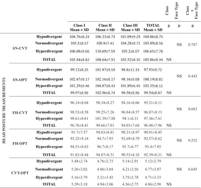
When interactive effects were examined, no significant relationship was observed between all subgroups and chronological age (p < 0.05). When the relationship between craniofacial pattern and gender was evaluated, no significant relationship was found between face type and gender (p > 0.05). When we examined the effects of different craniofacial growth patterns on head posture, no statistically significant difference was found between different malocclusion and face-type groups in all head posture measurements (p > 0.05) (Table 2).
Ta bl e 1 . D em og ra ph ic s ta tis tic s s tr at ifi ed b y c ra ni of ac ia l g ro w th p at te rn Class Face Type Class vs Face Type (Gr oup 1) (Gr oup 2) (Gr oup 3) Class III TOT AL p p p Class I Class II Chr onological Age (Mean ± SD) (n) Hypodivergent 14.61 ± 2.01 (n = 17) 14.39 ± 2.17 (n = 18) 14.58 ± 2.26 (n = 17) 14.52 ± 2.1 1 (n = 52) NS NS Normodivergent 14.36 ± 2.43 (n = 19) 14.43 ± 2.08 (n = 17) 14.64 ± 2.47 (n = 17) 14.47 ± 2.33 (n = 53) Hyperdivergent 14.39 ± 2.43 (n = 19) 14.48 ± 2.31 (n = 20) 14.24 ± 2.37 (n = 19) 14.37 ± 2.35 (n = 58) TOT AL 14.45 ± 2.35 (n = 55) 14.43 ± 2.19 (n = 55) 14.48 ± 2.37 (n = 53) 14.45 ± 2.34 (n = 163) NS GENDER * F M n (%) Hypodivergent 9 (52.94) 8 (47.06) 9 (50) 9 (50) 8 (47.06) 9 (52.94) 26 (50) 26 (50) NS NS Normodivergent 10 (52.63) 9 (47.37) 9 (52.94) 8 (47.06) 8 (47.06) 9 (52.94) 27 (50.95) 26 (49.05) Hyperdivergent 10 (52.63) 9 (47.37) 10 (50) 10 (50) 10 (52.63) 9 (47.37) 30 (51.73) 28 (48.27) TOT AL 29 (52.72) 26 (47.28) 28 (50.91) 27 (49.09) 26 (49.05) 27 (50.95) 83 (100) 80 (100) NS F= Fe m al e; M =M al e P †: T w o-w ay A N O VA ; P *: P ea rs on ’s ch i-s qu ar e t es t; N S: N ot s ig ni fic an t, p > 0 .0 5
DISCUSSION
When performing orthodontic diagnosis and planning the corresponding treatment, the head posture must be considered on account of its effects on the development of the tooth–jaw–face system and its relations with this system. Head posture may be an effective factor in ensuring and maintaining harmony between the jaws, which is one of the main goals of orthodontic treatment. During the selection of participants in our study, we sought to minimize factors that may affect the natural head position beyond the limits of change compared with normal individuals and cause positional changes
in these structures.22
Orthodontic, orthognathic, and functional treatments have been reported to be able to change the head
Table 2. Comparison of head posture measurements according to craniofacial growth pattern.
Class Face Type Class x Face Type Class I
Mean ± SD Mean ± SDClass II Mean ± SDClass III Mean ± SDTOTAL p p p
HEAD POSTURE MEASUREMENTS
SN-CVT Hypodivergent 104.76±8.24 106.33±8.73 101.09±9.29 104.06±8.75 NS 0.747 Normodivergent 105.5±8.57 108.9±7.41 104.28±9.71 105.89±8.56 Hyperdivergent 106.08±9.66 110.69±7.59 105.2±6.07 106.65±7.78 TOTAL 105.44±8.82 108.64±7.91 103.52±8.35 105.86±8.34 NS SN-OPT Hypodivergent 99.12±8.25 101.87±9.59 98.8±11.33 97.93±9.72 NS 0.443 Normodivergent 102.47±9.17 102.16±8.17 98.16±9.08 100.19±8.81 Hyperdivergent 101.29±9.46 104.87±8.43 101.89±6.45 103.35±8.11 TOTAL 99.97±8.96 102.96±8.74 98.58±8.96 99.54±8.87 NS FH-CVT Hypodivergent 96.14±8.04 98.34±8.27 94.16±8.06 95.21±8.11 NS 0.683 Normodivergent 98.53±8.58 99.25±7.26 96.84±8.57 96.87±8.13 Hyperdivergent 98.61±8.61 101.39±7.88 94.1±6.31 97.36±7.61 TOTAL 96.76±8.41 98.66±7.81 94.03±7.64 96.48±7.96 NS FH-OPT Hypodivergent 91.7±7.57 94.83±8.81 90.21±8.97 90.91±8.45 NS 0.552 Normodivergent 93.25±9.14 94.7±7.93 92.69±8.79 92.57±8.62 Hyperdivergent 94.51±8.63 96.7±8.17 91.7±6.77 93.4±7.85 TOTAL 91.82±8.44 94.87±8.31 90.53±8.18 92.39±8.31 NS CVT-OPT Hypodivergent 5.48±2.74 4.76±2.73 5.14±2.91 5.12±2.79 NS 0.645 Normodivergent 5.26±3.02 4.86±3.04 4.21±2.56 4.77±2.87 Hyperdivergent 5.16±3.79 5.21±3.42 3.75±2.78 4.71±3.33 TOTAL 5.29±3.18 4.94±3.06 4.36±2.75 4.86±2.98 NS
P: Two-way ANOVA; NS: not-significant p>0.05
posture. Structural and positional changes created by treatment of the chewing system, especially of the mandible and tongue, can change the head posture; thus, changes that may occur in functions such as chewing, swallowing, and breathing as a result of
treatment can directly affect these structures.18,22 Given
these findings, individuals with a history of previous orthodontic, orthopedic, or orthognathic treatment were not included in our study.
Attention must be paid to head posture not only in posture-related studies but also in routine cephalometric radiographs. Compared with other positions, taking radiographs in the natural head position during the cephalometric evaluation of teeth, jaws, head, and face is better able to reflect the true characteristics of the
used in orthodontic practice to evaluate individual growth patterns and skeletal, dental, and soft tissue morphologies to diagnose skeletal anomalies, plan treatment, predict future changes, and examine head and facial parameters in individuals or communities. The greatest benefit of cephalometry compared with other techniques is that it is a quantitative method that enables the evaluation and analysis of the relationship between skeletal and dental structures and soft tissue. While cephalometric films do not show three-dimensional details, they offer higher image resolution than any other imaging method, including computed tomography. In the present study, we used cephalometry as a research material because cephalometric films are the most widely used imaging tools in orthodontic studies.
In our study, no statistically significant difference in head posture measurements was observed among the skeletal Class I, Class II, and Class III groups and different face types. Cole and Ertürk divided patients into Class I, Class II, and Class III according to ANB angle and examined differences in head posture among
these groups.23,24 The team observed that SN/CVT
measurements determining the craniocervical posture are statistically different between the skeletal Class II and Class III groups and between the skeletal Class I
and Class II groups.24
Ertürk et al. argued that SN/CVT measurements do not differ significantly between skeletal Class I, Class
II, and Class III groups.23 The researchers further
determined that the head is positioned downward in the Class III group and upward in the Class II
group compared with that in the Class I group 23,24.
In our study, no significant difference in SN/CVT measurements was found between the skeletal Class I, Class II, and Class III groups. Therefore, head posture did not show a statistically significant difference among these groups. However, we also observed that the head was positioned lower in the Class III group and higher in the Class II group compared with that in the Class I group. Thus, our findings are similar to the findings of Ertürk et al. but differ from those of Cole. The difference between Cole’s findings and our results may be attributed to differences in the ANB angles used for grouping, as well as differences in the age, sex, and growth-development periods of the groups.
Soytarhan and Aras examined head position in different malocclusions and observed that although the head was not at a significant level in the Class
III group.25 In this respect, the findings of Soytarhan
and Aras are similar to those of our study. Hedeyati et al. investigated the characteristics of cranial base inclination and craniocervical posture in adults with skeletal Class I, Class II, and Class III malocclusions and found no significant difference in craniocervical posture and cervical colon curvature among the
groups.26 In a study of children aged 11–14 years, Liu
et al. did not observe a significant difference among groups in terms of craniocervical posture and cervical
colon curvature.11 Our findings on head posture,
craniocervical posture, and cervical colon curvature are in line with those of Liu et al.
The unique feature of our study is that, besides different malocclusions, we also examined different facial types. However, because previous studies examining nine subgroups are not available in the literature, exact comparisons of our findings with earlier results cannot be conducted. Thus, comparisons with studies presenting similar findings were made. The current study can help guide future research on the factors affecting head posture and contributes to the literature by examining head posture with a large sample size using different methods.
CONCLUSION
Among the groups studied, craniocervical angle measurements are lowest in Class III and hypodivergent individuals. Head posture is similar among subgroups of different malocclusion types separated by vertical growth pattern.
CONFLICT OF INTEREST
All authors declare no conflict of interest related to the publication of this research.
REFERENCES
1. Mariya Q, Mushtaq M. Cephalometric evaluation of cervical column curvature with respect to sagittal jaw position. Int J Appl Dent Sci. 2017;3(4):238-42. 2. Gomes Lde C, Horta KO, Gonçalves JR, Santos-Pinto AD. Systematic review: craniocervical posture and craniofacial morphology. Eur J Orthod. 2014 Feb;36(1):55-66.
3. Cerruto C, Di Vece L, Doldo T, Giovannetti A, Polimeni A, Goracci C. A computerized photographic method to evaluate changes in head posture and scapular position following rapid palatal expansion: a pilot study. J Clin Pediatr Dent. 2012;37(2):213-8.
4. Clavel L, Rémy-Neris S, Skalli W, Rouch P, Lespert Y, Similowski T, Sandoz B, Attali V. Cervical spine hyperextension and altered posturo-respiratory coupling in patients with obstructive sleep apnea syndrome. Front Med. 2020;(7):30.
5. Bass NM. Measurement of the profile angle and the aesthetic analysis of the facial profile. J Orthod. 2003 Mar;30(1):3-9.
6. Yagci A, Uysal T, Usumez S, Orhan M. Rapid maxillary expansion effects on dynamic
measurement of natural head position. Angle Orthod. 2011 Sep;81(5):850-5.
7. Linder-Aronson S, Woodside D. Factors affecting the facial and dental structures in excess face height: malocclusion, etiology, diagnosis, and treatment. Chicago: Quintessence Pub Co. 2000:1-33.
8. Schwarz A. Positions of the head and malrelations of the jaws. Int J Orthod, Oral Surg, Radiograph. 1928;14(1):56-68.
9. Rocabado M, Ben E. Johnston Jr, Mitchell G Blakney. Physical therapy and dentistry: an overview. J Craniomand Pract. 1982,1:1,46-9. 10. Woodside D, Linder-Aronson S, Stubbs DO:
relationship between mandibular incisor crowding and nasal mucosal swelling. Proc Finn Dent Soc. 1991;87:127-38.
11. Liu Y, Sun X, Chen Y, Hu M, Hou X, Liu C. Relationships of sagittal skeletal discrepancy, natural head position, and craniocervical posture in young Chinese children. Cranio. 2016;34(3):155-62. 12. Basheer B, Hegde KS, Bhat SS, Umar D, Baroudi
K. Influence of mouth breathing on the dentofacial growth of children: a cephalometric study. J Int Oral Health. 2014;6(6):50-5.
13. Afrand M, Ling CP, Khosrotehrani S, Flores-Mir C, Lagravère-Vich MO. Anterior cranial-base time-related changes: a systematic review. Am J Orthod Dentofacial Orthop. 2014;146(1):21-32.e6.
14. Meiyappan N, Tamizharasi S, Senthilkumar KP, Janardhanan K. Natural head position: an overview. J Pharm Bioallied Sci. 2015;7(Suppl 2):S424-7. 15. Shenoy U, Banerjee S, Wajekar P, Vasvani
V. Assessment and comparison of the head posture and craniofacial growth in vertical dimension—a cephalometric study. J Evol Med Dent Sci. 2020;9(15):1276-81.
16. Solow B, Tallgren A. Head posture and craniofacial morphology. Am J Phys Anthropol. 1976;44(3):417-35.
17. Tekale PD, Vakil KK, Nagmode SL. Hyoid bone position and head posture comparison in skeletal Class I and Class II subjects: a retrospective cephalometric study. APOS Trend Orthod. 2014;4(3):53.
18. Jose NP, Shetty S, Mogra S, Shetty VS, Rangarajan S, Mary L. Evaluation of hyoid bone position and its correlation with pharyngeal airway space in different types of skeletal malocclusion. Contemp Clin Dent. 2014;5(2):187-9.
19. Soheilifar S, Momeni MA. Cephalometric comparison of position of the hyoid bone in class I and class II patients. Iran J Ortho. 2017;12(1). 20. Torre C, Guilleminault C. Establishment of nasal
breathing should be the ultimate goal to secure adequate craniofacial and airway development in children. J Pediatr (Rio J). 2018;94(2):101-3. 21. Cassi D, De Biase C, Tonni I, Gandolfini M,
Di Blasio A, Piancino MG. Natural position of the head: review of two-dimensional and three-dimensional methods of recording. Br J Oral Maxillofac Surg. 2016;54(3):233-40.
22. Graber LW, Vanarsdall RL, Vig KW, Huang GJ. Orthodontics: current principles and techniques. Elsevier Health Sciences. 2016.
23. Ertürk N, Dogan S, Aras A. The investigation of the relationships between the natural head position, posture and sagittal jaw relations. Turk J Orthod. 1990;3:1-6.
24. Cole S. Natural head position, posture, and prognathism: the Chapman Prize Essay. Br J Orthod. 1988;15(4):227-39.
25. Soytarhan A, Aras A. Evaluation of the head posture in orthodontic malocclusions. Turk J Orthod. 1990;3:102-6.
26. Hedayati Z, Paknahad M, Zorriasatine F. Comparison of natural head position in different anteroposterior malocclusions. J Dent (Tehran, Iran). 2013;10(3):210.