125 Ankara Üniversitesi Tıp Fakültesi Mecmuası 2009, 62(3) DAHİLİ BİLİMLER / MEDICAL SCIENCES
Case Report / Olgu Sunumu
Makale türü olgu sunumu/ case report
Received: 07.10.2009 • Accepted: 16.04.2010 Corresponding author
Dr. Seçil Saral
Ankara Üniversitesi Tıp Fakültesi Deri ve Zührevi Hastalıkları Anabilim Dalı
Phone : 0 312 508 22 31 E-mail Address : [email protected]
Aktif psoriasis nedeniyle takip edilen bir hastanın kolunda saptanan dermatofibroma derma-toskopik olarak değerlendirildi. Dermaderma-toskopik muayenede lezyonda homojen yayılım gösteren noktasal damarlanma ve kahverengi noktalar izlendi. Lezyonun ortasında yer alan skar benzeri alanda, seyrek firkete benzeri damarlar gözlendi. Psoriasis tanısı olan bu hastada ortaya çıkan dermatofibromada da psoriazistekine benzer dermatoskopik özellikler bulunması dikkat çe-kiciydi. Noktasal damarlanma gösteren pembe lezyonların ayırıcı tanısında dermatofibroma da düşünülmelidir.
Anahtar Sözcükler: Dermatofibrom, Dermatoskopi, Noktasal damarlanma
A dermatofibroma of a patient with active psoriasis was evaluated dermatoscopically. Dermato-scopic examination of the lesion revealed dotted vessels with homogenous distribution through-out the lesion and brown dots and occasional hairpin-like vessels in the white scar-like center. The reported case is unique because of presentation of psoriasis and dermatofibroma simultaneously since both exhibit homogenous red dots dermatoscopically. In the dermatoscopic differential di-agnosis of pink lesions showing dotted vessels in a homogenous pattern dermatofibroma must be considered.
Key Words: Dermatofibroma, Dermatoscopy, Dotted vessel 1 Ankara Üniversitesi Tıp Fakültesi Deri ve Zührevi Hastalıkları
Anabilim Dalı
2 Ankara Üniversitesi Tıp Fakültesi Tıbbi Patoloji Anabilim Dalı
Dermatoscopy and Its Role In Diagnosing Homogenous Dotted
Vascular Pattern In a Case Of Dermatofibroma
Dermatofibroma Olgu Sunumu: Dermatoskopinin Homojen Noktasal Vasküler Yapılarda Tanısal Rolü
Bengü Nisa Akay
1, Seçil Saral
1, Ezgi Ünlü
1, Aylin Okçu Heper
2, Aynur Akyol
1The diagnostic accuracy of dermatoscopy for pink lesions lacking significant pigment is less than that of more pigmented lesi-ons. The recognition of distinctive vas-cular structures may be helpful for diag-nostic purposes, especially when the clas-sic pigmented dermatoscopic structu-res are lacking. Arborizing, hairpin-like, comma, linear, dotted, glomerular and irregular vessels are the most common vascular patterns evaluated by derma-toscopy. A dermatoscopic pattern cha-racterized by the presence of dotted ves-sels in a homogenous pattern are gene-rally seen in Spitz nevi, Clark nevi, me-lanoma, psoriasis and clear cell acantho-ma (CCA) but rarely reported in deracantho-ma- derma-tofibroma (1-4). We report a case of der-matofibroma, dermatosopically (exami-ned using the Dermlite Foto at 10 fold magnification; 3 Gen, LLC, Dana Point, CA, USA) showing dotted vessels with homogenous distribution throughout the lesion brown dots and hairpin-like vessels in a white scar-like center.
Case Report
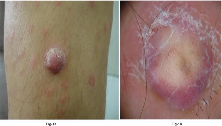
A 49-year-old man presented with a 3-year history of a slowly growing non-tender nodule on his left arm that had never bled or ulcerated. On examination of his left upper arm, there was a 2-cm, firm, pink exophytic nodule (Fig 1a). The patient had a history of sis for more than 23 years. His psoria-sis was previously treated with PUVA, methotrexate, cyclosporine and efali-zumab. Dermatoscopic analysis revea-led the presence of erythematous ho-mogeneous area surrounding a white patch. Dotted vessels were distributed throughout the entire lesion, whereas brown dots were observed only in the central white scar-like area. Occasio-nal hairpin like vessels were also obser-ved (Fig. 1b). The clinical differential diagnosis included melanoma, CCA, atypical fibroxanthoma and squamo-us cell carcinoma. Dermatoscopic dif-ferential diagnosis included melano-ma, Spitz nevi, Clark nevi, and CCA.
126 Dermatoscopy and Its Role In Diagnosing Homogenous Dotted Vascular Pattern In a Case Of Dermatofibroma
Ankara Üniversitesi Tıp Fakültesi Mecmuası 2009, 62(3)
The nodule was completely excised and histological examination showed a dermatofibroma (Fig. 2a,b,c).
Discussion
Dermatofibroma is a common, slow growing tumor of the skin derma-topathologically characterized by an increased number of fibrocytes in the dermis and occasionally subcutis and composed of a mixture of fibroblast-like spindle cells, histiocytes and blo-od vessels in varying proportions. Clinically they can appear with a co-lor varying from light brown to dark brown, purple-red or yellow, single or multiple hard papules, plaques or nodules. Their size ranges from 3mm to 2 cm. Our case presented with a 2 cm pink nodule on the left arm. He had active guttat psoriasis on admis-sion which had previously been tre-ated with PUVA, immunosuppressi-ves and efalizumab. Dermatoscopic analysis of the lesion revealed dot-ted vessels with homogenous
dist-ribution throughout the lesion, few hairpin-like vessels and brown dots in the white scar-like center. Dot-ted vessels histopathologically cor-relate with dilated and tortuous ca-pillaries in the middle reticular der-mis progressing to the top of the pa-pilla. Dotted vessels in a homogeno-us pattern are generally seen in Spitz nevi, Clark nevi, melanoma psoriasis and CCA (1-3). In a study of Argen-ziano et al. vascular structures like dotted vessels are found in 77.8% of Spitz nevi, 25.7% of Clark nevi, and 22.7% melanomas (1). This pat-tern of vessels can also be seen in se-borrheic keratoses and Bowen’s dise-ase but they are not distributed over the entire surface with a regular reti-cular pattern (1,5).
The most frequent dermatoscopic pat-tern associated with dermatofibromas is the central white scar like patch and peripheral delicate pigment network (34.7%-79%) (6-8). On the other hand dermatofibromas can show a
va-riety of dermatoscopic patterns contri-buting difficulties in the dermatosco-pic diagnosis. One of these is vascular structures (4,6-9) (Table 1). Agero et al found blood vessels in 82% of derma-tofibromas with polarized non-contact dermatoscopy (7). Zaballos et al ob-served vascular structures in 49.5% of dermatofibromas of which the most common structures are erythema (31.5%) and dotted vessels (30.6%) (6). However, the distribution of the dotted vessels (homogenous or limi-ted) were not discussed in the previo-us reports. Besides dotted vessels, com-ma vessels, hairpin vessels, glomerular vessels, telangiectasias and linear irre-gular vessels were observed in a decre-asing frequency. Ferrari et al reported 2 dermatofibromas with a dotted ves-sels pattern, one of these in the absen-ce of other pigmented dermatoscopic structures (4).
The dotted vascular pattern is also seen in psoriasis and CCA (2,3). CCA frequ-ently occurs as a smooth, red, solitary
Figure 1a: A 2-cm, firm, pink exophytic nodule with a collarette scaling and erythematous, squamous papules on the left arm.
Figure 1b: Erythematous homogeneous area surrounding the white patch. Dotted vessels are distributed throughout the entire lesion, whereas
brown dots observed only in the central white scar-like area. There are few hairpin like vessels (documented by DermLite foto)
Figure 1a. A 2-cm, firm, pink exophytic nodule with a collarette scaling and erythematous, squamous papules on the left arm.
127 Journal Of Ankara University Faculty of Medicine 2009, 62(3)
Bengü Nisa Akay, Seçil Saral, Ezgi Ünlü, Aylin Okçu Heper, Aynur Akyol
nodule affecting the lower limbs of ol-der individuals. Some authors suggest that CCA is a localized form of psori-asis (2) Concurrent cases of CCA and psoriasis are also reported (2). CCA and psoriasis have close dermatoscopic resemblance consisting in homogeno-us, symmetrically or bunch-like arran-ged pinpoint capillaries (2,3). The sig-nificant features identified for psoria-sis are a homogenous vascular pattern, red dots, and light-red background, yi-elding a diagnostic probability of 99% if all 3 features are present (2,3). The distribution of the dotted vessels in psoriasis is not so reticular, nor even
annular as seen in CCA. The associa-tion of psoriasis with CCA and derma-toscopic resemblance of both diseases led us to consider CCA in the differen-tial diagnosis of the lesion. Presentati-on of psoriasis and dermatofibroma si-multaneously is noteworthy in our pa-tient, since both exhibit homogenous red dots dermatoscopically. Further in-vestigations of psoriasis patients with dermatofibromas are needed to iden-tify if this observation is coinciden-tal or a common pathogenetic mec-hanism that effects dermatoscopic fe-atures of dermatofibromas on psoria-sis patients.
Similar to our finding Ferrari et al repor-ted light to brown globules and dots within the central white scar-like patch and reddish coloration around the central white scar-like patch (10). Ho-wever they did not observe homogeno-us vascular dots as in our case.
In conclusion we report a patient with dermatofibroma showing diffuse vas-cular dots on pinkish coloration surro-unding the central white scar like area with central brown dots on dermatos-copy. These findings suggest that der-matoscopic findings of dermatofibro-mas may be less than previously repor-ted.
Alt yazı uzun olduğu için bu bölümden yüklenemedi. Metnin içinde var.
Fig-2a
Fig-2b Fig-2c
Figure 2a: The tumor infiltrates the whole thickness of the dermis with a narrow Grenz zone under the hyperplastic epidermis, H&Ex12,5. Figure 2b: The tumor consists of spindle cell fascicles with storiform areas, H&Ex100.
128 Dermatoscopy and Its Role In Diagnosing Homogenous Dotted Vascular Pattern In a Case Of Dermatofibroma
Ankara Üniversitesi Tıp Fakültesi Mecmuası 2009, 62(3)
REFERENCES
1. Argenziano G, Zalaudek I, Corona R, et al. Vascular structures in skin tumors. Arch Dermatol. 2004;140:1485-89.
2. Bugatti L, Filosa G Broganelli P, et al. Pso-riasis like dermoscopic pattern of clear cell acanthoma. J Eur Acad Dermatol Venereol. 2003;17:452-55.
3. Blum a, Metzler G, Bauer J, et al. The der-matoscopic pattern of clear cell acanthoma resembles psoriasis vulgaris. Dermatology. 2001; 203: 50-52.
4. Ferrari A, Piccolo D, Concetta F, et al. Cutane-ous amelanotic melanoma metastasis and der-matofibromas showing a dotted vascular pat-tern. Acta Derm Venereol. 2003;84:164-65.
5. Bugatti L, Filosa G, De Angelis R. Dermos-copic observation of Bowen’s disease. J Eur Acad Dermatol Venereol. 2004;18: 572-74. 6. Zaballos P, Puig S, Llambrich A, et al.
Der-moscopy of dermatofibromas: a prospective morphological study of 412 cases. Arch Der-matol. 2008;144: 75-83.
7. Agero AL, Taliercio S, Dusza SW, et al. Con-ventional and polarized dermoscopy fea-tures of dermatofibroma. Arch Dermatol. 2006;142:1431-37.
8. Karaarslan I, Gencoglan G, Akain T, et al. Different dermoscopic faces of dermato-fibromas. J Am Acad Dermatol. 2007; 57: 401-06.
9. Zaballos P, Llambrich A, Ara M, et al. Der-moscopic findings of haemosiderotic and aneurysmal dermatofibroma: report of six patients. Br J Dermatol. 2006;154:244-50. 10. Ferrari A, Soyer HP, Peris K, et al. Central
white scarlike patch: a dermatoscopic clue for the diagnosis of dermatofibroma. J Am Acad Dermatol. 2000;43:1123-1125. Table 1: The reported vascular structures in dermatofibroma
Number of
Dermatofibromas Pattern of vascular structures Number of Vascular structures (n/%) Ferrari A
(5) 2 Presence of dotted vessels in the absence of any pigmented structure (case 1)
Presence of dotted vessels distributed at the periphery of a white scar-like area (case 2)
Zaballos P
(6) 412 Consisting mainly of erythema and dotted vessels. In 204 lesions (49.5%); • Erythema 130 (31.5%) • Dotted vessels125 (30.6%) • Comma vessels71(17.3%) • Hairpin vessels 63 (15.3%) • Glomerular vessels 3 (0.7%) • Telangiectasias 5 (1.2%) • Linear irregular vessels 9 (2.2%) Polymorphous/atypical vessels 10 (2.4%) Agero AL
(7) 50 Consisting mainly of dotted vessels within the central scar-like area In 41 (%82) lesions with polarized non-contact dermatoscopy. Karaaslan I
(8) 52 Mainly red globules, dotted, comma-like and linear irregular vessels In 5 lesions (%9) Zaballos P
(9) 6 Vascular structures were found at the periphery of the lesions mainly consisting of dotted veseels
• Dotted vessels 3 (66.6%) • Comma vessels 2 (33.3%)
• Isolated linear, irregular, dilated vessels (16.6%)