1986
1
1
20
Appearance of Fabry Disease
AB
Ruya Ozelsancak
BD
Bulent Uyar
Corresponding Author: Ruya Ozelsancak, e-mail: [email protected]
Conflict of interest: None declared
Patient: Male, 39
Final Diagnosis: Fabry disease
Symptoms: Acropareshesia • fatique
Medication: —
Clinical Procedure: Gene analysis
Specialty: Metabolic Disorders and Diabetics
Objective: Rare disease
Background: Fabry disease is an X-linked disorder. Due to deficiency of the enzyme a-galactosidase A, neutral glycosphin-golipids (primarily globotriaosylceramide) progressively accumulate within lysosomes of cells in various organ systems, resulting in a multi-system disorder, affecting both men and women. Misdiagnosis and delayed diag-nosis are common because of the nature of Fabry disease.
Case Report: We report a case of Fabry disease with a p.R301X (c.901 C>T) mutation in a 39-year-old man who was being treated for chronic sclerosing glomerulonephritis for 2 years. Family screening tests showed that the proband’s mother, sister, and daughter had the same mutation with different phenotypes. Levels of a-galactosidase A were low in the proband and his mother and sister. Cornea verticillata and heart involvement were present in multiple family members. Agalsidase alfa treatment was started in patients where indicated.
Conclusions: Pedigree analysis is still a powerful, readily available tool to identify individuals at risk for genetic diseases and allows earlier detection and management of disease.
MeSH Keywords: Codon, Nonsense • Fabry Disease • Heart Diseases • Kidney Failure, Chronic
Full-text PDF: http://www.amjcaserep.com/abstract/index/idArt/897024 Authors’ Contribution: Study Design A Data Collection B Statistical Analysis C Data Interpretation D Manuscript Preparation E Literature Search F Funds Collection G
Department of Nephrology, Baskent University Faculty of Medicine Adana Medical and Research Center, Adana, Turkey
Background
Fabry disease (FD) (OMIM 301500) (http://www.ncbi.nlm.nih.
gov/entrez/query.fcgi?db=OMIM) is an X-linked disorder. It is
caused by deficiency of the enzyme a-galactosidase A (EC 3.2.1.22) (http://www.chem.qmul.ac.uk/iubmb/enzyme/). The GLA gene produces enzyme a-galactosidase A and is present on the long arm of chromosome Xq22.1. Due to deficiency of the enzyme a-galactosidase A, neutral glycosphingolipids (primari-ly globotriaosylceramide) progressive(primari-ly accumulate within (primari- lyso-somes of cells in various organ systems. Globotriaosylceramide gradually accumulates in endothelial cells, vascular smooth muscle cells, pericytes, renal epithelial cells, myocardial cells, and neurons, which leads progressively to cellular dysfunction, necrosis, apoptosis, inflammation, and fibrosis [1].
Demonstration of deficient activity of a-galactosidase in plas-ma or leukocytes and showing GLA mutation are the priplas-mary diagnostic tools in men [1,2]. Female patients may have en-zyme activity within the normal range; therefore, they should have their status determined by genotyping (analysis of the GLA gene mutation) [3].
The first clinical manifestations of the disease appear in child-hood, and renal, cardiac, and cerebrovascular complications usually arise in adulthood. The clinical features of FD are ac-roparesthesia and pain crises, angiokeratomas, gastrointesti-nal symptoms, and corneal dystrophy. In later stages of dis-ease, proteinuria, renal failure, left ventricular hypertrophy, arrhythmias, and stroke develop [1].
Misdiagnosis of the FD is an important problem. Sometimes it takes years between beginning of the symptoms and correct diagnosis. The Fabry Outcome Survey study showed that the mean time was 14.5 years for men and 16.8 years for wom-en [4]. In another study, the recognition of the underlying diagno-sis was delayed by 14 years in men and 19 years in women [5]. Therefore, it is important to examine the index cases. There are cases in which the clinical indicators of FD overlap with those of some rheumatic disorders, such as familial Mediterranean fe-ver [6,7]. Retrospective studies found a significant delay in diag-nosis of Fabry disease in ~40% of men and ~70% of women [7]. Phenotypic variability in the same family, in terms of involved organs and severity, was previously defined for M51I muta-tion [8]. It has been defined in 8 Italian patients aged 22–58 years. Six patients were female. The proband was a 22-year-old female patient who had been suffering from recurrent fe-ver of unknown origin, burning pain located in the hands and feet, and gastrointestinal disturbances. It took 10 years after the appearance of the first clinical manifestations to diagnose FD. The other female patient had cardiac involvement with dys-pnea and arrhythmias. A 28-year-old man with no enzymatic
activity showed serious cerebrovascular involvement, although the other patient, a 58-year-old man, was completely asymp-tomatic. One of the daughters of the 58-year-old man had no enzymatic activity. She showed cerebral involvement and was also diagnosed with multiple sclerosis based on the presence of lesions of the corpus callosum on MRI.
This study reports the clinical, biochemical, and molecular char-acterization of 4 members of the same family. The proband was being treated for chronic sclerosing glomerulonephritis for 2 years. Four members of the family showed the exonic p.R301X (c.901 C>T) nonsense GLA gene mutation, which is a disease causing mutation associated with the atypical phe-notype (Figure 1). The p.R301X (c.901 C>T) mutation has pre-viously been reported in families with FD, but here we report the clinical history of a family that highlights a phenotypic vari-ability in terms of involved organs and severity [9].
Case Report
The proband was a 39-year-old man who was treated for chronic sclerosing glomerulonephritis over 2 years. Recently, he began to complain of fatigue, hypohidrosis, and Raynaud’s phenom-enon. His physical examination showed angiokeratoma, grade 3/6 systolic murmur on apex, and cornea verticillata. Laboratory evaluation showed hemoglobin 13.9 g/dl, white blood cell count 6.73×10³/µL, platelet count 1.96×10⁵/µL, erythrocyte
sedimen-tation rate 6 mm/h, blood urea nitrogen 51 mg/dL, creatinine 3.21 mg/dL, 24-h urinary protein 3.3 g/day, and other parame-ters were in the normal ranges. Echocardiography showed left ventricular hypertrophy, mitral regurgitation (grade 1–2/4), and tricuspid regurgitation (grade 1/4). Electrocardiography was in normal range. At the time of intake his family history was un-remarkable. We evaluated the patient with suspicion of FD. The patient had a pathogenic p.R301X (c.901 C>T) mutation and showed enzymatic activity of a-galactosidase 6.56 nmol/mg·h (normal range: 32–60 nmol/mg·h), which was below the nor-mal value. Agalsidase alfa treatment was started.
Genetic and biochemical tests were extended to the proband’s family members. These analyses showed that the proband inherited the p.R301X (c.901 C>T) mutation from his moth-er. She was a 63-year-old woman with enzymatic activity of 5.8 nmol/mg·h (normal range: 32–60 nmol/mg·h) and with symptoms not clearly related to FD. She was taking 50 mg/day L-thyroxine for hypothyroidism and 50 mg/day metoprolol for palpitations. Her past medical history showed that she had acroparesthesia and neuropathic pain in her thirties. She be-gan to have cardiac symptoms in the third decade. Physical examination showed blood pressure of 160/90 mmHg, grade 2/6 systolic murmur on apex, angiokeratoma, and cornea ver-ticillata. Echocardiography showed significant left ventricular
hypertrophy, electrocardiography showed normal sinus rhythm, and 24-h urinary protein was 31 mg/day. There was no neu-rological or renal involvement.
The proband’s sister’s enzymatic activity was 12.8 nmol/mg·h (normal range: 32–60 nmol/mg·h). She began to complain of hyperhidrosis, palpitations, and neuropathic pain in her third decade of life. Physical examination showed blood pressure of 140/90 mmHg, grade 2/6 systolic murmur on aorta, angio-keratoma, and cornea verticillata. Echocardiography showed significant left ventricular hypertrophy and mitral regurgita-tion, and 24-h urinary protein was 712 mg/day.
Cerebral magnetic resonance imaging (MRI) was normal in all patients. Agalsidase alfa treatment were started in the moth-er because of heart involvement, and in the sistmoth-er for heart and kidney involvement in addition to low enzymatic activity. The proband’s 6-year-old daughter’s genetic test showed that she had the same mutation and her enzymatic activity was in
the normal range – 46.20 nmol/mg·h (32–60 nmol/mg·h). She does not have any symptoms associated with FD.
All patients gave written informed consent to participate in this study.
Peripheral blood samples were collected, using EDTA as an an-ticoagulant, for detection of a-galactosidase A activity and ge-netic analysis. a-Galactosidase A activity was measured by fluo-rometric assay, as described by Blau et al. (2008) in an external laboratory (Duzen Laboratory, Ankara) [10]. Fabry disease was indicated when a-galactosidase A activity in blood was <32 nmol/mg·h (normal range 32–60 nmol/mg·h). We also performed mutation analysis with an automated sequencing method that screened all 7 exons of GLA. The MiSeq next generation sequenc-ing (NGS) platform (Illumina, San Diego, CA, USA) was used for GLA gene sequencing analysis. The manufacturer’s standard procedure was used while extracting genomic DNA using the QIAamp DNA Blood Midi Kit (Qiagen, Hilden, Germany). IGV 2.3 (Broad Institute) software was used for visualization of the data. Figure 1. Exonic p.R301X (c.901 C>T) nonsense GLA gene mutation.
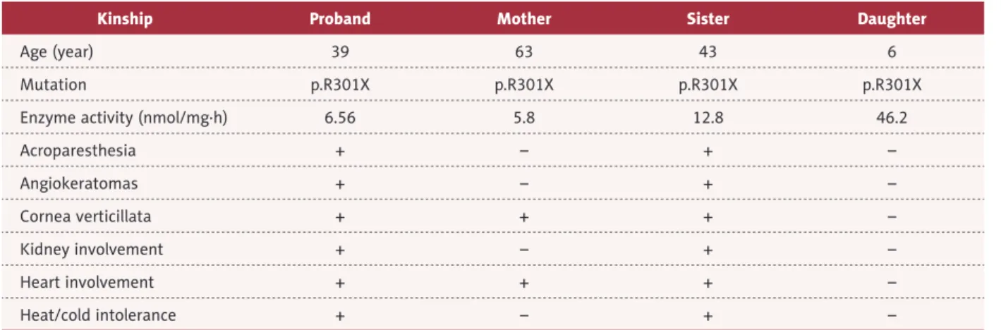
Relevant enzymatic and molecular data of the family are giv-en in Table 1.
Discussion
The estimated incidence of FD in the general population is 1 in 117 000 [11]. Newborn screening initiatives have found a high prevalence of the disease, as high as 1 in ~3100 new-borns in Italy [12].
To date, >600 mutations have been described in the exons and introns of the GLA gene, and discrimination between patho-logical and neutral mutations is difficult
(http://fabry-data-base.org/mutants/september 2015). Classically, clinical
mani-festations are more severe in male hemizygous subjects than in female heterozygotes. Recently, it was found that FD can also severely affect women, but progression of the disease to organ failure generally takes longer and the severity of symp-toms are more variable than in men [4,5]. For this reason, ac-curate follow-up is required for female patients, independent of enzymatic activity levels.
Misdiagnosis of the FD is common; it takes years (14–19 years) between beginning of the symptoms and correct diagnosis [4,5]. In our case, the patient was being treated for chronic scleros-ing glomerulonephritis for 2 years, which is a relatively short time. Fabry disease may not be diagnosed until development of end-stage renal failure in patients with proteinuria. The fre-quency of FD was 0.04% (2/5408) in men and 0% (0/3139) in women, and then 0.02% (2/8547) in all patients undergo-ing dialysis in the Japan Fabry Disease Screenundergo-ing Study [13]. In Spain, 3650 samples from hemodialysis patients (18.5% of all patients undergoing hemodialysis in Spain) were tested and 11 new unrelated cases of Fabry disease (4 males and 7 females, 0.003%) were diagnosed [14]. The prevalence of FD was detected as 0.17% in Turkish hemodialysis patients [15].
Although the average presentation age is 6–8 years in males, the proband’s symptoms began in his late thirties. His moth-er’s and sistmoth-er’s symptoms also began at about the same age. His 6-year-old daughter does not have symptoms. Suspicion, careful physical examination, and past medical history (includ-ing acroparesthesia, heat and cold intolerance, and hypohidro-sis) are very valuable.
In the M51I mutation, there were reports of cardiac, cerebral, and gastrointestinal involvement, but the kidneys are pro-tected [8]. Another study evaluating intrafamilial phenotypic variability in 4 families (GLA gene mutations; p.R220X, p.C52Y, p.C172Y, p.R342X) with FD showed that renal and cardiac in-volvements, angiokeratoma, and acroparesthesia are common findings [16]. Cerebral involvements are found only in p.C52Y and p.R342X mutations. In our patient’s p.R301X mutation, ce-rebral and gastrointestinal involvement were absent. European renal best practice recommends screening patients for Fabry disease when there is unexplained chronic kidney disease in men younger than 50 years and women of any age. In men, the activity of a-galactosidase A can be measured in plasma, blood cells, or dried blood spots. In women, mutation analysis is necessary, as enzyme measurement alone can miss over one-third of female Fabry patients [17]. Our proband pa-tient was a 39-year-old man who was being treated for chronic sclerosing glomerulonephritis. His sister had renal involvement. Cardiac symptoms, including left ventricular hypertrophy, ar-rhythmia, angina, and dyspnea, are reported in 40–60% of pa-tients with FD [1]. The prevalence of Fabry disease in primary cardiology practice with strictly defined otherwise unexplained LVH was 4% in males. Systematic screening is recommended in all men older than 30 years with LVH of unknown etiology for FD, even in the absence of obvious extracardiac manifesta-tions [18]. Many characteristics of Fabry disease cardiomyop-athy, with regard to electrocardiography and cardiac imaging,
Kinship Proband Mother Sister Daughter
Age (year) 39 63 43 6
Mutation p.R301X p.R301X p.R301X p.R301X
Enzyme activity (nmol/mg·h) 6.56 5.8 12.8 46.2
Acroparesthesia + – + – Angiokeratomas + – + – Cornea verticillata + + + – Kidney involvement + – + – Heart involvement + + + – Heat/cold intolerance + – + –
have been claimed. None of the criteria were specific enough (90%) to be used for definitive diagnosis of Fabry disease. Cerebrovascular manifestations result from multifocal small-vessel involvement and may include thromboses, seizures, paroxysmal hemiplegia, or hemianesthesia. Sensory neurons in spinal ganglia and small myelinated and unmyelinated fi-bers are preferentially affected. As a result of these features and the frequent presence of lesions in MRI scans, FD is of-ten misdiagnosed as multiple sclerosis [19]. Fabry disease may explain ~1% of all strokes in young people, including 3–5% of cryptogenic strokes. Early recognition of FD may help to initi-ate appropriiniti-ate treatment to decrease the risk of subsequent complications [20].
The European Fabry Working Group consensus recommends enzyme replacement therapy as soon as there are early clini-cal signs of kidney, heart, or brain involvement for classiclini-cally affected men. Classically affected women and men with non-classical FD should be treated as soon as there are early clin-ical signs of kidney, heart, or brain involvement, while treat-ment may be considered in women with non-classical FD with early clinical signs that are considered to be due to FD [2].
Conclusions
In conclusion, our findings confirm the intrafamilial phenotyp-ic variability in patients who have the same mutation. In the light of previous studies and results of the present report, it appears that heterozygous women could be affected as se-verely as man and should not be considered as asymptomat-ic carriers. Pedigree analysis remains a powerful, readily avail-able tool to identify individuals at risk for genetic diseases. Acknowledgements
a-Galactosidase A activity was measured by Dr. Murat Oktem at Duzen Laboratory, Ankara. Genetic analysis was performed by Serdar Ceylaner, MD, Associate Professor Medical Geneticist at Intergen Genetic Centre, Ankara, Turkey.
Conflict of interest
Dr. Ruya Ozelsancakhas declared no competing interest. Dr. Bulent Uyar has declared no competing interest.
Statement
Financial Support: No. No experimental investigation on hu-man subjects.
References:
1. Germain DP: Fabry disease. Orphanet J Rare Dis, 2010; 5: 30
2. Biegstraaten M, Arngrímsson R, Barbey F et al: Recommendations for initi-ation and cessiniti-ation of enzyme replacement therapy in patients with Fabry disease: The European Fabry Working Group consensus document. Orphanet J Rare Dis, 2015; 10: 36
3. Linthorst GE, Vedder AC, Aerts JM, Hollak CE: Screening for Fabry disease using whole blood spots fails to identify one-third of female carriers. Clin Chim Acta, 2005; 353: 201–3
4. Mehta A, Ricci R, Widmer U et al: Fabry disease defined: baseline clinical manifestations of 366 patients in the Fabry Outcome Survey. Eur J Clin Invest, 2004; 34: 236–42
5. Eng CM, Fletcher J, Wilcox WR et al: Fabry disease: baseline medical char-acteristics of a cohort of 1765 males and females in the Fabry Registry. J Inherit Metab Dis, 2007; 30: 184–92
6. Zizzo C, Colomba P, Albeggiani G et al: Misdiagnosis of familial Mediterranean fever in patients with Anderson-Fabry disease. Clin Genet, 2013; 83: 576–81 7. Lidove O, Kaminsky P, Hachulla E et al: FIMeD investigators. Fabry disease
‘The New Great Imposter’: Results of the French Observatoire in Internal Medicine Departments (FIMeD). Clin Genet, 2012; 81: 571–77
8. Cammarata G, Fatuzzo P, Rodolico MS et al: High variability of Fabry dis-ease manifestations in an extended Italian family. Biomed Res Int, 2015; 2015: 504784
9. Eng CM, Niehaus DJ, Enriquez AL et al: Fabry disease: twenty-three muta-tions including sense and antisense CpG alteramuta-tions and identification of a deletional hot-spot in the alpha-galactosidase A gene. Hum Mol Genet, 1994; 3: 1795–99
10. Blau N, Duran M, Gibson KM: Laboratory guide to the methods in biochem-ical genetics. Berlin Heidelberg: Springer-Verlag, 2008
11. Meikle PJ, Hopwood JJ, Clague AE, Carey WF: Prevalence of lysosomal stor-age disorders. JAMA, 1999; 281: 249–54
12. Spada M, Pagliardini S, Yasuda M et al. High incidence of later-onset fabry disease revealed by newborn screening. Am J Hum Genet, 2006; 79: 31–40 13. Saito O, Kusano E, Akimoto T et al: Prevalence of Fabry disease in dialysis
patients: Japan Fabry disease screening study (J-FAST). Clin Exp Nephrol, 2015 [Epub ahead of print]
14. Herrera J, Miranda CS: Prevalence of Fabry’s disease within hemodialysis patients in Spain. Clin Nephrol, 2014; 81: 112–20
15. Okur I, Ezgu F, Biberoglu G et al: Screening for Fabry disease in patients un-dergoing dialysis for chronic renal failure in Turkey: Identification of new case with novel mutation. Gene, 2013; 527: 42–47
16. Rigoldi M, Concolino D, Morrone A et al: Intrafamilial phenotypic variability in four families with Anderson-Fabry disease. Clin Genet, 2014; 86: 258–63 17. Terryn W, Cochat P, Froissart R et al: Fabry nephropathy: indications for
screening and guidance for diagnosis and treatment by the European Renal Best Practice. Nephrol Dial Transplant, 2013; 28: 505–17
18. Palecek T, Honzikova J, Poupetova H et al: Prevalence of Fabry disease in male patients with unexplained left ventricular hypertrophy in prima-ry cardiology practice: prospective Fabprima-ry cardiomyopathy screening study (FACSS). J Inherit Metab Dis, 2014; 37: 455–60
19. Callegaro D, Kaimen-Maciel DR: Fabry’s disease as a differential diagnosis of MS. Int MS J, 2006; 13: 27–30
20. Shi Q, Chen J, Pongmoragot J et al: Prevalence of Fabry disease in stroke patients – a systematic review and meta-analysis. J Stroke Cerebrovasc Dis, 2014; 23: 985–92