Mustafa Cosan Terek,
Levent Akman, Behiye Seda Hursitoglu, Ulus Ali Sanli1, Zeynep Ozsaran2, Mustafa Agah Tekindal3, Yilmaz Dikmen, Osman Zekioglu4, Ahmet Aydin Ozsaran Departments of Obstetrics and Gynecology and 4Pathology, 1Division of Medical Oncology, 2Division of Radiation Oncology, Ege University Medical School, Izmir, 3Department of Biostatistics, Baskent University School of Medicine, Ankara, Turkey For correspondence: Dr. Levent Akman, Department of Obstetrics and Gynecology, Ege University Medical School, 35100 Bornova, Izmir, Turkey. E‑mail: leventakman@ gmail.com
The retrospective analysis of patients with
uterine sarcomas: A single‑center experience
ABSTRACT
Background: Uterine sarcomas are rare, malignant, gynecological tumors and show diverse histopathological features. Therefore, there is no consensus on risk factors for poor outcome and optimal treatment. The aim of this retrospective analysis is to report the clinical outcome of patients with uterine sarcoma treated at a single center.
Materials and Methods: The data was obtained regarding the patient’s demographic characteristics, pathological results, treatments given, survival, and complications of all uterine sarcoma patients treated in a single center between the years 2000 and 2012. The 80‑month overall survival (OS) was determined with respect to prognostic factors including age, stage of disease, histopathological type, and adjuvant treatment.
Results: A total of 57 case records are retrieved for this retrospective analysis. The mean age of the patients is 62.5 ± 11.2 years. International Federation of Gynecology and Obstetrics (FIGO) stage distribution is stage I: 29; stage II: 13; stage III: 9; stage IV: 6. Fifty‑seven patients underwent surgery, 33 received postoperative radiotherapy (PORT), and 32 received chemotherapy. Median follow‑up period was 25 months (range 2–85 months). The 80‑month OS for the entire group of patients was 36.7%. The significant prognostic factors for survival are age under 50 years, stage of disease, and adjuvant chemotherapy.
Conclusion: Although limited by small sample size and retrospective nature, age under 50 years, stage of disease, and adjuvant chemotherapy are significant prognostic factors for survival for uterine sarcomas.
KEY WORDS: Chemotherapy, radiotherapy, survival, uterine sarcoma
INTRODUCTION
Uterine sarcomas are rare tumors and previous reports have been estimated to comprise 4–9% of all
invasive uterine cancers.[1‑3] Uterine sarcomas usually
have aggressive clinical behavior with propensity to local recurrence and distant metastasis. Because of the rarity and histopathological diversity of uterine sarcomas, there is lack of consensus on risk
factors for poor outcome and optimal treatment.[4,5]
Histologically, uterine sarcomas are classified into carcinosarcomas (40%), leiomyosarcomas (40%), endometrial stromal sarcomas (10–15%), and undifferentiated sarcomas (5–10%).
Recently, carcinosarcoma has been reclassified as a dedifferentiated or metaplastic form of endometrial
carcinoma.[5] A new International Federation of
Gynecology and Obstetrics (FIGO) classification and staging system is designed for uterine sarcomas reflecting their different biologic behavior in 2009. Stage I leiomyosarcomas and endometrial stromal sarcomas are subdivided according to size. The subdivision of stage I adenosarcomas take into account myometrial invasion. Carcinosarcomas are
still staged as endometrial carcinomas.[5] Uterine
sarcomas occur in women aged 40–60 years. Abnormal uterine bleeding, abdominal or pelvic mass, and pain are the most common symptoms of patients. The most frequently reported prognostic factors include tumor stage, histological subtype, grade, lymphovascular invasion, menopausal
status, and adjuvant radiotherapy.[4,6,7]
The aim of the present study is to analyze the treatment results of the uterine sarcomas at a single center.
MATERIALS AND METHODS
We retrospectively retrieved the case records of the all uterine sarcoma patients treated at Ege University Hospital between the years 2000 and 2012. The case records were analyzed regarding the patient’s demographic characteristics, pathological results, treatments given, survival, and complications [Table 1]. Each patient was subjected to clinical examination, routine biochemical analysis, and computed tomography (CT) imaging of the thorax and abdominopelvic region. Staging was done according to FIGO 2009 criteria.[8]
Access this article online Website: www.cancerjournal.net DOI: 10.4103/0973-1482.148698
PMID: ***
Surgery in the form of total abdominal hysterectomy (TAH) and bilateral salpingo‑oophorectomy (BSO) with or without pelvic lymph node dissection was performed for all the operable patients [Table 2]. Neoadjuvant chemotherapy was performed in four patients and surgery was performed afterwards. Depending upon clinical and pathological factors obtained from surgical staging, adjuvant therapy with radiation therapy (RT) or chemotherapy is planned by the members of tumor board comprised of pathologist, gynecological oncologist, radiation oncologist, and medical oncologist.
Adjuvant RT consisted of combination of external beam RT to whole pelvis followed by intravaginal brachytherapy to vault. The prescribed dose of external beam radiation was 45–50 Gy with conventional fractionation (1.8–2.0 by per fraction, 5 days a week) with four‑field box technique (two anterioposterior‑posteroanterior (AP‑PA) and two lateral fields). Treatment was carried out on linear accelerator. After the completion of external beam radiation, intravaginal brachytherapy was performed to treat vaginal cuff using intravaginal cylinders. The dose of intravaginal brachytherapy was prescribed at depth of 0.5 cm from the surface of ovoids.
A dose of 6 Gy × 3 (weekly) by high dose rate was delivered using remote after loading unit.
Follow‑up was done every 3 months for the 1st year. Then,
the patients were followed‑up every 6 months. Clinical examination was performed at every visit with abdominopelvic ultrasonography and chest X‑ray to rule out lung metastases. Positron emission tomography (PET) scan was also done if there was a suspicious disease on clinical/radiological examination.
Statistical analysis
First, descriptive statistics of the continuous values were given. Descriptive statistics are defined as median ± standard deviation and median (minimum and maximum values). Log rank statistical analyses are used when comparing life times of cases with categories of variables. Α =0.05 is considered as statistically significant. The overall survival (OS) was calculated by Kaplan–Meier survival method. OS was determined with respect to histopathological type, stage of disease, and adjuvant treatment. Statistical analysis was performed using the software Statistical Package for Social Sciences (SPSS; Chicago IL, Version 17). P < 0.005 was considered significant.
RESULTS
A total of 57 case records were retrieved for this retrospective analysis. Patient’s characteristics are shown in Table 1. The mean age of the patients was 62.5 ± 11.2 years. Carcinosarcoma [Figure 1] and endometrial stromal sarcoma [Figure 2] were the most common histopathological types. Most patients had a stage I disease. One patient had a liver metastasis. The most common presenting complaint among the patients is abnormal vaginal bleeding (72.7%). The other complaints are pain (22%) and vaginal discharge (4.3%). Treatment details are shown in Table 2. All patients underwent surgery with or without adjuvant treatment.
Table 1: Clinical features of patients
Age (years)
Histopathological type 62.5±11.2
Carcinosarcoma 30
Leiomyosarcoma 8
Endometrial stromal sarcoma 15
Adenosarcoma 3 Undifferentiated sarcoma 1 FIGO stage I 29 II 13 III 9 IV 6 Treatment Surgery alone 9
Surgery and PORT 16
Surgery and PORT and chemotherapy 17
Surgery and chemotherapy 15
Follow-up period (months) 25 (2-85)
PORT=Postoperative radiotherapy, FIGO=International federation of gynecology and obstetrics
Table 2: Treatment modalities
Surgery (n=57) TAH 4 Vaginal hysterectomy 1 Subtotal hysterectomy 1 TAH, BSO 13 TAH, BSO, PLND 38 Adjuvant chemotherapy (n=32)
Carboplatin and paclitaxel 4
Ifosfamide and adriamycin 6
Ifosfamide and carboplatin 13
Cisplatin and adriamycin 9
Adjuvant radiotherapy (n=33)
External radiotherapy 11
External radiotherapy and brachytherapy 22
TAH=Total abdominal hysterectomy, BSO=Bilateral salpingo-oophorectomy, PLND=Pelvic lymph node dissection
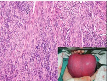
Figure 1: Endometrial stromal sarcoma. Tumor of fusiform cells with
uniform, eosinophilic cytoplasm with oval/round cells with small nuclei with dispersed chromatin pattern is shown. Macroscopic view of the tumor (inset)
Most common surgical procedure was TAH and BSO and pelvic lymph node dissection (38 of 57 patients). The biopsy of tumor mass was performed in only one patient because of clinically inoperable situation and she received palliative chemotherapy. Vaginal hysterectomy was performed in a patient because of stage III uterine prolapse and subtotal hysterectomy was performed in a patient because of dense
pelvic adhesions. Thirty‑three patients received radiotherapy. Thirty‑two patients received chemotherapy and most common regimens were ifosfamide plus carboplatin and cisplatin plus adriamycin.
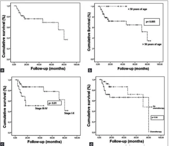
The mean follow‑up period was 25 months (range 2–85months). All patients were followed‑up for 80 months. In this period, 15 patients died. The 80‑month OS for all patients was 36.7% [Figure 3a]. The mean OS was 61.17 ± 5.101 months. The 80‑month OS for patients over 50 years of age was 34.5%. However, no deaths occurred in patients under 50 years of age (P < 0.05). The patients are evaluated with respect to being under 50 years of age or over 50 years of age [Figure 3b]. The OS for patients under 50 and over 50 years of age are 100 and 34.5%, respectively.
The 80‑month OS of patients who has received postoperative radiotherapy (PORT) was 34.6%. The 17‑month OS of patients with has not received PORT was 70%. The mean OS of patients who received PORT was 62.02 ± 6.04 months as compared to 53.22 ± 7.13 month of patients who has not received PORT. The 80‑month OS in stage I–II was 41.6%. However, 17‑month OS in stage III–IV was 50.9%. The mean OS in stage I–II and III–IV was 68.15 ± 5.17 and 28.42 ± 4.93, respectively (P < 0.05) [Figure 3c].
Figure 2: Uterine carcinosarcoma. Malign epithelial and mesenchymal
components are shown together (hematoxylin and eosin (H and E), ×20). Intraoperative view of the enlarged uterus (inset)
Figure 3: (a) The 80-month overall survival of all the patients. (b) The 80-month overall survival of patients >50 years of age vs <50 years age.
(c) The 80-month overall survival of patients with stage I–II vs stage III–IV. (d) The 80-month overall survival of patients who received chemotherapy
vs no chemotherapy
d c
b a
Chemotherapy had significant impact on OS. The 80‑month OS of patients who has received chemotherapy was 76.2%. However, 71‑month OS of patients who did not receive chemotherapy was 47.9%. The mean OS of patients who received chemotherapy was 55.91 ± 7.04 months as compared to 65.90 ± 7.08 months of patients who did not receive chemotherapy (P < 0.05) [Figure 3d].
At the end of 80 months, OS for carcinosarcoma was 41.2%. Forty‑six months OS for leiomyosarcomas was 37.5% and 16‑months OS for endometrial stromal sarcoma was 65.3%. The different histological types did not affect OS significantly.
DISCUSSION
Uterine sarcomas are rare malignant gynecological tumors with poor prognosis. In the present study, the 80‑month OS for all patients was 36.7% and the mean OS was 61.7 ± 5.101 months. Our results have demonstrated that age under 50 years, stage of disease, and adjuvant chemotherapy are significant prognostic factors for survival for uterine sarcomas.
Carcinosarcoma is a biphasic neoplasm composed of epithelial and mesenchymal elements. It typically occurs in postmenopausal women and most women present with abnormal vaginal bleeding and uterine enlargement. At presentation, extrauterine spread is found in one‑third of cases. Carcinosarcomas are typically large bulky polypoid masses, filling the uterine cavity, and prolapsing. On microscopic examination, the carcinomatous component is serous in two‑third of cases and endometrioid in one‑third of
cases.[5] Ferguson et al., reported that 10% of carcinomatous
components were FIGO grade I, 10% grade II, and 80% grade III. The sarcomatous components are heterogeneous
and almost all are high‑grade sarcomas.[9] The overall 5‑year
survival for patients with carcinosarcoma is around 30% and
for those with stage I, approximately 50%.[9,10] Surgical stage
and especially depth of myometrial invasion are the most important prognostic indicators. Tumors containing serous and clear cell carcinoma are associated with higher frequency of metastases. Appropriate treatment includes TAH and BSO with or without pelvic lymphadenectomy. The role of adjuvant radiotherapy and chemotherapy are uncertain, but some studies have demonstrated the advantage of radiotherapy for local control. Taxanes and cisplatin‑based chemotherapy with pelvic radiotherapy may increase the survival in patients
with metastatic carcinosarcomas.[11‑13] In the present study,
chemotherapy had significant impact on OS. The 80‑month OS of patients who has received chemotherapy was 76.2%. However, 71‑month OS of patients who did not receive chemotherapy was 47.9%.
Leiomyosarcoma occurs in women over 40 years of age who usually present with abnormal vaginal bleeding, palpable pelvic mass, and pelvic pain. Rarely, a leiomyosarcoma
originates from a leiomyoma. Histopathologic diagnosis of uterine leiomyosarcoma is done by microscopic appearance of hypercellularity, severe nuclear atypia, and high mitotic rate exceeding 15 mitotic figures per 10 high power fields. Supportive clinicopathologic features are peri‑ or postmenopausal age, extrauterine extension, large size (over 10 cm), infiltrating border, necrosis, and atypical mitotic figures.[10]
Leiomyosarcomas are very aggressive tumors even when confined to the uterus and even when diagnosed at an
early stage. The recurrence rate ranges from 53 to 71%.[13,14]
Tumor size and mitotic index are reported to be a major
prognostic factors.[13,15] However, stage is stillaccepted as
the most significant prognostic factor for uterine sarcomas. Treatment of leiomyosarcomas includes TAH and debulking of any extrauterine tumor. Removal of the ovaries and lymph node dissection remain controversial as metastases to these organs occur in a small percentage of cases and are
frequently associated with intra‑abdominal disease.[15] The
influence adjuvant radiotherapy on survival is uncertain. Radiotherapy may be useful in controlling local recurrences and chemotherapy with doxorubicin or docetaxel/gemcitabine
is used for advanced or recurrent disease.[16,17]
The principal treatment of uterine sarcoma is surgery and
adjuvant radiotherapy is controversial.[17] The only phase III
randomized trial assessing the efficacy of adjuvant pelvic RT in uterine sarcoma was published by the European Organization for the Research and Treatment of Cancer‑Gynecological Cancer
Group.[18] Patients with stage I–II uterine sarcomas were
randomized to adjuvant radiotherapy (50 Gy) to the pelvis or observation after TAH and BSO. Pelvic and paraaortic lymph node dissection was optional. With a median follow‑up 6 years adjuvant radiotherapy resulted in a significant reduction in crude local failures in all patients (22 versus 40%, P = 0.004). Moreover, the rate of isolated local failure dropped from 14 to 2%. Subgroup analysis showed significant local benefit for carcinosarcoma, but not for leiomyosarcoma. In the present study, at the end of 80 months OS for carcinosarcoma was 41.2%. Forty‑six months OS for leiomyosarcomas was 37.5% and 16‑months OS for endometrial stromal sarcoma was 65.3%. We did not find any survival difference between histological subtypes.
In Gynecologic Oncology Group (GOG) 150 trial, stage I–IV carcinosarcoma patients were randomized to whole abdominal radiotherapy or adjuvant chemotherapy consisting cisplatin,
ifosfamide, and MESNA.[19] Analyses of initial sites of failure
showed a nonsignificant lower rate of vaginal failures in the radiation arm versus the chemotherapy arm (3.8 versus 9.9%), but similar rate of isolated pelvic failures (13%). Adjusting for stage and age the recurrence rate was 21% lower for chemotherapy patients than for whole abdominal radiotherapy patients. The estimated death rate was 29% lower among the chemotherapy groups.
Endometrial stromal tumors are composed of cells that resemble endometrial stromal cells of the proliferative endometrium. They are divided into endometrial stromal nodule, low grade endometrial stromal sarcoma, and
undifferentiated endometrial sarcoma.[5] Endometrial stromal
nodules are well‑circumscribed tumors ranging from 1 to 22 cm. They have expansive, noninfiltrating margin without any vascular invasion. The prognosis is good and patients
are cured by hysterectomy.[20] Low‑grade endometrial stromal
sarcomas occur in women between 40 and 55 years of age. Extrauterine pelvic extension, most commonly involving ovary, is found in one‑third of cases. Tumor mass penetrates the myometrium and tumor cells distend the veins. Endometrial stromal sarcomas are indolent tumors, but late recurrences may occur in pelvis and abdomen. Stage I tumors have a
good prognosis.[21] In the series by Abeler et al., prognosis
of endometrial stromal sarcoma confined to the uterus was
related to mitotic index and tumor cell necrosis.[13]
Undifferentiated endometrial sarcoma exhibit myometrial invasion, severe nuclear pleomorphism, high mitotic activity (>10 mitotic figures/10 high power fields), tumor cell necrosis,
and smooth muscle or endometrial stromal differentiation.[5]
They have poor prognosis and most patients die of disease
within 2 years of the diagnosis.[13] In the present study, there
is only one case of undifferentiated sarcoma.
Adenosarcomas have low malignant potential; the incidence of distant metastases is 5% and the have a more favorable prognosis than other uterine sarcomas. However, they have
a tendency for late local recurrence in about 20% of cases.[13,18]
The standard treatment consists of TAH with BSO. Tumor cell necrosis was the synergist prognostic factor in adenosarcomas.
[13] In the present study, there are three cases of adenosarcomas.
In conclusion, age under 50 years, stage of disease, and adjuvant chemotherapy are significant prognostic factors for survival in uterine sarcomas. In addition, PORT does not have significant effect on survival. Further, clinical studies are needed for the treatment decisions of uterine sarcomas.
REFERENCES
1. Nordal RR, Thoresen SO. Uterine sarcomas in Norway 1956‑1992: Incidence, survival and mortality. Eur J Cancer 1997;33:907‑11. 2. Harlow BL, Weiss NS, Lofton S. The epidemiology of sarcomas of the
uterus. J Natl Cancer Inst 1986;76:399‑402.
3. Brooks SE, Zhan M, Cote T, Baquet CR. Surveillance, epidemiology, and endresults analysis of 2677 cases of uterine sarcoma 1989‑1999. Gynecol Oncol 2004;93:204‑8.
4. Sharma DN, Rath GK, Kumar S, Kumar L, Bhatla N, Gandhi AK, et al. Clinical outcome of patients with uterine sarcomas. J Cancer Res Ther 2011;7:270‑4.
5. D’Angelo E, Prat J. Uterine sarcomas: A review. Gynecol Oncol 2010;116:131‑9.
6. Livi L, Paiar F, Shah N, Blake P, Villanucci A, Amunni G, et al. Uterine sarcoma: Twenty‑seven years of experience. Int J Radiat Oncol Biol Phys 2003;57:1366‑73.
7. Park JY, Kim DY, Suh DS, Kim JH, Kim YM, Kim YT, et al. Prognostic factors and treatment outcomes of patients with uterine sarcoma: Analysis of 127 patients at a single institution, 1989‑2007. J Cancer Res Clin Oncol 2008;134:1277‑87.
8. Pecorelli S. Revised FIGO staging for carcinoma of the vulva, cervix, and endometrium. Int J Gynaecol Obstet 2009;105:103‑4.
9. Ferguson SE, Tornos C, Hummer A, Barakat RR, Soslow RA. Prognostic features of surgical stage I uterine carcinosarcoma. Am J Surg Pathol 2007;31:1653‑61.
10. Yamada SD, Burger RA, Brewster WR, Anton D, Kohler MF, Monk BJ. Pathologic variables and adjuvant therapy as predictors of recurrence and survival for patients with surgically evaluated carcinosarcoma of the uterus. Cancer 2000;88:2782‑6.
11. Callister M, Ramondetta LM, Jhingran A, Burke TW, Eifel PJ. Malignant mixed Müllerian tumors of the uterus: Analysis of patterns of failure, prognostic factors, and treatment outcome. Int J Radiat Oncol Biol Phys 2004;58:786‑96.
12. Villena‑Heinsen C, Diesing D, Fischer D, Griesinger G, Maas N, Diedrich K, et al. Carcinosarcomas‑a retrospective analysis of 21 patients. Anticancer Res 2006;26:4817‑23.
13. Abeler VM, Røyne O, Thoresen S, Danielsen HE, Nesland JM, Kristensen GB. Uterine sarcomas in Norway. A histopathological and prognostic survey of a total population from 1970 to 2000 including 419 patients. Histopathology 2009;54:355‑64.
14. D’Angelo E, Spagnoli LG, Prat J. Comparative clinicopathologic and immunohistochemical analysis of uterine sarcomas diagnosed using the World Health Organization classification system. Hum Pathol 2009;40:1571‑85.
15. Giuntoli RL 2nd, Metzinger DS, DiMarco CS, Cha SS, Sloan JA, Keeney GL, et al. Retrospective review of 208 patients with leiomyosarcoma of the uterus: Prognostic indicators, surgical management, and adjuvant therapy. Gynecol Oncol 2003;89:460‑9.
16. Hensley ML, Ishill N, Soslow R, Larkin J, Abu‑Rustum N, Sabbatini P, et al. Adjuvant gemcitabine plus docetaxel for completely resected stages I‑IV high grade uterine leiomyosarcoma: Results of a prospective study. Gynecol Oncol 2009;112:563‑7.
17. García‑Martínez E, Prefasi LE, García‑Donas J, Escolar‑Pérez PP, Pastor F, González‑Martín A. Current management of uterine sarcomas. Clin Transl Oncol 2011;13:307‑14.
18. Reed NS, Mangioni C, Malmström H, Scarfone G, Poveda A, Pecorelli S, et al.; European Organisation for Research and Treatment of Cancer Gynaecological Cancer Group. Phase III randomised study to evaluate the role of adjuvant pelvic radiotherapy in the treatment of uterine sarcomas stages I and II: An European Organisation for Research and Treatment of Cancer Gynaecological Cancer Group Study (protocol 55874). Eur J Cancer 2008;44:808‑18.
19. Wolfson AH, Brady MF, Rocereto T, Mannel RS, Lee YC, Futoran RJ, et al. A gynecologic oncology group randomized phase III trial of whole abdominal irradiation (WAI) vs. cisplatin‑ifosfamide and mesna (CIM) as post‑surgical therapy in stage I‑IV carcinosarcoma (CS) of the uterus. Gynecol Oncol 2007;107:177‑85.
20. Dionigi A, Oliva E, Clement PB, Young RH. Endometrial stromal nodules and endometrial stromal tumors with limited infiltration: A clinicopathologic study of 50 cases. Am J Surg Pathol 2002;26:567‑81.
21. Chang KL, Crabtree GS, Lim‑Tan SK, Kempson RL, Hendrickson MR. Primary uterine endometrial stromal neoplasms. A clinicopathologic study of 117 cases. Am J Surg Pathol 1990;14:415‑38.
Cite this article as: Terek MC, Akman L, Hursitoglu BS, Sanli UA, Ozsaran Z, Tekindal MA, et al. The retrospective analysis of patients with uterine sarcomas: A single-center experience. J Can Res Ther 2016;12:309-13.