ENZYME ELECTRODES FABRICATED BY
DAD TYPE Poly
(2,5-di(furan-2-yl)thiazolo[5,4-d]thiazole) CONDUCTING POLYMER
2020
THESIS OF MASTER OF SCIENCE
DEPARTMENT OF CHEMISTRY
Nadia Mohammed Salama KURZAMA
Thesis Advisor
ENZYME ELECTRODES FABRICATED BY DAD TYPE Poly (2,5-di(furan-2-yl)thiazolo[5,4-d]thiazole)
CONDUCTING POLYMER
A THESIS SUBMITTED TO
THE INSTITUTE OF GRADUATE PROGRAMS OF KARABUK UNIVERSITY
BY
Nadia Mohammed Salama KURZAMA
IN PARTIAL FULFILLMENT OF THE REQUIREMENTS FOR THE DEGREE OF MASTER OF SCIENCE IN
DEPARTMENT OF CHEMISTRY
“I declare that all the information within this thesis has been gathered and presented in accordance with academic regulations and ethical principles and I have according to the requirements of these regulations and principles cited all those which do not originate in this work as well.”
ABSTRACT
M. Sc. Thesis
ENZYME ELECTRODES FABRICATED BY DAD TYPE Poly (2,5-di(furan-2-yl)thiazolo[5,4-d]thiazole)
CONDUCTING POLYMER
Nadia Mohammed Salama KURZAMA
Karabük University Institute of Graduate Programs
The Department of Chemistry
Thesis Advisor:
Assist. Prof. Dr. A. Elif BÖYÜKBAYRAM February 2020, 65 pages
In this study, DAD type polymer was used as a new electrode material and enzyme electrodes were prepared by immobilizing polyphenol oxidase enzyme in this matrix. 2,5-di(furan-2-il)tiyazolo[5,4-d]tiyazol monomer was polymerized electrochemically and coated to platinum electrodes and polyphenol oxidase enzyme was immobilized on the polymer surface with cross-linker. Kinetic characterization and optimization of enzyme electrodes formed was made and the study was completed with real sample analysis. Kinetic parameters were assessed for Vmax (maximum reaction rate) and Km
(enzyme affinity of substrate) respectively as 0.028 ± 0.001umol min1- electrode1- and
669.68 ± 64.73 mM. The effect of reaction conditions was examined and the highest enzyme activity was obtained at pH 7.5 and 45 ℃. Linear operation interval determined in optimum conditions is between 1.0 – 90.0 mg mL1-. And LOQ value was found as 7.827 mg mL1- The stability of enzyme electrodes was investigated with
consecutive activity measurements. After 50 consecutive measurements, it was observed that activity of enzyme electrode was at 70% value. Meanwhile, in the shelf-life study, it was determined that activity fell to 70% after 50 days. Enzyme electrodes application was designed in the form of analysis of polyphenolic substances in waste water samples. Total polyphenolic substance amount in sample was found as 268.48 ± 6.62 mg mL1- by enzyme electrodes. This result was verified using Folin-Ciocalteau
analysis method as the control method with a result of 262.86 ± 19.54 mg mL1-.
Key Words : Electrochemical polymerization, conducting polymer, enzyme
immobilization, polyphenol oxidase, enzyme electrode.
ÖZET Yüksek Lisans Tezi
DAD TİPİ Poli (2,5-di(furan-2-il)tiyazolo[5,4-d]tiyazol) İLETKEN POLİMERİYLE OLUŞTURULAN
ENZİM ELEKTROTLARI
Nadia Mohammed Salama KURZAMA
Karabük Üniversitesi Lisansüstü Eğitim Enstitüsü
Kimya Anabilim Dalı Tez Danışmanı:
Dr. Öğr. Üyesi A. Elif BÖYÜKBAYRAM Şubat 2020, 65 sayfa
Bu çalışmada yeni bir elektrot malzemesi olarak DAD tipi polimerde polifenol oksidaz enzimi tutuklanarak enzim elektrotları hazırlanmıştır. 2,5-di(furan-2-il)tiyazolo[5,4-d]tiyazol monomeri elektrokimyasal yolla polimerleştirilerek platin elektrotlara kaplanmış ve polifenol oksidaz enzimi çapraz bağlayıcı ile polimer yüzeyine yerleştirilmiştir. Oluşturulan enzim elektrotlarının kinetik karakterizasyonu ve optimizasyonları yapılmış ve numune analizi ile çalışma sonuçlandırılmıştır. Kinetik parametreler, Vmax (maksimum reaksiyon hızı) ve Km (substratın enzim ilgisi)
tutuklanmış enzim için sırasıyla 0.028 ± 0.001umol dak-1 elektrot-1 ve 669.68 ± 64.73
mM olarak tayin edilmiştir. Reaksiyon koşullarının etkisi incelenmiş ve en yüksek enzim aktivitesi pH 7.5 ve 45 ℃ sıcaklıkta elde edilmiştir. Optimum koşullarda tespit edilen doğrusal çalışma aralığı 1.0 – 90.0 mg mL1- arasındadır. LOQ değeri ise 7.827
enzim elektrotlarının stabilitesi incelenmiştir. 50 ardışık ölçümden sonra enzim elektrodunun aktivitesinin %70 değerinde olduğu gözlenmiştir. Raf ömrü çalışmasında ise 50 günün sonunda aktivitenin %70’e düştüğü saptanmıştır. Enzim elektrotlarının uygulaması atık su numunelerinde polifenolik maddelerin analizi şeklinde tasarlanmıştır. Toplam polifenolik madde miktarı enzim elektrotlarıyla 268,48 ± 6,62 mg mL1- olarak saptanmıştır. Bu sonuç kontrol yöntemi olarak Folin-Ciocalteau analiz metodu kullanılarak doğrulanmış ve 262,86 ± 19,54 mg mL1- bulunmuştur.
Anahtar Kelimeler : Elektrokimyasal polimerizasyon, iletken polimer, enzim
tutuklaması, polifenol oksidaz, enzim elektrodu.
ACKNOWLEDGEMENT
I would like to express my greatest appreciation to my supervisor Assist. Prof. Dr. Ayşe Elif BÖYÜKBAYRAM for her encouragements and patience during my study.
I owe Prof. Dr. Şadi Şen a great debt of gratitude due to his support throughout my study.
I would like to give thanks to the academic staff of Department of Chemistry in Karabük University for their guidance.
I would like to extend my thanks to the government of Libya and the Libyan Embassy in Turkey, especially to Academic Office for providing me financial support and all the expenses in order to obtain MSc.
I also would like to express my thanks to my beloved father Mohammed Salama KURZAMA and my beloved mother Fatima Moftah ALDAWDI who helped me to achieve my goals with their encouragement during my journey.
I also wish to present endless thanks to my family, especially to my husband Saleh Ali SALEH, for his patience, moral support and for always being there for me whenever I need. Very special thanks to my dear children for being a very important part of my life.
This master study was supported by Karabük University Coordinatorship of Scientific Research Projects with project number: FYL-2019-2101.
CONTENTS Page APPROVAL ... ii ABSTRACT ... iv ÖZET... vi ACKNOWLEDGEMENT ... viii CONTENTS ... ix
LIST OF FIGURES ... xiii
LIST OF TABLES ... xv
ABBREVIATIONS ... xvi
PART 1 ... 1
INTRODUCTION ... 1
PART 2 ... 4
THEORETICAL BACKGROUND AND LITERATURE SURVEY ... 4
2.1. CONDUCTING POLYMERS ... 4
2.1.1. Conductivity Theory in Polymers ... 4
2.1.1.1. Band Theory ... 5
2.1.1.2. Doping in Conducting Polymers ... 7
2.1.1.3. Hopping in Conducting Polymers ... 9
2.1.2. Synthesis of Conducting Polymers ... 10
2.1.2.1. Chemical Synthesis ... 10
2.1.2.2. Electrochemical synthesis ... 10
2.1.3. Usage Areas of Conducting Polymers ... 12
2.2. ENZYMES ... 13
2.2.1. Classification of Enzymes ... 15
2.2.2. Enzyme Activity... 15
2.2.2.1. Effect of Substrate Concentration ... 16
Page
2.2.2.3. Effect of Temperature ... 17
2.2.2.4. pH Effect ... 17
2.2.2.5. Inhibitor Effect ... 18
2.2.3. Enzyme Kinetics ... 19
2.2.4. Enzyme Immobilization Methods ... 24
2.2.4.1. Binding to a Support ... 24
2.2.4.2. Cross-linking ... 25
2.2.4.3. Entrapment ... 25
2.2.5. Poliphenol Oxidase ... 26
2.2.5.1. Working Mechanism of Polyphenol Oxidase ... 27
2.2.5.2. Usage Areas of Polyphenol Oxidase ... 29
2.2.5.3. Use of Polyphenol Oxidase in Biosensors ... 30
2.3. SAMPLE ANALYSIS ... 30
PART 3 ... 32
MATERIALS AND METHODS ... 32
3.1. CHEMICAL MATERIALS ... 32 3.2. INSTRUMENTS ... 32 3.2.1. Potentiostat ... 32 3.2.2. UV-Visible Spectrometry... 32 3.2.3. SEM ... 33 3.2.4. pH meter ... 33
3.2.5. Magnetic Mixer with Heater ... 33
3.2.6. Vortex Tube Mixer ... 33
3.2.7. Shaking Water Bath ... 34
3.3. METHODS ... 34
3.3.1. Electrochemical Synthesis of Poly(2,5-di(furan-2-yl)thiazolo[5,4-d]thiazol) ... 34
3.3.2. Enzyme Immobilization in PTTzFr Matrix... 36
3.3.3. Besthorn’s Hydrazone Method ... 37
3.3.4. Determination of the Enzyme Activity ... 37
Page
3.4. OPTIMIZATIONS ... 39
3.4.1. Optimum pH Determination ... 39
3.4.2. Optimum Temperature Determination ... 40
3.4.3. Optimization of Immobilized Enzyme Amount ... 40
3.4.4. Optimization of Glutaraldehyde Amount... 40
3.4.5. Assessment of Stability ... 40
3.4.6. Determination of Shelf Life ... 40
3.5. PHENOLIC SUBSTANCE DETERMINATION IN WASTE WATER SAMPLE ... 41
3.5.1. Analysis with Pt/PTTzFr/PPO Enzyme Electrode ... 41
3.5.2. Analysis with Folin-Ciocalteau Method ... 42
PART 4 ... 43
RESULTS AND DISCUSSION ... 43
4.1. ELECTROPOLYMERIZATION OF TTzFr MONOMER ... 43
4.2. SEM ANALYSIS ... 44
4.3. OPTIMIZATION OF ENZYME ELECTRODES ... 47
4.3.1. Effect of pH on Enzyme Electrodes ... 47
4.3.2. Effect of Temperature on Enzyme Electrodes ... 48
4.3.3. Optimization of Immobilized Enzyme Amount ... 49
4.3.4. Optimization of GA Amount Applied... 50
4.4. DETERMINATION OF KINETIC PARAMETERS ... 51
4.5. STABILITY STUDIES ... 54
4.5.1. Operational Stability of Enzyme Electrodes ... 54
4.5.2. Shelf Life of Enzyme Electrodes ... 55
4.6. ANALYSIS OF WASTE WATER SAMPLES ... 55
4.6.1. Determination of Polyphenolics with Pt/PTTzFr/PPO Enzyme Electrode .. 56
4.6.2. Determination of Polyphenolics with Folin-Ciocalteau Method ... 57
PART 5 ... 59
CONCLUSION AND SUGGESTIONS ... 59
Page
LIST OF FIGURES
Page
Figure 2.1. Formation of bond and anti-bond orbitals in the polymer molecule. ... 5
Figure 2.2. Band structures for insulators, semi-conductors and conductors. ... 6
Figure 2.3. Polaron, bipolaron structures and band diagrams. a) neutralpolymer b) polaron structure c) bipolaron structure. ... 8
Figure 2.4. Polyacetylene’s soliton, polaron and bipolaron structures. ... 9
Figure 2.5. Transportation of charge, a) transportation of charge along the chain, b) transportation of charge between chains, c) transportation of charge between chain blocks [3]. ... 10
Figure 2.6. Electropolymerization mechanism of heterocyclics (X= N-H, S, O). ... 11
Figure 2.7. Effect of enzyme in activation energy ... 14
Figure 2.8. Key-lock model proposed for the enzyme-substrate complex. ... 15
Figure 2.9. Effect of substrate concentration over reaction rate [12]... 16
Figure 2.10. Effect of enzyme concentration over reaction rate [12]. ... 16
Figure 2.11. Effect of temperature over reaction rate. ... 17
Figure 2.12. Effect of pH over reaction rate. ... 18
Figure 2.13. Enzyme-inhibitor relationship [23]... 18
Figure 2.14. Substrate concentration versus reaction rate graph [13]. ... 19
Figure 2.15. Lineweaver-Burk graph [13]. ... 23
Figure 2.16. Enzyme immobilization methods. ... 24
Figure 2.17. Enzyme immobilization methods. ... 25
Figure 2.18. Polyphenol oxidase catalysis reaction. ... 26
Figure 2.19. Cyclical transitions of PPO’s active site between oxy and deoxy forms during catalysis. Blue, red and green circles represent copper, oxygen and histidine [26]…...………..………..27
Page
Figure 3.1. Polimerization of TTzFr. ... 34
Figure 3.2. Three electrode system. ... 35
Figure 3.3. Electropolymerization cell. ... 36
Figure 3.4. Besthorn’s Hydrazone method. ... 37
Figure 3.5. Pyrocatechol. ... 38
Figure 3.6. PPO’s cresolase and catecholase activities: A) monophenol’s o-hydroxylation, B) oxidation of o-diphenol to o-quinone. ... 38
Figure 3.7. o-Quinone formed to establish colored compound with MBTH reactive [36]……...………..39
Figure 3.8. Process steps of Folin-Ciocalteau method. ... 42
Figure 4.1. Polymerization of TTzFr monomer against Ag/AgCl with cyclic voltametry. ... 44
Figure 4.2. a) SEM, polymer surface (x5000), b) SEM, polymer surface (x10000). ... 45
Figure 4.3. a) SEM, after enzyme immobilization (x5000), b) SEM, after enzyme immobilization (x10000). ... 46
Figure 4.4. Effect of pH on Pt/PTTzFr/PPO electrode activity. ... 48
Figure 4.5. Effect of temperature on Pt/PTTzFr/PPO electrode activity. ... 49
Figure 4.6. Effect of enzyme amount on activity. ... 49
Figure 4.7. Effect of GA amount on activity. ... 50
Figure 4.8. Michaelis-Menten graph of Pt/PTTzFr/PPO electrode... 52
Figure 4.9. Lineweaver-Burk graph of Pt/PTTzFr/PPO electrode. ... 52
Figure 4.10. Operational stability of Pt/PTTzFr/PPO electrode. ... 54
Figure 4.11. Operational stability of Pt/PPO electrode. ... 55
Figure 4.12. Calibration graph of Pt/PTTzFr/PPO electrode (a) 8 catechol solutions (b) 11 catechol solutions. ... 57
LIST OF TABLES
Page
Table 4.1. Kinetic parameters of free and immobilized PPO enzyme. ... 53 Table 4.2. Total polyphenolic material amounts determined as mg.mL1- catechol
equivalent in waste water samples with Pt/PTTzFr/PPO enzyme
ABBREVIATIONS
Km : Michaelis-Menten constant
Vmax : Maximum reaction rate
LOQ : Limit of Quantitation mL : Mililiter m : Micrometer μL : Microliter μmol : Micromole nm : Nanometer ℃ : Degree Celcius g : Gram mg : Miligram min : Minute Å : Ångström A : Amper V : Volt VB : Valence Band CB : Conductance Band CV : Cyclic Voltamogram
SEM : Scanning Electron Microscope
UV : Ultraviolet
PA : Polyacetylene
PAN : Polyacrylonitrile
PPy : Polypyrrole
PPO : Polyphenole oxidase
MBTH : 3-Methyl-2-benzodiazolinon hydrazon hydrochloride monohydrate
DAD : Donor-Acceptor-Donor
PTTzFr : poly(2,5-di(furan-2-yl)thiazolo[5,4-d]thiazol) polymer Pt/Ppy/PPO : Enzyme electrode composed of polyphenol oxidase enzyme
immobilized into PPy coated on platinum electrode
Pt/PTTzFr/PPO : Enzyme electrode composed of polyphenol oxidase enzyme immobilized into PTTzFr coated on platinum electrode
PART 1
INTRODUCTION
It is an extensive research field that enzymes are immobilized in many natural or synthetic organic polymers and inorganic materials. Enzymes are materials that are both expensive and lose their activity easily. For this reason, it is important to immobilize and regain the enzyme back and use it more and more instead of using them one time in a free form. An immobilized enzyme can be received back from the reaction environment and this is an important factor in reducing cost. Besides, by receiving the enzyme back from the reaction environment, a green production can be obtained.
Enzymes are specific catalyzers and specific analysis of the enzyme’s substrate can be possible by the entrapped enzyme. Beside the advantage of enzyme being specific to its substrate, the high sensitivity that is created by electrochemical methods resulted in more researches to be made with enzyme electrodes for analysis purposes. Enzyme electrodes are electrode structures where the enzyme is entrapped within or on the surface of a conductive matrix and it is the main component of enzymatic biosensors. Electrode materials are gold (plate or wire), platinum (plate or wire) and carbon-based materials (paper, rod, paste, metalized carbon, glassy carbon, carbon fiber). Enzyme electrodes are basically composed of the enzyme and a composite structure covered on the electrode material. The performance of the enzyme electrode is much more due to the used composite materials. These materials are conductive polymers, functional polymers, metal complexes, sol-gel materials and Nano-materials (carbon Nano-tube, Nano-particulates). Researches in the field of biosensors are in the direction to develop new matrices and composite materials and obtain more sensitive, easy-to-use and cheaper methods.
Electroactive conductive polymers are used widely in the field of bioanalysis with their charge carrying capacity and biocompatibility. Two important reasons that they are used in enzyme electrodes are their active enzyme binding capacity and helping electron exit from the enzyme and carrying the electrons to the surface of the electrode, which are important factors in amperometric biosensors. For this reason, conductive polymers have sensitivity increasing effects. Besides this, due to their stabile structure, the enzyme lengthens the life of the electrode and increases its repeatability. With these advantages of them, conductive polymers have attracted more attention recently and they were coated to the surface of electrodes from many different materials. These coated electroactive films are used to entrape enzymes or proteins.
The lifetime of the entrapped biomolecule depends on both the composite material and the entrapment technique of the enzyme. Generally, enzyme immobilization methods are adsorption, covalent bonding, cross-linking and entrapment. As the adsorption method consists of weak bonds between the electrode and enzyme, it can’t hold the enzyme in a manner that is strict enough. Entrapment method maintains a strict closeness between the enzyme and the matrice but it doesn’t contain covalent bonds. In covalent bonding, a cross-linking material such as glutaraldehyde is used in order to bond the enzyme to polymer or enzyme molecules with each other. In this method, bonds forming between enzyme molecules give stability to the entrapped enzyme and it lengthens the life time of the enzyme electrode.
Polyphenol oxidase is a metaloprotein that contains copper and it is an enzyme used commonly in animals, plants and microorganisms. The substrates of polyphenol oxidase are phenolic materials and it is an important biomaterial that can be used in the analysis of phenolic compounds. Recent studies are focused on polyphenolic materials which exist in waste waters.
Phenolic compounds in nature are sourced from natural ways and industrial waste drained to rivers and these compunds in wastes cause contamination in the water and soil. Phenols are used commonly in the industry, the most important of which are plastics and textile production, drugs, petroleum and paper industries and insecticide and fungicide productions. Phenols in rivers poison water life and it is one of the
important problems of environmental chemistry since they persist longtime in the nature. Phenolic compounds are life-threatening even in small concentrations. Analysis and follow up of phenolic compounds which are among the most important contaminants of water is important to protect public health and environment. Traditional methods used for this purpose require complicated preparation processes and they are not suitable for analysis in the field. In spite of this, analysis method by enzyme electrodes is effective and simple. It attracts interest as an alternative method with the advantages to be more practical compared to conventional methods, receiving fast responses, not requiring long sample preparations, being easy-to-use and cheap.
In this study, polymerization was performed by cyclic voltammetry. The morphology of the polymer was examined with SEM images. After polymerization, polyphenol oxidase enzyme was immobilized on the polymer surface by cross-linking method. First, activity determination and optimization of the enzyme electrodes were performed. Optimum enzyme amount and glutaraldehyde amounts were determined and its optimization and then characterization of it was made by pH, temperature and strength tests. Then, following the examination of enzyme electrodes, the study was completed by sample analyses.
PART 2
THEORETICAL BACKGROUND AND LITERATURE SURVEY
2.1. CONDUCTING POLYMERS
Polymers were previously known as insulating organic materials that have very low conductivity at room temperature. Therefore, they have been used as electrically insulating materials for long years. It has been understood first with studies made on polyacetylene (PA) that polymers can gain conductivity by transmitting electricity directly over electrons.
While polyacetylene which is also known as the first conductive polymer was produced as a black powder material, Natta et al. developed the system related to polymerization of acetylene by using a Ziegler type catalyzer. And in 1963, Karl Ziegler and Giulio Natta were jointly awarded the Nobel Prize due to their discoveries in the field of polymer chemistry and technology. In 1974, Hideki Shirakawa produced polyacetylene films in silver color which don’t have the required conductivity by using Ti (O-n-But)4 known as the Ziegler-Natta catalyzer. In 1977, H.
Shirakawa, A.J. Heeger and A.G. MacDiarmid discovered that the conductivity of polyacetylene films increase 109 times when they are subjected to Iodine, Chlorine or Fluorine vapor, that is to say, the polymer is oxidized and its conductivity reaches to around 105 S.m1- [1]. This discovery related to the electrical conductivity system of polymers led them be awarded with the Nobel Prize for chemistry in 2000.
2.1.1. Conductivity Theory in Polymers
Conductive polymers have π conjugation which occurs as a result of ordering of single and double bonds in their chains sequentially. Due to the conjugated double bonds in the main chain of polymers electrons are carried along the chain and electrical
conduc-tivity is maintained. For a high conducconduc-tivity, conjugation is not sufficient alone. For this reason, electrons that will maintain conductivity are given to or taken from the polymer structure and negatively or positively charged holes are formed in the polymer network. An electron which jumps to the positively charged hole from another place will also leave a positively charged hole in the place it came from. And the electrons are transmitted when this process is repeated sequentially along the polymers chain [2].
2.1.1.1. Band Theory
During bond formation, two new energy levels appear; the bonding orbital where there are two electrons and the anti-bonding orbital which is the anti-bonding energy level in empty condition. Electrons in the bonding orbital may ascend to the anti-bonding orbital with higher energy when they receive sufficient energy with the effect of heat or light. It is also possible to explain bond formation between complex molecules which have more than one electron. When a new atom joins a molecule, a new bonding and anti-bonding energy levels are formed in the electronic structure of the molecule. This condition is shown in Figure 2.1 for a medium-sized molecule.
Figure 2.1. Formation of bonding and anti-bonding orbitals in polymer molecule [2].
When the size of molecule increases, number of bonding orbitals also increases. Therefore, the difference between orbital energy levels decreases. After a point, instead of energy levels that are separated clearly from each other, a continous merged
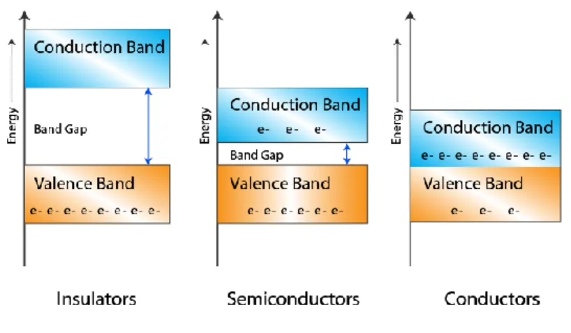
energy band is formed. This band is called as valence band (Figure 2.2). Electrons within the valence band can easily change their places and move easily inside the band. And the anti-bonding orbitals having number close to infinity form another energy band with the approaching anti-bonding orbitals named as the conductivity band. The interval between the valence band and the conductivity band is named as band gap and the energy required to pass this interval is named as band gap energy. In grouping materials as insulator, semi-conductor, conductor, the magnitude of band gap energy is considered [2].
Figure 2.2. Band structures for insulators, semi-conductors and conductors.
The undoubled electrons are responsible from electrical conductivity which exists at the valence band, conduction band or at a new energy level in the band gap. This type of free electrons moves to the desired direction according to the applied potential. When the valence band energy levels are totally full of electrons, it is hard to maintain the flow of electrons to one direction. In such a system, free electrons may be formed with heat or light excitation. Electrons which have sufficient energy on the top level of valence band pass the band gap and they are situated to the energy level at the bottommost level of the conduction band and they maintain conductivity. In insulators, the band gap is wide enough not to let this pass [2].
In semi-conductors, band threshold gap is smaller than insulators and their conductivity varies between 10-6 - 102 S.cm1-. Although the electrical conductivity seems to be low at this level, it is at sufficient size to maintain sufficient amount of electric current. Conjugated polymers that include single and double bonds
sequentially may exhibit conductance. When the energy level in semi-conducting polymers between valence band and conduction band is sufficiently small, free electrons may pass to the lowest energy level of the conduction band. These electrons move within the conductance band and carry charges and they are directed towards positively charged direction by moving along the chain. Meanwhile, the positively charged hole remaining inside the valence band moves in opposite direction to the electron along the polymer chain. Their conductivity increases with an increase in temperature or light intensity [2].
Most metal atoms have a single electron and it can’t make a covalent bond with another metal atom near it. So, valence band of metals are partially full and conductance band is empty. Metal electrons exist at the low energy orbitals of the valence band with a great possibility. There are always empty places with higher energy levels where they may pass within the same band or the conductance band matching the same band. They maintain transmission of their electrons over partially full valence or conductance band or by band gap transition [2].
2.1.1.2 Doping in Conducting Polymers
If electrons in the valence band of the polymer are removed with an oxidizing agent the polymer is loaded with positive charge, it is called as p-type doping and if this process happens by giving one electron to the empty conductance band with a reducing agent, it is called as n-type doping. With this doping process, dopant molecules don’t exchange places with polymer atoms, dopants only help electrons pass from energy shells. As the band theory isn’t sufficient to explain conductivity of conducting polymers, it is made use of neutral polymer, polaron and bipolaron structures as it is seen in Figure 2.3. Electric charge given to make doping to the skeleton structure of the polymer causes a change in the electronic condition of the polymer [2].
S S S S S S S S S S S S S S S S S S S S S S S A S -A -A -• -1e -+A --1e -+A -b) q=+1, s=1/2 a) q=0, s=0 c) q=+2, s=0
Figure 2.3. Polaron, bipolaron structures and band diagrams. a) neutral polymer b) b) polaron structure c) bipolaron structure.
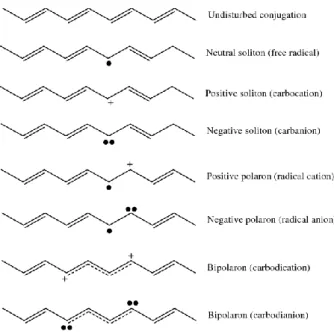
By the oxidation of uncharged polymer having conjugated bonds (Figure 2.3 (a)), the double bond is destroyed and a positively charged radical is formed over the polymer chain (Figure 2.3 (b)). These charge carriers formed on the conducting polymer chain are named as ‘polaron’ or ‘radical cation’. Joining of two radicals coming from polaron creates a new π bond. And with the oxidation of the free radical of polaron, a new positive hole without spin is formed named as ‘bipolaron’ (Figure 2.3 (c)). Here, two radicals combine and form a new π bond. There are no undoubled electrons in bipolaron and by this way conductance is maintained without requiring free electrons [3]. Polarons and bipolarons can move along the chain according to the mobilization capability of counter ions. For the ions to be mobile enough, satisfactory amount of counter ions should be provided by doping. As it is shown in Figure 2.4, an electron is removed with controlled doping of polyacethylene and a neutral or charged ‘soliton’
is obtained. Soliton which is formed within the structures makes the charge distribution along the polymer chain and carbenium (carbocation) stable. In a similar way, if the polymer is processed with an electron-giving or n-doping material, one electron is added to the medium-level energy gaps and a negative soliton is formed [3].
Figure 2.4. Polyacetylene’s soliton, polaron and bipolaron structures.
2.1.1.3. Hopping in Conducting Polymers
Besides the charge carriers along the chain, transfer of electrical charge between different polymer chains is made by hopping. A neutral soliton interacts with the charged soliton in a chain close to itself and the electron of the charged soliton jumps to the neutral soliton. In the hopping rule, mobility of the electronic charge is maintained by transfer between chains and transfer between blocks along the polymer chain (Figure 2.5) [3].
Figure 2.5. Transportation of charge, a) transportation of charge along the chain, b) transportation of charge between chains, c) transportation of charge between chain blocks [3].
2.1.2. Synthesis of Conducting Polymers
2.1.2.1. Chemical Synthesis
In the chemical method, a monomer that is dissolved in a suitable solvent is polymerized by interacting with a chemical substance used as an oxidation or reduction agent. This method has disadvantages like oxidation step can’t be controlled and the product not being pure and advantages like obtaining products in the desired amount with a reasonable cost. The doping material and catalyst that will be used in the chemical method has an important effect on the electrical conductivity of the conducting polymer to be obtained [4]. In the study of synthesizing poly(p-phenylene) made by Toshima, the polymer which is obtained by using CuCl2 as doping material
and AlCl3 as catalyzer didn’t exhibit electrical conductivity. However, by using
materials such as AsF5 or Li, conductivity that varies between 0.3 S.cm-1 - 500 S.cm-1
has been observed. All of conjugated polymers can be synthesized with the chemical method. In another study, the polymer of pyrrole was prepared by the chemical method using methanol as solvent and 2.5 M FeCl3 as doping material and it has been
determined to reach a conductivity of 190 S.cm-1 [4].
2.1.2.2. Electrochemical synthesis
Electronically conductive polymers are obtained by electrochemical oxidation of aromatic molecules in resonance stability such as pyrrole, thiophene, aniline and furan.
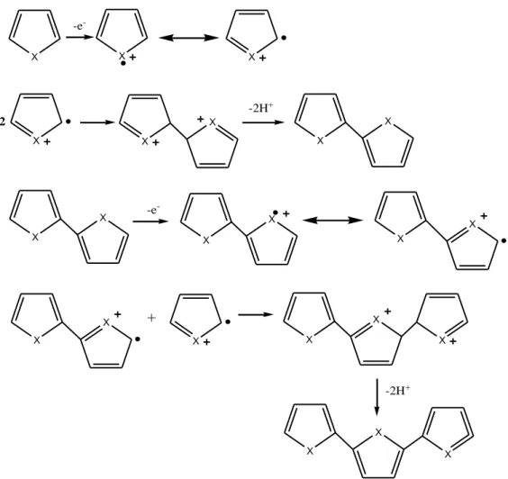
Electropolymerization is made by putting monomers into solvent/electrolyte medium and applying a potential to this system. With the neutral polymer and radical cation reaction or radical cation/radical cation reaction, polymerization occurs. In figure 2.6, polymerization mechanism of heterocyclic compounds that happened in the anode is given [5]. X X X X -e -X X X 2 X -2H+ X X X X -e -X X X X + X X X X X X X -2H+
Figure 2.6. Electropolymerization mechanism of heterocyclics (X= N-H, S, O).
Electrochemical stage begins with the monomer to be oxidized over the electrode with a suitable potential and form a radical cation. Electron transfer reaction is faster than the diffusion of the monomer and the solution around the electrode surface has a high radical concentration. This first stage is followed by the coupling reaction that happens with two radical cations couple each other. In case of radical-radical coupling, dihydro dimer cation is formed by two hydrogens separating from one dimer. This anti-aromatic condition represents the chemical stage. As potential is applied, dimer is
oxidized and subjected to a coupling reaction again with a monomeric radical. From this coupling reaction, a trimer forms then it loses two protons. Dimers, trimers and oligomers continue to go to coupling reaction with the radical cation of the monomer and they lose their protons and become anti-aromatic. Electrochemical and chemical stages continue this way following one after another. Until oligomers remain undissolved within the electrolyte solution, this reaction continues on the electrode surface [6].
In electrochemical polymerization, cyclic voltammetry (CV) technique can be used. Cyclic voltammetry provides information related to the oxidation potential of the monomer, its growth on the film, redox behavior of the polymer and surface concentration (charge spent by the polymer). In the specific area, CV can be used about the interaction between the polimer and solvent molecules and ions, charge transportation process and the ratio of charge transporters. The potential of an electrode is used to shed light to electrode reactions as a result of examining current-voltage relationship by changing it linearly between the working electrode and the opposite electrode. In this technique, first scanning is made in a certain potential interval and then it is continued to scan in the opposite way (anodic-cathodic). When the potential applied to the working electrode reaches the oxidation or reduction potential of the electro-active material inside the electrolysis cell, the material on the surface of the electrode is consumed rapidly. Measured current between working and opposite electrodes increases. As a result of this, concentration difference occurring between electrode surface and the solution causes mass transfer from the solution to electrode surface. As mass transfer rate is lower than transfer rate of electrons, a current decrease is observed and a peak is obtained. The potential which corresponds to the peak point of this peak is called as ‘oxidation or reduction peak potential’ [2].
2.1.3. Usage Areas of Conducting Polymers
Conductive polymers have extensive usage areas due to that they can be synthesized with electrochemical methods, show electrical conductivity and they can be used with insulator polymers. Conductive polymers are used as electrodes in batteries such as
heart batteries as they are rechargeable and long life due to that they produce low currents and they have reversible doping characteristics.
Chemical signals which are formed as a result of reduction-oxidation reactions of the polymer are converted into electric signals and electronic components such as diodes and transistors are made. Besides this, they are used in production of materials working with electrochromic principle like smart windows, high technology eye-glasses and military camouflage dresses. Besides, it is used in pH, gas and humidity sensor, corrosion inhibitor, photoelectrochemical cells, light emitting diodes (LED-OLED), field effective transistors, photovoltaic cells and supercapacitors [7,8].
Another important usage area of conducting polymers is biosensors and enzyme electrodes. Biosensors that are analytical instruments which are formed by a selective biological element and the detector that produces signals proportional to the concentration of the analyte have the potential to be used in analyzing many materials as enzyme sensor due to their high selectivity, quick response time, simple design and cheapness [9,10,11].
2.2. ENZYMES
Enzymes are protein-structured biocatalyzers which increase the rate of almost all biochemical reactions in a ratio of about 1010 and 1012 like synthesis and degradation of organic molecules, which are not consumed during the reaction and which can be reused as they are not altered after the reaction [12].
Enzymes are only synthesized by living organisms and their difference from other chemical catalyzers is that they are specific. Their catalyzing the molecule entering the reaction, that is to say, its substrate expresses absolute specificity, the activity of an enzyme to a specific substrate group shows its group specificity and their activity to a certain bond shows its bond specificity. This property of specificity maintains all unwanted side reactions and products to be prevented and the selected reaction to be realized and to gain more efficiency. Therefore, as the desired product will be
produced with fewer steps and as energy requirement is low and it can be worked in mild conditions, cost is also reduced.
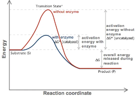
The minimum energy required for a reaction to start is called as activation energy. And an alternative path towards lowering the activation energy occurs by the help of enzymes as it is seen in Figure 2.7 [12].
Figure 2.7. Effect of enzyme in activation energy
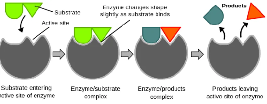
Enzymes stimulate their substrates in the appropriate direction and maintain them to bind to their active sites consisting of certain amino acid sequences. Thus, enzyme-substrate complex is formed that is named as active complex or transition complex. The first hypothesis related to this formation mechanism is the key-lock model proposed by Emil Fischer the first time (Figure 2.8).
Figure 2.8. Key-lock model proposed for the enzyme-substrate complex.
2.2.1. Classification of Enzymes
Simple enzymes are enzymes formed only from protein. The part of simple enzymes that show catalytic effect is polypeptide chain of which pepsin and urease enzymes are examples.
On the other hand, complex enzymes are enzymes that contain its protein part and a much smaller organic molecule or metal with this. The part of enzymes that composes only from protein is named as ‘apoenzyme’ and the non protein group providing it to show catalytic effect is named as ‘cofactor’. Cofactors can be composed of a metal ion and they may also compose of an organic group named as “coenzyme” [12,13].
Today, enzymes are divided into 7 basic classes by the International Union of Biochemistry and Molecular Biology (IUBMB). These are: oxyreductases, transferases, hydrolases, liases, isomerases, ligases and translocases [12].
2.2.2. Enzyme Activity
The activity of an enzyme is defined according to the substrate amount spent in unit time and converted to product at optimum conditions. Its unit is IU. 1 IU enzyme activity is the enzyme amount that catalyzes 1 umol of substrate in 1 minute when optimum conditions are present. There are many factors that influence rate of enzymatic reactions. Some of these are substrate concentration, enzyme concentration, temperature, pH and inhibitor effect [12].
2.2.2.1. Effect of Substrate Concentration
In enzyme catalyzed reactions, reaction rate at optimum conditions increases proportionally with increase in substrate concentration. When the entire enzyme molecules form the enzyme-substrate complex, reaction rate reaches maximum (Vmax).
Although substrate concentration continues increasing, no increase in reaction rate is observed anymore. This means that enzyme is saturated with substrate (Figure 2.9) [12].
Figure 2.9. Effect of substrate concentration over reaction rate [12].
2.2.2.2. Effect of Enzyme Concentration
When unlimited substrate is present in the environment, reaction rate increases proportionally with enzyme concentration. The more enzyme amount is added, the more reaction rate increases (Figure 2.10) [12].
2.2.2.3. Effect of Temperature
The collision rate of molecules that enter reaction increases when temperature increases. Rate of reactions show 1 to 3 times of increase with each 10 ℃ of temperature increase. But this increase continues up to a certain value. This value is the optimum temperature of that reaction as it is in Figure 2.11. Afterwards, some changes which occur in enzyme molecules either slow down the reaction or stop it completely. As enzymes are protein-structured, the temperature causes a deterioration in their three-dimensional structures, that is to say, denaturation. This denaturation can be reversed at low temperatures, that is to say, they may regain their activity by a decrease in temperature but they can’t be reversed at high temperatures. In this condition, activity is totally lost [12].
Figure 2.11. Effect of temperature over reaction rate [12].
2.2.2.4. pH Effect
Enzymes are amphoteric molecules which contain both acidic and basic groups. The pH of the environment changes the charge of these groups and affects the charge distribution on the surface of the enzyme and net charge. Therefore, a change occurs in the activity of the enzyme. The pH at which enzymes exhibit maximum activity is named as optimum pH as seen in Figure 2.12. This pH value is different for each enzyme [12,13].
Figure 2.12. Effect of pH over reaction rate [12].
2.2.2.5. Inhibitor Effect
Materials which lower the reaction rate or stop it are named as inhibitors. As it is shown in Figure 2.13, some inhibitors form intermediate compounds with the enzyme and lower the reaction rate and some of them are bonded to the active region of enzyme and inhibit the enzyme activity [13].
Figure 2.13. Enzyme-inhibitor relationship [23].
Other than these, there are many other factors that influence enzyme activity like water amount, product concentration and time.
2.2.3. Enzyme Kinetics
The first studies related to enzymatic reactions were made by Michaelis-Menten in 1913 and they are known with the theory titled by their names. According to this rule, the enzyme (E) first combines with the substrate (S) and forms the enzyme-substrate (ES) complex. Afterwards, this complex is converted to the product (P) and free enzyme [14,15]. k1 k2 k3 k4 E+S ES E+P
Here, k1, k2, k3, k4 shows the reaction rate constants that with k1 ES complex forms,
with k2 ES complex is separated to the enzyme and substrate, with k3 ES complex
forms the enzyme and product and k4 forms the ES complex from enzyme and product.
The rate of free enzyme and substrate to be converted into ES complex (k4) is very low
and ignored and the total reaction is shown as below [14,15]. ES → E + P
When the reaction rate is plotted versus substrate concentration, the hyperbolic curve in Figure 2.14 is obtained.
As it is also seen in the reaction rate vs. substrate concentration graph, rate is directly proportional with substrate concentration. The consumption of [ES] is two types here. A part of ES is separated to enzyme and substrate by k2 rate constant. And the other
part of it is converted to enzyme and product with k3 rate constant [14,15].
E + S ⇌ ES ⇌ E + P
The rate of formation of [ES] is given by Equation 2.1.
𝑉 = k1. ES (2.1)
The consumption rate of [ES] is given in Equation 2.2.
𝑉 = k2. ES + k3. ES = ES (k2+ k3) (2.2)
According to the steady state approach theory, as the formation rate of [ES] is equal to the consumption rate of [ES], equation 2.3 is obtained [14,15].
k1. ES = (k2 + k3) ES (2.3)
If Equation 2.3 is solved, Equation 2.4 is obtained.
ES
ES = 𝐾m (2.4)
When concentrations of substances E and S is divided by ES, a standard value is obtained. As part of total enzymes in the environment (Et) is in free form and part of
is in ES form, the equation below is obtained (Equation 2.5) [14,15].
E = E𝑡 − ES (2.5)
Equation 2.5 is written in its place at Equation 2.4 and the new equation is as below (Equation 2.6).
ES = E𝑡S
Km+S (2.6)
As ES is equal to E+P in certain conditions, rate of enzymatic reaction is expressed by Equation 2.7.
𝑉 = k3 ES (2.7)
If Equation 2.6 is written in Equation 2.7, the new rate obtained is as shown below (Equation 2.8).
𝑉 = k3 E𝑡S
Km+S (2.8)
As a result of the reaction, as the entire enzyme will be binded with the substrate, maximum rate is reached and it is given by Equation 2.9.
𝑉max = k3 E𝑡 (2.9)
Equation 2.9 is written in its place at Equation 2.8 and Michaelis-Menten Equation is obtained (Equation 2.10).
𝑉 = 𝑉maxS
𝐾m+S (2.10)
Km shows the interest of the enzyme to the substrate and it is substrate concentration
that makes half of the maximum rate. If the interest of the enzyme to substrate is high, Km is small. It means that even in low S’s, enzyme makes ES] complex with the
substrate. If the interest of enzyme to the substrate is weak, Km becomes high [14,15].
As it is seen in Michaelis-Menten equation, the rate of an enzymatic reaction depends on S and Km.
• If S is too smaller than Km, adding [S] to Km changes its value very few
anymore. So, S is removed from the denominator (Equation 2.11). In this condition, rate will depend on S.
𝑉 = 𝑉maxS 𝐾m+S
𝑉maxS
𝐾m KS (2.11)
• If S is too bigger than Km, adding Km to [S] changes its value very few
anymore. So, Km is removed from the denominator (Equation 2.12). The
beginning rate is equal to Vmax.
𝑉 = 𝑉maxS
𝐾m+S ≅
𝑉maxS
S = 𝑉max (2.12)
• When S equals to Km, Equation 2.13 is obtained. In this condition, beginning
rate is half of maximum rate.
𝑉 = 𝑉maxS 𝐾m+S ≅ 𝑉maxS S+S = 𝑉max 2 (2.13)
If Michaelis-Menten Equation which is a hyperbolic curve is reversed to be converted into a linear equation for practicality and separated to its multiples, Lineweaver-Burk equation is obtained (Equality 2.14) [14,15].
1 𝑉 = 𝐾m 𝑉max 1 S+ S 𝑉maxS (2.14)
The expression that gives a straight line equation is shown by Equation 2.15.
y = ax + b (2.15)
By using the similarity given in Equation 2.14 as y = 1
𝑉 ; a = 𝐾m 𝑉max ; x = 1 S ; b = S 𝑉maxS when y = 0, x = (−b)
a and therefore Equation 2.16 is obtained.
x =(−1)
Value of Km is calculated from here. And from the b value of Lineweaver-Burk graph,
Vmax is obtained. And kinetic parameters are assigned in this manner (Figure 2.15).
2.2.4. Enzyme Immobilization Methods
Enzyme immobilization is the process of immobilization or bonding of the free enzyme to a carrying substance [9]. As a result of this process, enzymes can be easily separated from the product and reused. And this eliminates the processes applied to prevent contamination of the product and provides advantages such as saving from time and cost. Besides, they have many more advantages such as being more stable and having longer life time and enzymes in different optimum conditions can be used together in the same process [9].
Enzyme immobilization methods are divided into three as binding, cross-linking and encapsulation as it is seen in Figure 2.16.
Figure 2.16. Enzyme immobilization methods.
2.2.4.1. Binding to a Support
In this process, new bonds are established between the enzyme and functional groups of support and enzyme is modified. Binding to the transporter is made by enzymes to be binded to the supporting material with physical adsorption or making ionic or covalent bonds (Figure 2.17). These supports are inorganic materials such as silica and porous glass, organic supports such as polyacrylamide including weak activity –OH group or biological supports such as cellulose [16].
Figure 2.17. Enzyme immobilization methods.
2.2.4.2 Cross-linking
It is a method that enzyme molecules form nonsoluble complexes in water by cross-linking with each other with multiple covalent bonds as a result of their reaction with a cross-linker that contains a functional group like glutaraldehyde (Figure 2.17) [17]. In this method, it can be seen a decrease in enzyme activity due to other structure be bonded to the enzyme structure with covalent bonds or active region can not reach to the substrate staying at the center of the cross-linked structure. On the other hand, cross-linking increases the stability of the enzyme and maintains immobilization to be long lasting.
2.2.4.3. Entrapment (Encapsulation)
It is the placing of the enzyme in an appropriate porous polymer or a membrane for the diffusion of substrate in and diffusion of product out (Figure 2.17). Entrapment method is good for smaller substrate or product molecules compared to other methods. The difference of the entrapment method is that there are no bonds between the enzyme and matrix. As there is no bond between, enzyme losses through matrix pores can be
possible. Electrochemical polymerization method in physical immobilization of enzymes is an attractive one as it is faster, reliable, economical and simple. [18,19].
2.2.5. Poliphenol Oxidase
Enzymes have a distinguished place as biocatalysts. Many reactions that enzymes catalyze do not occur without these catalysts and many of these effective synthetic catalysts that will replace these biological catalysts couldn’t be synthesized yet.
Polyphenol oxidase (PPO, also known as tyrosinase; EC 1.14.18.1) is an enzyme which was discovered by Schoenbein in 1856 in mushrooms. Polyphenol oxidases are oxidoreductases containing copper and in the presence of molecular oxygen, they catalyze hydroxylation and oxidation of phenolic compounds. Polyphenol oxidases are divided into three groups as tyrosinase, catechol oxidase and laccase. Tyrosinase catalyzes (EC. 1.14.18.1) hydroxylation of monophenols to o-diphenols (monophenolase or cresolase activity) and oxidation of o-diphenols to o-quinones (diphenolase or catecholase activity) (Figure 2.18). Catechol oxidase (EC. 1.10.3.1) catalyzes oxidation of only o-diphenols. And laccase (EC. 1.10.3.2) catalyze oxidation of both o-diphenols and p-diphenols to corresponding quinones. Laccase and catechol oxidases can’t catalyze hydroxylation reactions [20,21].
Figure 2.18. Polyphenol oxidase catalysis reaction.
Reaction catalyzed by polyphenol oxidase is a biotransformation that occurs in almost all organisms and it is only catalyzed by tyrosinase enzyme [22]. Tyrosinase enzyme is responsible from the formation of pigments in skin, hair and eye and the color darkenings occuring in fruits, vegetables and mushrooms as a result of contact with oxygen.
This enzyme that is commonly found in plants oxidizes its substrates to dark colored quinonoic compounds in the presence of oxygen. These compounds polymerize and form brown pigments and due to this, these reactions are named as browning reactions. These browning reactions that tyrosinase form are unwanted in the food industry [23]. Fruits and vegetables darkening when they are cut and peeled decreases the food quality and value of these products. This is known as enzymatic darkening in the food industry. On the other hand, this color change is something desired in some foods. For example, these browning products give taste and odor to foods such as tea, coffee, cacao, dry grapes and dry prune. The good color and taste of dry fig and date is as a result of enzymatic browning [24].
2.2.5.1. Working Mechanism of Polyphenol Oxidase
PPO does not need coenzyme for its activity. They are enzymes that use all phenolic compounds as substrate which show group specificity. Generally, PPO oxidizes phenolic substrates in the presence of oxygen. Active sits of PPO pass through transitions between metoxy, oxy and deoxy forms in a cyclical manner (Figure 2.19).
Figure 2.19. Cyclical transitions of PPO’s active site between oxy and deoxy forms during catalysis. Blue, red and green circles represent copper, oxygen and histidine [26].
In each cycle, two catechol molecules are oxidized. One molecule oxygen is reduced to water and two quinone products are formed. Dioxygen (O2) replaces the solvent
molecule (H2O) that is bonded to CuA in reduced enzyme form (reduced deoxy form)
and it is bonded to the copper metal center of the enzyme [25].
UV spectroscopy results support the idea that molecular oxygen is first bonded as peroxide and then catechol is bonded. One of two hydroxyl groups of catechol molecule goes under deprotonation and catechol is bonded to CuB (oxy form) (Figure 2.20). After two electrons are transferred from the substrate to peroxide, peroxide group is protonated and O-O bond is broken [25].
The second hydroxyl group that didn’t enter coordination gives one proton and maintains formation of water loss and formation of o-quinone product. Protoning of the bridge making group with solvent makes active sit hydroxyl bridged with two coppers form (metoxy form). Another catechol molecule performs a co-substrate function and reduces hydroxyl bridged Cu (II) to Cu (I) form. This step of the proposed reaction path is supported by data about o-diphenol oxidase activity of tyrosinase (Figure 2.20). Cu(I)-Cu(I) form of the active site repeats catalytic cycle again [25].
NHIS Cu(II) A NHIS O Cu(II) B NHIS NHIS NHIS NHIS NHIS _ H OH HO O O H2O + 2H+ H+ Cu(II) A NHIS O Cu(II) B NHIS NHIS NHIS NHIS O O HO Cu(II) A NHIS O Cu(II) B NHIS NHIS NHIS NHIS O HO H OH HO O O + O2 H2O + H+ Cu(I)A Cu(I) B NHIS NHIS NHIS NHIS NHIS H2O Metoksi Form Oksi Form Deoksi Form
Figure 2.20. Reaction mechanism of PPO.
2.2.5.2. Usage Areas of Polyphenol Oxidase
Industrial use and applications of tyrosinase attract the interest of scientists and researchers more recently. Tyrosinase enzyme is used in health and cosmetics industry, in cleaning of waste waters and determination of phenolic materials. An important usage area of tyrosinase is the food industry. It is used in eliminating the sour and bitter taste in tea, coffee and cacao beans and developing the taste of these drinks. On the other hand, tyrosinase enzyme should be active for products like dry grape and dry prune to become desired in view and taste [26]. Another use of it is that it is an alternative material for transglutaminase enzyme used in enzymatic cross-linking of proteins. Besides this, tyrosinase is used in synthesis of some drug active materials and in the synthesis of melanin added to cosmetic products to prevent ultraviolet lights from the sun.
On the other hand, phenolic substances that contaminate environmental waters with industrial and domestic waste create pollution and it is hazardous for living species in
Metoxy Form
Oxy Form
water. In order to prevent phenolic compounds that show toxicity to contaminate nature, it is better to give waste materials to the environment after being refined from phenolic compounds and for this purpose, enzymes are used as an efficient and easy way. For the use of tyrosinase to remove toxic phenolic compounds, studies continue in the field of engineering. When the enzyme is used in waste water, it oxidizes selectively phenolic substances and converts them to quinones. The reactive quinones that are formed precipitate by polymerizing and polyphenols are removed from water by this way.
2.2.5.3. Use of Polyphenol Oxidase in Biosensors
Like all pollutants, it is also important to determine phenolics in environmental waters and industrial waste water. Besides this, it is required to analyze phenolic compounds in vegetables and fruits or determine these materials in industrial processes. But methods used for this purpose are expensive and require long processes. As an alternative method, biosensors lead in this area and another use of tyrosinase is that it exists as biomaterial in biosensors used in determining phenolic compounds. Biosensors which have sensitive and fast determining quality are obtained as a result of combination of sensor systems with biological materials and they are used for analysis purposes in many areas like medicine, defense, food industry etc. [27]. In enzymatic biosensors, the biological material are enzymes and selective determination of polyphenols can be made by tyrosinase based amperometric biosensors due to the enzyme being a catalyzer specific to phenolics [28-32].
2.3. SAMPLE ANALYSIS
In this study, total polyphenol material content of a waste water sample was measured using polyphenol oxidase enzyme electrodes. Folin-Ciocalteau method was also used as control method to validate the analysis by enzyme electrodes.
Folin-Ciocalteau method developed by Singleton et al is used to measure total phenolic content. This method is based on the fact that phenolic compound dissolved in water forms a colored compound with the Folin reactive in alkali environment [33] and
measurement of absorbance according to this color intensity. Folin-Ciocalteau reactive (FCR) which gives its name to the method is a molibdophospotungstic heteropolyacid and its assumed active center is Mo (VI) (3H2O.P2O5.13WO3.5MoO3.10H2O).
Mo (VI) (yellow) + e- (antioxidant) → Mo (V) (blue)
Phenolic compounds enter into reaction with FCR only under basic conditions and pH of the environment is set to 10 by sodium carbonate solution. Removal of a phenolic proton causes formation of a phenolate anion that has reducing capability of FCR and blue colored compounds are formed between phenolate and FCR. FCR can also be reduced by many non-phenolic compounds and it is not specific to phenolic compounds. In fact, this method measures the reduction capacity of a sample. It may interact with reductant materials in the environment such as ascorbic acid [34].
In spite of this disadvantage, this method is used very frequently due to its simplicity, repeatability and the correlation it shows with other methods. Method results are given as a standard phenolic material or usually as gallic acid equivalent.
PART 3
MATERIALS AND METHODS
3.1. CHEMICAL MATERIALS
Tyrosinase from mushroom (EC 1.14.18.1) and 25% glutaraldehyde solution was purchased from Sigma-Aldrich. Phosphate buffer components, NaH2PO4 and
Na2HPO4 were from Merck. For MBTH solution, 3-Methyl-2-benzodiazolinon
hydrazon hydrochloride monohydrate (Aldrich) was dissolved in ethanol (Carlo Erba). Acetone and catechol were provided from Sigma-Aldrich and sulfuric acid was obtained from Merck Company.
3.2. INSTRUMENTS
3.2.1. Potentiostat
Polymerization was made by cyclic voltammetry using GAMRY Instruments Interface 1000 Potentiostat/Galvanostat ZRA. The voltage was changed in the scanning interval cyclically and the current was measured. Polymerization was performed in 60 cycles.
3.2.2. UV-Visible Spectrometry
In measurements, to determine the activity of immobilized enzyme, Shimadzu UV– 1201V model spectrophotometer was used. Colored complexes of polyphenol oxidase products formed after biotransformation were obtained using Besthorn’s Hydrazone method and their absorbance in 495 nm were measured. Enzyme activities were determined with this method.
3.2.3. SEM
SEM images were taken with Carl ZEISS ULTRA PLUS GEMINI FESEM scanning electron microscope in Karabük University Iron & Steel Institute MARGEM laboratories. Poly(2,5-di(furan-2-yl)thiazolo[5,4-d]thiazol) conducting polymer was coated over platinum electrode. 5000 times and 10000 times magnified pictures of surface of polymer coated electrode were taken before and after enzyme immobilization.
3.2.4. pH meter
Hanna Instruments HI 221 brand pH meter was used. This instrument uses an electrode that produces an electrical signal to provide a potentiometric measurement converting the signal into pH units. The signal is measured as potential. What is required to make pH measurement is the voltage that sensing electrode provides proportional to the logarithm of hydrogen ion activity within the product and the voltage that reference electrode provides ideally that is independent, fixed and continuous. Electrical signal is formed with the difference between these two voltages. Voltage difference between the reference and sensing electrode is measured by pH meter and converted into pH value.
3.2.5. Magnetic Mixer with Heater
It was performed to mix prepared solutions with the help of a magnet and with the effect of magnetic field at the same number of cycles and temperature by a MTOPS MS300 HS brand magnetic mixer with heater.
3.2.6. Vortex Tube Mixer
Dlab MX-S Brand mixer was used in constant speed. It was used for the purpose of providing an effective and efficient mixing vortex to the solutions in experiment tubes and obtaining a homogeneous mixing.
3.2.7. Shaking Water Bath
Nuve ST 30 brand shaking water bath was used for keeping solutions inside shaking water bath at 25 ℃ constant temperature and by maintaining homogeneous mix of them by shaking during the reaction of enzyme and substrate. In temperature optimization, water bath is brought to temperatures between 4 ℃ and 80 ℃ and activity measurements were made to determine the temperature at which enzyme electrode works best.
3.3. METHODS
3.3.1. Electrochemical Synthesis of Poly(2,5-di(furan-2-yl)thiazolo[5,4-d]thiazol)
Polymerization of 2,5-di(furan-2-yl)thiazolo[5,4-d]thiazol (TTzFr) monomer was performed electrochemically as it is seen in Figure 3.1. Monomer was synthesized by Söylemez et al. [35].
Figure 3.1. Polimerization of TTzFr.
Electropolymerization was made by cyclic voltammetry using a 3-electrode system (Figure 3.2) and poly(2,5-di(furan-2-yl)thiazolo[5,4-d]thiazol) (PTTzFr) polymer was obtained. Platinum was used as working and counter electrode and silver (Ag/Ag+) electrode was used as reference electrode.
Figure 3.2. Three electrode system.
Electropolymerization cell was prepared by adding monomer and electrolyte to the solvent (deposition solution) (Figure 3.3). As solvent, 95:5 ratio acetonitrile (ACN) and dichloromethane (DCM) was used and as electrolyte, 0.1 M lithium perchlorate/sodium perchlorate was used. Monomer concentration was taken as 10-3
M and polymerization window was determined by scanning with cyclic voltammetry in (-2) – (+2) V potential interval, then by using this interval, polymerization of the monomer was maintained [10]. After this operation, each enzyme electrode was washed a few times with distilled water to remove free supporting electrolyte remaining on the surface. When it is not used, it was kept at +4 °C in the pH 7.5 phosphate tampon.
Figure 3.3. Electropolymerization cell.
3.3.2. Enzyme Immobilization in PTTzFr Matrix
In this study, enzyme immobilization was made with cross-linking method. In enzyme immobilization, adsorption, covalent bonding, cross-linking and entrapment methods are used. Adsorption method causes the enzyme flow to the environment faster and disappear and enzyme electrode loses its activity after a short time. Entrapment method is more advantageous than adsorption. It maintains the enzyme to remain stable for longer time. And in cross-linking and covalent bonding, stability of the enzyme and therefore the electrode increases remarkably. In this immobilization method, more enzyme is immobilized, enzyme loss is less and higher signals are received. In this study, enzyme solution was prepared at 0.2 g mL1- concentration in pH 7.5 phosphate buffer (Enzyme Unit: 2687 U/mg solid). 6 uL enzyme solution was dropped to the surface of polymer coated on platinum electrode and it was left to dry for 30 minutes at room temperature conditions. 6 uL 1% gluteraldehyde solution was added above and it was dried for 2 hours at room temperature and after this it was left for one night at +4 ℃. Therefore, enzyme electrodes coated with polymer over platinum and which PPO enzyme was immobilized were prepared (Pt/PTTzFr/PPO).
3.3.3. Besthorn’s Hydrazone Method
Catechol solutions are prepared in various concentrations and Besthorn’s Hydrazone method is applied with its process steps in the literature [36]. Firstly, MBTH reactive is added to the solutions. Enzyme electrode is dipped for certain periods. After product formation, H2SO4 and acetone is added. The colored complexes which are
formed are measured with respect to their absorption in 495 nm and enzyme activities are determined (Figure 3.4.).
Figure 3.4. Besthorn’s Hydrazone method.
MBTH solution used in this method was prepared as the concentration of 3-methyl-2-benzothiazolinone hydrazone hydrochloride monohydrate (C8H9N3S.HCl.H2O) was
3.0 mg mL1- dissolving in ethyl alcohol.
3.3.4. Determination of the Enzyme Activity
In the reaction where polyphenol oxidase enzyme is the catalyzer, the substrate of the enzyme is phenolic materials. In the substrate analysis, polyphenol oxidase enzyme procedure was applied taking as a basis Besthorn’s Hydrazone method and pyrocatechol that is one of the substrates of the enzyme was used (Figure 3.5).
Figure 3.5. Pyrocatechol.
Solutions of pyrocatechol in different concentrations were prepared in the pH 7.5 buffer and after adding 1 mL MBTH reactive to each solution with different concentration, polyphenol oxidase enzyme electrode was dipped for 0, 5, 10, 15 minutes and transformation reaction of phenols to o-quinone was performed as it is seen in Figure 3.6.
Figure 3.6. PPO’s cresolase and catecholase activities: A) monophenol’s o-hydroxylation, B) oxidation of o-diphenol to o-quinone.
Afterwards, complex formation of quinones with MBTH was occurred as it is seen in Figure 3.7. 1 mL sulfuric acid, 1 mL acetone solutions were added and mixed then absorbance measurements were performed at 495 nm. Absorbance versus time graphs were plotted and reaction rate was calculated from the slope of these graphs. Therefore, for each concentration, polyphenol oxidase enzyme electrode activities were determined.
Figure 3.7. o-Quinone formed to establish colored compound with MBTH reactive [36].
3.3.5. Assessment of Kinetic Parameters
With measurements made, kinetic characterization of enzyme electrode was made for immobilized polyphenol oxidase enzyme. Enzyme activity V vs. substrate concentration [S] graph was plotted and from this graph, Lineweaver-Burk graph was obtained (1/V vs. 1/[S]) [38,39]. From Lineweaver-Burk graph, with Michaelis-Menten method [40], enzyme electrode’s kinetic parameters maximum enzyme activity (Vmax) and Michaelis-Menten constant (Km) were detected.
3.4. OPTIMIZATIONS
3.4.1. Optimum pH Determination
For immobilized polyphenol oxidase enzyme, pH optimization was made. For this purpose, activity measurements of enzyme electrodes were taken at pH values between 3.0 and 10.5 and the optimum pH value at the enzyme electrode that operates best was determined.
![Figure 2.1. Formation of bonding and anti-bonding orbitals in polymer molecule [2].](https://thumb-eu.123doks.com/thumbv2/9libnet/5396811.101872/23.892.187.767.719.973/figure-formation-bonding-anti-bonding-orbitals-polymer-molecule.webp)

![Figure 2.5. Transportation of charge, a) transportation of charge along the chain, b) transportation of charge between chains, c) transportation of charge between chain blocks [3]](https://thumb-eu.123doks.com/thumbv2/9libnet/5396811.101872/28.892.192.736.142.281/figure-transportation-charge-transportation-charge-transportation-charge-transportation.webp)
![Figure 2.13. Enzyme-inhibitor relationship [23].](https://thumb-eu.123doks.com/thumbv2/9libnet/5396811.101872/36.892.244.710.650.960/figure-enzyme-inhibitor-relationship.webp)

