ORIGINAL ARTICLE
Evidence of association of Vitamin D receptor
Apa I gene
polymorphism with bone mineral density in postmenopausal
women with osteoporosis
Umit Dundar&Mustafa Solak&Vural Kavuncu&
Mujgan Ozdemir&Tuncay Cakir&Handan Yildiz&
Deniz Evcik
Received: 7 March 2009 / Revised: 8 June 2009 / Accepted: 12 June 2009 / Published online: 17 July 2009
# Clinical Rheumatology 2009
Abstract The vitamin D receptor (VDR) was the first candidate gene to be studied in relation to osteoporosis, and most attention has focused on polymorphisms situated near the 3′ flank of VDR. The aim of this study was to investigate the association about VDR gene Apa I poly-morphism with bone mineral density (BMD) in postmen-opausal women with osteoporosis. We studied a total of 136 postmenopausal women with a mean age of 56.36 ± 10.29 years. Among them, a total of 75 had osteoporosis, 37 had osteopenia, and 24 had normal BMD. Venous blood samples were obtained for evaluation of bone metabolism and genotyping. The VDRApa I genotype was determined by polymerase chain reaction-restriction fragment length polymorphism. BMDs at the lumbar spine and hip were measured by dual-energy X-ray absorptiometry. Postmeno-pausal women with aa genotype had significantly lower BMD values (grams per centimeter square) at lumbar spines compared to persons with AA genotype. Also, postmeno-pausal women with AA genotype had significantly higher serum Ca level than the subjects with aa genotype. In
conclusion, our result may indicate that VDR Apa I gene polymorphism may be responsible for a important part of the heritable component of lumbar spine BMD in postmen-opausal women, possibly related to impaired calcium absorption from the bowel.
Keywords Bone mineral density . Osteoporosis . Vitamin D receptorApa I gene polymorphism
Introduction
Osteoporosis is a common disease characterized by low bone mass, disturbed microarchitecture of bone tissue, and increased fracture risk. Osteoporosis is defined to exist when bone mineral density (BMD) values at the spine or hip fall 2.5 standard deviations (SD; T-score values) or more below the population average in young adults [1]. Peak bone mass is attained in early adult life but declines in postmenopausal women due to a reduction in estrogen production, with effects on bone and intestinal and renal calcium handling [2].
Previous studies in twins and families show that genetic factors play an important role in the formation of BMD. The heritability of BMD has been estimated to be between 50% and 85% in twin studies, with the strongest effects in the axial skeleton [3–6]. It also shows that 27–68% of the
variance in osteoporotic fracture is heritable [7].
Osteoporosis is a polygenic disorder, determined by the effects of several genes, each with relatively modest effects on bone mass and other determinants of fracture risk. Population-based studies and case–control studies have similarly identified polymorphisms in several candidate genes that have been associated with bone mass or
U. Dundar (*)
:
V. Kavuncu:
T. CakirDepartment of Physical Medicine and Rehabilitation, Afyon Kocatepe University Faculty of Medicine, Mavi Hastane,
03200 Afyonkarahisar, Turkey e-mail: [email protected] M. Solak
:
M. Ozdemir:
H. YildizMedical Biology, Kocatepe University Faculty of Medicine, Afyonkarahisar, Turkey
D. Evcik
Physical Medicine and Rehabilitation, Ufuk University Faculty of Medicine, Ankara, Turkey
osteoporotic fracture, including the vitamin D receptor (VDR), estrogen receptor, and collagen type IαI gene [8].
Vitamin D, through its principal bioactive form 1,25-dihydroxyvitamin D3(1,25-(OH)2D3) plays a crucial role in bone metabolism. The action of 1,25-(OH)2D3 is mediated through a specific hormone-receptor [9]. Muta-tions in VDR cause the syndrome of vitamin D-resistant rickets, which is a recessive condition characterized by alopecia, hypocalcaemia, hypophosphatemia, and severe rickets, and is resistant to treatment with vitamin D and its active metabolites [9,10]. The VDR was the first candidate gene to be studied in relation to osteoporosis, and most attention has focused on polymorphisms situated near the 3′ flank of VDR recognized by the restriction enzymesBsm I, Apa I, and Taq I [9]. However, few studies were performed to identify the relationship of candidate gene polymorphism underlying peak bone mass variation.
The objective of this study was to investigate the association about VDR gene Apa I polymorphism with BMD in postmenopausal women with osteoporosis.
Materials and methods
We studied a total of 136 postmenopausal women with a mean age of 56.36±10.29 years. Among them, a total of 75 had osteoporosis (fulfilled criteria for osteoporosis accord-ing to WHO guidelines and had a BMD T-score of lesser than −2.5 SD), 37 had osteopenia (T-score between −1.0 and −2.5 SD), and 24 had normal BMD (T-score greater than−1 SD) [1].
Exclusion criteria for this study were diseases well known to affect bone metabolism (thyroid disease, parathyroid disease, renal disease, liver disease, and malignancy), any medication known to affect bone turnover such as glucocorti-coids, and anticonvulsant drugs. Patients were also excluded from study if they had taken anti-osteoporotic treatments including calcium supplementation >1,000 mg/day, vitamin D supplementation >400 IU/day, parathyroid hormone (PTH), calcitonin, estrogen, bisphosphonates, or strontium ranelate within 12 months previous to study entry.
Bone mineral density measurements
BMD was measured in all patients and controls by dual-energy X-ray absorptiometry (Hologic QDR 4,500 W) both at the lumbar spine (antero-posterior projection of L1-L4) and the proximal femur (total score). The instrument was calibrated daily according to the manufacturer’s instruc-tions. BMD data were expressed as grams per centimeter square and SD scores which compare individual BMD determinations to those of young (T) and age/sex-matched (Z) normal populations [1].
Biochemical analyses
Following on overnight fasting (at least 8 h), venous blood samples were obtained for evaluation of bone metabolism. Bone metabolism was evaluated by serum calcium (Ca), phosphorus, alkaline phosphatase, PTH, 25 (OH) vitamin D, osteocalcin (OC), and serum C-telopeptide cross-linked collagen type I (CTX) levels. Serum PTH was measured by immunoradiometric assay (Allegro Intact PTH, Nichols Institute, San Juan, Capistrano, CA, USA). Serum CTX was measured by electrochemiluminescence immunoassay (Roche Diagnostics, Mannheim, Germany). Serum OC was measured with a radioimmunoassay (RIA) technique (DiaSorin, Saluggia, Italy). The level of 25-(OH) vitamin D, was measured by RIA (Nichols Institute Diagnostics). The level of alkaline phosphatase was measured by standardized colorimetric method. The level of phosphorus was measured by ammonium phosphomolybdate colori-metric method. The level of serum calcium was measured by o-cresolphthalein endpoint colorimetric method. Genotyping
Genomic DNA was extracted and purified from ethyl-enediaminetetraacetic acid blood samples using routine procedure. Genotypic analysis of VDR gene Apa I poly-morphisms was determined by polymerase chain reaction (PCR)-restriction fragment length polymorphism. VDR gene fragment including theApa I polymorphism site was amplified using primers 5′-CAG AGC ATG GAC AGG GAG CAA-3′ and 5′-GCA ACT CCT CAT GGC TGA GGT CTC-3′ [11]. The PCR was carried out in 30μL of a buffer solution: Tris–HCl 10 mmol/L, KCl 50 mmol/L, MgCl2 1.5 mmol/L, 200 μmol/L each of the four deoxyribonucleotides, 2.5 U of Taq polymerase, and 0.25 μmol/L of each primer. PCR was performed with the following steps: at 94 C for 3 min and then at 94 C for 1 min, at 63 C for 20 s, at 72 C for 20 s, for 35 cycles, and at 72 C for 7 min. After amplification, VDR gene fragment was digested with Apa I restriction endonuclease and electrophoresed in 2.0% agarose gel. The Apa I genotype was named as follows: AA (absence of the restriction), aa (presence of the restriction site), Aa (heterozygous for the restriction site).
Statistical analyses
All parametric results were expressed as mean ±SD. Differ-ences were considered significant if thep values were less or equal to a level of 5%. Chi-square (χ2) test was used for comparison of frequencies distribution of genotypes for VDR Apa I between postmenopausal women with osteo-porosis, osteopenia, and normal BMD. The association
between VDR Apa I genotype and demographic features, bone metabolism markers, and BMD values of postmeno-pausal women were tested using analysis of covariance.
Informed consent was obtained before the examination, and approval for the study was granted by the local ethical committee of the university.
Results
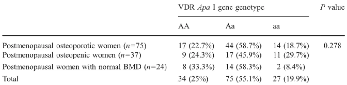
The distribution of VDR Apa I genotype of the 136 postmenopausal women evaluated were as follows: AA 34 (25%), Aa 75 (55.1%), and aa 27 (19.9%), respectively. Comparison of frequencies distribution of genotypes for VDR Apa I between postmenopausal women with osteo-porosis, osteopenia, and normal BMD did not show any significant differences (Table1).
Comparison of bone metabolism markers and BMD values of 136 postmenopausal women on the basis of VDR Apa I genotype showed that postmenopausal women with aa genotype had significantly lower BMD values (grams per centimeter square) at lumbar spines compared to persons with AA genotype. Also, postmenopausal women with AA genotype had significantly higher serum Ca level
than the subjects with aa genotype. However, comparison of BMD values at proximal femur and other bone metabolism markers did not show any significant differ-ences between postmenopausal women with AA, Aa, and aa genotype (Table2).
Discussion
Several studies have since been carried out on the relation-ships between VDR genotype, bone density, and other aspects of calcium metabolism. However, the results of the previous studies are conflicting. Zhang et al. studied the frequencies distribution of VDR gene Fok I, Apa I, Bsm I, and Taq I polymorphisms and their association with BMD in postmenopausal women, and they found that cross-genotyping Apa I and Bsm I or Taq I polymorphisms was not associated with BMD in postmenopausal women [12]. In another study, Qin et al. studied association of VDRapa I and estrogen receptor-α gene polymorphisms with peak bone mass in premenopausal Chinese women. They did not find any significant association with BMD at lumbar spine and proximal femur [13]. However, Gennari et al. showed that there was some evidence of 13% higher lumbar BMD
VDRApa I gene genotype P value
AA Aa aa
Postmenopausal osteoporotic women (n=75) 17 (22.7%) 44 (58.7%) 14 (18.7%) 0.278 Postmenopausal osteopenic women (n=37) 9 (24.3%) 17 (45.9%) 11 (29.7%)
Postmenopausal women with normal BMD (n=24) 8 (33.3%) 14 (58.3%) 2 (8.4%)
Total 34 (25%) 75 (55.1%) 27 (19.9%)
Table 1 Comparison of fre-quencies distribution of geno-types for VDRApa I between postmenopausal women with osteoporosis, osteopenia, and normal BMD
VDR vitamin D receptor, BMD bone mineral density
n=136 AA Aa aa P value
(n=34) (n=75) (n=27)
Age (years) 54.28±10.71 57.17±9.75 56.61±11.30 0.412 Body mass index (kg/m2) 28.17±3.1 27.98±4.2 28.35±3.3 0.542 Lumbar spine BMD (g/cm2) 0.950±0.16a 0.904±0.18 0.828±0.15a 0.026 Lumbar spine BMD (T-score) −1.93±1.53 −2.35±1.46 −2.81±1.12 0.060 Lumbar spine BMD (Z-score) −1.23±1.55 −1.50±1.40 −2.07±1.08 0.063 Proximal femur BMD (g/cm2) 0.858±0.13 0.818±0.13 0.804±0.14 0.190 Proximal femur BMD (T-score) −1.54±0.86 −1.81±1.12 −1.90±1.15 0.355 Proximal femur BMD (Z-score) −0.52±0.91 −0.56±0.86 −0.75±1.09 0.594 Osteocalcin (ng/ml) 18.04±7.39 20.39±9.07 21.40±12.12 0.455 ALP (U/l) 202.99±83.9 185.53±72.89 167.19±70.40 0.228 CTX (ng/ml) 0.50±0.22 0.55±0.32 0.57±0.29 0.576 Calcium (mg/dl) 9.64±0.47a 9.47±0.46 9.21±0.47a 0.005 Phosphorus (mg/dl) 3.81±0.61 3.63±0.55 3.67±0.59 0.340 PTH (pg/ml) 40.45±15.5 42.62±24.1 45.09±16.06 0.171 25-OH-vitamin D (ng/ml) 33.25±19.5 29.17±11.3 32.50±15.9 0.375 Table 2 Comparison of
demo-graphic features, bone metabo-lism markers, and BMD values of 136 postmenopausal women on the basis of VDRApa I genotype
BMD bone mineral density, VDR vitamin D receptor, ALP alkaline phosphatase,CTX se-rum C-telopeptide cross-linked collagen type I,PTH parathy-roid hormone
a <0.05
values in aabbTT genotype with respect to AABBtt genotype in postmenopausal women, but this difference of approximately 0.1 g/cm2 did not reach statistical signifi-cance [14]. Also, Zambrano-Morales et al. studied the association of Bsm I, Apa I, and Taq I VDR gene polymorphism with osteoporosis in 147 postmenopausal women. They found that BBAAtt haplotype was a risk factor for osteoporosis and BbaaTT was a protection factor [15]. In contrast, we found that postmenopausal women with aa genotype had significantly lower BMD values (grams per centimeter square) at lumbar spines compared to persons with AA genotype. Consistent with our result, Huang et al. investigated association of Apa I polymor-phism of VDR gene with bone mass in 388 healthy men. They found that AA genotype had higher bone mass [16].
Vitamin D interacts with its receptor to play an important role in calcium homeostasis by regulating bone cell growth and differentiation, intestinal calcium absorption, and PTH secretion [17]. VDR Apa I gene polymorphism may be responsible for some part of the heritable component of bone density in women, possibly related to impaired calcium absorption from the bowel. In our study, postmen-opausal women with aa genotype had significantly lower serum Ca level than the women with AA genotype. This finding may show that persons with aa genotype for VDR Apa I gene may have a poor intestinal Ca absorption. This abnormality may lead to secondary hyperparathyroidism, which is characterized by high serum PTH and an increase in bone resorption. Also in our study, postmenopausal women with aa genotype had higher PTH level compared to AA and Aa genotype; however, this difference did not reach statistical significance. Yokoyama et al. showed that Japanese patients with end-stage renal disease with aa genotype for VDR Apa I had significantly higher concen-tration of blood PTH level than the patients with the AA and Aa genotypes [18]. Impaired intestinal Ca absorption and secondary hyperparathyroidism may explain the sig-nificantly low BMD values at lumbar spine in our postmenopausal women with aa genotype for VDRApa I gene. However, the mechanisms by which this polymor-phism modulates VDR function remain unclear: it may influence RNA stability, and isoforms of VDR encoded by different alleles may possess different functions [19].
It is interesting to observe that although the genotypic distribution was consistent with the Hardy–Weinberg’s equilibrium law in most studies, the relative distribution of VDR Apa I genotypes varied remarkably between populations. For example, the AA genotype was 7.8% in Chinese women [20], 15.1% in Japanese women [21], 36% in Indian population [22], or 27% in Greek women [23]. In our study population, the AA genotype was 25%. It is not clear why there was such a major difference in the genotypic distributions; however, population stratification
and/or mixed ethnicities could be the underlying responsi-ble factors. Ethnicity may interact to the influence of VDR Apa I polymorphism in association to BMD. We also found that postmenopausal osteoporotic women had higher aa genotype and lesser AA genotype than the postmenopausal women with normal BMD. However, comparison of frequencies distribution of genotypes for VDR Apa I between postmenopausal women with osteoporosis, osteo-penia, and normal BMD did not show any significant differences. Our study groups consisted of a small number of individuals, so our results may not reach statistical significance.
Our result also showed that Apa I genotype is not associated with BMD at proximal femur. Apa I polymor-phism in VDR gene possibly influence loss of trabecular bone mass of the lumbar spine. Also, a recent meta-analysis showed that there was evidence of an association between spine BMD and VDR Bsm I polymorphism, and no association with femoral BMD was observed [24].
Low number of patients included is the main limitation of our study, which should be an argue that decreased the effect of statistical analysis.
In conclusion, our result showed that postmenopausal women with aa genotype had significantly lower BMD values (grams per centimeter square) at lumbar spines compared to subjects with AA genotype. VDR Apa I gene polymorphism may be responsible for a important part of the heritable component of BMD in women, possibly related to impaired calcium absorption from the bowel.
Disclosures None.
References
1. Kanis JA, Melton LJ, Christiansen C, Johnston CC, Khaltaev N (1994) The diagnosis of osteoporosis. J Bone Miner Res 9:1137– 1141
2. Prince RL, Dick I (1997) Oestrogen effects on calcium membrane transport: a new view of the inter-relationship between oestrogen deficiency and age-related osteoporosis. Osteoporos Int 7(suppl 3):S150–S154
3. Pocock NA, Eisman JA, Hopper JL, Yeates MG, Sambrook PN, Eberl S (1987) Genetic determinants of bone mass in adults: a twin study. J Clin Invest 80:706–710
4. Christian JC, Yu PL, Slemenda CW, Johnston CC (1989) Heritability of bone mass: a longitudinal study in aging male twins. Am J Hum Genet 44:429–433
5. Slemenda CW, Christian JC, Williams CJ, Norton JA, Johnston CC (1991) Genetic determinants of bone mass in adult women: a reevaluation of the twin model and the potential importance of gene interaction on heritability estimates. J Bone Miner Res 6:561–567
6. Flicker L, Hopper JL, Rodgers L, Kaymakci B, Green RM, Wark JD (1995) Bone density in elderly women: a twin study. J Bone Miner Res 10:1607–1613
7. Michaelsson K, Melhus H, Ferm H, Ahlbom A, Pedersen NL (2005) Genetic liability to fractures in the elderly. Arch Intern Med 165:1825–1830
8. Stewart TL, Ralston SH (2000) Role of genetic factors in the pathogenesis of osteoporosis. J Endocrinol 166:235–245 9. Ralston SH, de Crombrugghe B (2006) Genetic regulation of
bone mass and susceptibility to osteoporosis. Genes Dev 20:2492–2506
10. Kristjansson K, Rut AR, Hewison M, O’Riordan JL, Hughes MR (1993) Two mutations in the hormone binding domain of the vitamin D receptor cause tissue resistance to 1,25 dihydroxyvita-min D3. J Clin Invest 92:12–16
11. Zmuda JM, Cauley JA, Danielson ME, Wolf RL, Ferrell RE (1997) Vitamin D receptor gene polymorphisms, bone turnover, and rates of bone loss in older African-American women. J Bone Miner Res 12:1446–1452
12. Zhang ZL, Zhao JX, Meng XW, Zhou XY, Xing XP, Xia WB (2003) Association of polymorphisms of vitamin D receptor gene start codon and 3′-end region with bone mineral density in postmenopausal women. Zhonghua Yi Xue Yi Chuan Xue Za Zhi 20(1):5–8
13. Qin YJ, Zhang ZL, Huang QR, He JM, Hu YQ, Zhao Q, Lu JH, Li M, Liu YJ (2004) Association of vitamin D receptor and estrogen receptor-alpha gene polymorphism with peak bone mass and bone size in Chinese women. Acta Pharmacol Sin 25(4):462– 468
14. Gennari L, Becherini L, Masi L, Gonnelli S, Cepollaro C, Martini S, Mansani R, Brandi ML (1997) Vitamin D receptor genotypes
and ıntestinal calcium absorption in postmenopausal women.
Calcif Tissue Int 61:460–463
15. Zambrano-Morales M, Borjas L, Fernández E, Zabala W, de Romero P, Pineda L, Morales-Machín A (2008) Association of the vitamin D receptor gene BBAAtt haplotype with osteoporosis in post-menopausic women. Invest Clin 49(1):29–38
16. Huang QR, Zhang ZL, Qin YJ, He JW, Lu JH, Zhou Q, Hu YQ, Li M, Liu YJ (2003) Association of Apa I polymorphism of
vitamin D receptor gene with bone mass in men. Zhongguo Yi Xue Ke Xue Yuan Xue Bao 25(3):254–257
17. Williams FMK, Spector TD (2007) The genetics of osteoporosis. Acta Reumatol Port 32:231–240
18. Yokoyama K, Shigematsu T, Tsukada T, Ogura Y, Takemoto F, Hara S, Yamada A, Kawaguchi Y, Hosoya T (1998) Apa I polymorphism in the vitamin D receptor gene may affect the parathyroid response in Japanese with end-stage renal disease. Kidney Int 53(2):454–458
19. Arai H, Miyamoto K, Taketani Y, Yamamoto H, Iemori Y, Morita K, Tonai T, Nishisho T, Mori S, Takeda E (1997) A vitamin D receptor gene polymorphism in the translation initiation codon: effect on protein activity and relation to bone mineral density in Japanese women. J Bone Miner Res 12:915–921
20. Zhang ZL, Qin YJ, Huang QR, He JW, Li M, Zhou Q, Hu YQ, Liu YJ (2004) Association of estrogen receptor-alpha and vitamin D receptor genotypes with therapeutic response to calcium in postmenopausal Chinese women. Acta Pharmacol Sin 25 (12):1690–1697
21. Iki M, Saito Y, Dohi Y, Kajita E, Nishino H, Yonemasu K, Kusaka Y (2002) Greater trunk muscle torque reduces postmenopausal bone loss at the spine independently of age, body size and vitamin D receptor genotype in Japanese women. Calcif Tissue Int 71:300–307
22. Bid HK, Mishra DK, Mittal R (2005) Vitamin-D receptor (VDR) gene (Fok-I, Taq-I and Apa-I) polymorphisms in healthy individuals from north Indian population. Asian Pac J Cancer Prev 6(2):147–152
23. Fountas L, Moutsatsou P, Kastanias I, Tamouridis N, Tzanela M, Anapliotou M, Sekeris CE (1999) The contribution of vitamin D receptor gene polymorphisms in osteoporosis and familial osteoporosis. Osteoporos Int 10(5):392–398
24. Thakkinstian A, D’Este C, Eisman J, Nguyen T, Attia J (2004) Meta-analysis of molecular association studies: vitamin D receptor gene polymorphisms and BMD as a case study. J Bone Miner Res 19:419–428