The effect of magnetic field strength on shoot
regeneration and Agrobacterium tumefaciens-mediated
gene transfer in flax (Linum usitatissimum L.)
Murat Aycan
1, Ramazan Beyaz
2, Anzel Bahadir
3, Mustafa Yildiz
4*
1Department of Field Crops, Graduate School of Natural and Applied Sciences,Ankara University, Diskapi, Ankara, Turkey
2Department of Soil and Plant Nutrition, Faculty of Agriculture, Ahi Evran University, Bağbaşı, Kırşehir, Turkey
3Department of Biophysics, Faculty of Medicine, Duzce University, Konuralp, Duzce, Turkey 4Department of Field Crops, Faculty of Agriculture, Ankara University, Diskapi, Ankara, Turkey *Corresponding author: [email protected]
Citation: Aycan M., Beyaz R., Bahadir A., Yildiz M. (2019): The effect of magnetic field strength on shoot regeneration
and Agrobacterium tumefaciens-mediated gene transfer in flax (Linum usitatissimum L.). Czech J. Genet. Plant Breed., 55: 20−27.
Abstract: This study was conducted to determine the effects of magnetic field (MF) strength on shoot regenera-tion and Agrobacterium tumefaciens-mediated gene transfer in flax (Linum usitatissimum L.). Seeds of flax cv. Madaras were exposed to different MF strengths (0 – control, 75, 150, and 300 millitesla (mT)) for 24 h by using an electromagnetic generator system fabricated in laboratory conditions. After sterilization, seeds were germinated on MS (Murashige and Skoog) medium in Magenta vessels. Hypocotyl explants excised from 7-days-old seedlings were used for regeneration. GV2260 strain of Agrobacterium tumefaciens was used in transformation studies. In-oculated hypocotyls were cultured on MS medium containing 1 mg/l BAP (6-benzylaminopurine) and 0.02 mg/l NAA (naphthaleneacetic acid) for 2 days by co-cultivation. Then, they were transferred to MS medium containing the same growth regulators, 100 mg/l kanamycin and 500 mg/l Duocid for selection. The presence of the nptII gene was verified by PCR (polymerase chain reaction) analysis in putative transgenic plants. The highest results with respect to shoot regeneration and transformation frequency were obtained from treatments of 75 mT MF strength. Keywords: Agrobacterium tumefaciens; Linum usitatissimum; magnetic field; regeneration efficiency; transformation
Agrobacterium-mediated transformation has been widely used for the introduction of foreign genes into plants and consequent regeneration of trans-genic plants (Hooykaas & Schilperoort 1992). This bacterium naturally infects the wound sites in dicotyledonous plants and induces diseases known as crown gall (Nester et al. 1984). The production of transformed flax plants from transformed cal-lus is very low. In particular, after inoculation with Agrobacterium, the regeneration capacity of explants is somehow decreased dramatically. Possible expla-nations for this phenomenon are: plant cells may
perceive Agrobacterium infection as a pathogenic attack, and the inoculation process may influence plant regeneration negatively (Jordan & Mc Hughen 1988; Heberle-Bors et al. 1990; Yildiz et al. 2002). It was reported that seed germination and plant development within previous studies were increased by exposing seeds of the various plant species to dif-ferent magnetic field (MF) strengths (Soltani et al. 2006; Florez et al. 2007; Carbonell et al. 2008). The MF increased seed germination by faster water assimilation and higher photosynthesis (Podlesny et al. 2004). Also, Yinan et al. (2005) reported that
the MF pretreatment had a positive effect on cu-cumber seedlings by stimulating seedling growth and development. Furthermore, Dayal and Singh (1986) exposed tomato seeds to different MF strengths varying from 15 to 155 millitesla (mT) for different exposure times, and they noted an increase in height and number of primary branches in treated plants compared to controls. Besides, Florez et al. (2004) also observed an increase in the initial growth stages and an early sprouting of rice seeds when exposed to 125 and 250 mT MFs.
This study was conducted to evaluate the effects of different MF strengths on shoot regeneration and transformation frequencies by Agrobacterium tumefaciens in flax (Linum usitatissimum L.).
MATERIAL AND METHODS
Plant material. Flax (Linum usitatissimum L., cv. Madaras) seeds were obtained from Northern Crop Science Laboratories in North Dakota, USA. Flax seeds were exposed to different MF strengths (0 – control, 75, 150, and 300 mT) for 24 h and then they were surface sterilized with 40% commercial bleach containing 5% sodium hypochlorite at 10°C for 20 min with continuous stirring and then were washed three times with sterile distilled water at the same temperature according to the protocol described by Yildiz and Er (2002). Sterilized seeds were germinated on a basal medium containing the mineral salts and vitamins of Murashige and Skoog (MS) (Murashige & Skoog 1962), 3% (w/v) sucrose and 0.7% (w/v) agar.
Magnetic field generation. The magnetic field sys-tem was integrated with an electromagnet consisting of two Helmholtz coils of copper wire (cross-sections: 0.5 mm2) in each, mounted on a wooden frame. The
pole pieces were cylindrical in shape with a diameter of 9 cm and a length of 8 cm. The number of turns per coil was 3000 and the coil resistance was 16 Ω. The induced mean MF in the centre of the coils could range from 50 to 500 mT. Each of the coils was located in a horizontal position. These coils are connected to a tunable power supply (0–12 A, ref. 13506-93, PHYWE, Germany) to produce a ho-mogeneous MF in the horizontal direction in the central area near the axis of the coils. Additionally, an ampere meter is used to measure the current intensity through the coils, which is proportional to the applied MF strength. The coil nuclei are con-fronted and separated by a distance of about 12 cm
to place the GD anode between them, both coils are connected in parallel. The accuracy and uniformity of these magnetic field strengths produced in the middle of the gap between the nuclei (inside the GD anode) were measured by using a digital teslameter (ref. 13610-93, PHYWE) combined with a tangential flat-electrode Hall probe (ref. 13610-02, PHYWE). The probe was fitted with connecting cable and diode plug to the teslameter and its dimensions were 1.2 × 4 × 70 mm. Also, an electrolytic capacitor (22 000 µF, ref. 06211-00, PHYWE) was connected parallelly to the power supply to minimize instabilities.
Magnetic field treatment. The flax seeds were exposed to three different MF strengths containing 0 (control), 75, 150, and 300 mT produced in the middle of the gap between the coil nuclei by using an electromagnetic generator system fabricated in laboratory conditions. One hundred visibly sound, mature and healthy seeds held in the plastic container were located in the region within the coils of the electromagnet under a homogeneous MF and treated for 24 h. Static continuous MF between the poles of the coils was measured as 75, 150, and 300 mT with a digital teslameter. Moreover, the control samples were kept far enough (at least 30 cm away from each other) from the MF-producing device to avoid any potential exposure to the magnetic field. The differ-ently MF-treated flax variants were compared with untreated flax seeds (control group) in the same growing conditions. The local geomagnetic field in the laboratory was less than 60 µT and the direction of the field was north to south. In all experiments, temperature levels were monitored and controlled by using a temperature control unit. The temperature during the course of seed exposure was kept at 25 ± 0.5°C. All treatments in the experiments were run simultaneously along with the control under similar conditions. The same procedure was applied to the control group except for the MF exposure.
Culture conditions. All cultures were incubated at 25 ± 1°C under cool white fluorescent light (27 μmol per m2/s) with a 16 h light/8 h dark photoperiod.
The pH of the medium was adjusted to 5.8 prior to autoclaving.
Tissue culture response. The hypocotyl sections (20 explants per Petri dish) of the 7-days-old flax seedlings were cultured on MS medium containing 1 mg/l BAP (6-benzylaminopurine) and 0.02 mg/l NAA (naphthaleneacetic acid) in a Petri dish at 1.0 × 1.0 cm distances for 4 weeks. After four weeks, cul-ture initiation, regeneration percentage, number
of shoots per explant, the highest shoot length per explant, total shoot number per Petri dish, number of rooted explants transferred to the soil, number of plantlets growing in soil and length of each plantlet were recorded to determine the effect of different MF strengths on the tissue culture response of flax hypocotyls.
Agrobacterium tumefaciens strain. Agrobacterium tumefaciens strain GV2260 harbouring the plasmid p35S GUS-INT was used for inoculation. The binary plasmid p35S GUS-INT contains neomycin phos-photransferase II (nptII) gene driven by nopaline synthase (NOS) promoter and β-glucuronidase (GUS) gene controlled by cauliflower mosaic virus (CaMV) 35S promoter. A single colony of A. tumefaciens strain GV2260 was grown overnight in a liquid NB (Nutri-ent Broth) medium containing 50 mg/l kanamycin and 50 mg/l rifampicin at 28°C in a rotary shaker (180 rpm) (OD600nm = 0.6). Then, 100 µl of this cul-ture was added to 10 ml NB containing antibiotics and incubated overnight at 28°C in a rotary shaker (180 rpm) (OD600nm = 0.6).
Transformation procedure. A. tumefaciens strain GV2260 was grown overnight and diluted with a liquid NB medium to 1 × 10⁸ cells/ml. Hypocotyl
explants, 0.5 cm in length, excised from 7-days-old sterile seedlings were inoculated in a liquid regenera-tion MS medium containing 1 mg/l BAP, 0.02 mg/l NAA. After inoculation, hypocotyl explants were transferred to solid MS medium containing 1 mg/l BAP and 0.02 mg/lNAA for co-cultivation for 2 days in culture room at a temperature of 24 ± 1°C. Explants were then transferred to the medium which had the same content as that for co-cultivation, supplemented with 100 mg/l kanamycin and 500 mg/l Duocid for selection during 4 weeks.
Rooting of shoots and recovery of transgenic plants. Regenerated shoots were then transferred to a rooting medium containing 3 mg/l indole-butyric acid (IBA) and 100 mg/l kanamycin in Magenta vessels (60 × 60 mm) with three replications (clonally propagated regenerants) and incubated at 24 ± 1°C for 3 weeks to induce root formation. Plantlets having roots were then transferred to pots with three replications again (plantlets from each Magenta vessel were transferred to pots) in a growth room for 3 weeks where light (27 µmol/m2/s), temperature (24 ± 1°C) and humidity
were controlled. Humidity was decreased gradually from 100% to 40% during three weeks.
Genomic DNA extraction and polymerase chain reaction (PCR). The confirmation of gene transfer
was performed by PCR method. According to Della-porta et al. (1983) total DNA was isolated from 100 mg of fresh tissue of leaves from plants grown in pots as described in GeneJET Plant Genomic DNA purification mini kit (No. K0791; Thermo Fisher Scientific, Lithuania) for each MF strength. The percentage of the transgene was determined by PCR amplification and agarose gel electrophoresis. The PCR primers were chosen to amplify the coding se-quence of the transgenes: 5'-TTGCTCCTGCCGAGA-AAG-3' and 5'-GAAGGCGATAGAAGGCGA-3' for a 0.46 kb portion of the nptII gene.
Verification of the putative transgenic plants was checked with the chv gene of Agrobacterium genomic DNA. The existence of the chv virulence gene in-cluded in the chromosome of A. tumefaciens was investigated by means of primers, using the protocol, reported by Yang et al. (2013) to be available in the samples taken during the PCR reaction. If the band belonging to the chv virulence gene was not seen in gel, it was understood that there was no bacte-rial contamination. chv control PCR primers were chosen to amplify the coding sequence: 5'-CGAAC-CGCTGTTCGGCCTGTGG-3' and 5'-GTTCAG-GCCGGCGGCATCCTGG-3' for a 0.85 kb portion of the chv gene.
PCR reaction mixtures were prepared using 100 ng/µl DNA, 2 mmol MgCl2, 0.25 mmol dNTP, 0.5 pmol forward (sense) primer, 0.5 pmol reverse (anti-sense) primers and 0.625 U Taq DNA Polymerase (No. EP0402; Thermo Fisher Scientific, USA). Reac-tions were incubated in a programmable thermocycler (Techne-Prime Thermal Cycler) for 35 cycles. Each PCR cycle consisted of denaturation at 95°C for 1 min, annealing at 58°C (nptII gene), 62°C (chv gene) for 30 s, and extension at 72°C for 1 min. The PCR products were separated by electrophoresis in 1.5% agarose in TAE (tris-acetate EDTA) buffer followed by ethidium bromide staining. The bands were ob-served under UV after electrophoresis.
Statistical analysis. Three replications were tested. Petri dishes (100 × 10 mm) containing 20 explants, Magenta vessels containing 20 plantlets and pots containing 20 plans were considered the units of replication. One-way analysis of variance (ANOVA) was used to test the effect of different MF strengths on shoot regeneration from flax hypocotyls and A. tumefaciens-mediated transformation. All ex-periments were repeated twice. Data were statisti-cally analysed by IBM SPSS Statistics 22 computer program. Duncan’s multiple range test was used to
compare the means. Data presented in percentages were subjected to arcsine (√—X) transformation before statistical analysis (Snedecor & Cochran 1989).
RESULTS AND DISCUSSION
It is quite difficult to assess the effects of electromagnet (EM) or MFs the organisms are exposed to. The main reason for this is the complex structure of biological systems. Many experiments with the effects of electrical or MFs on both complex structured and simple struc-tured organisms have been conducted. Since most of the biological structures are non-homogeneous, when they are exposed to electrical or MFs, they show important cellular changes. This situation can be explained by the examination of the responses of molecules, ions, and membranes under electrical and MFs. The cell level effects of electrical and MFs can be listed as membrane changes, ionic effects (Ca2+, Na+, K+), nucleic acid and
gene expression, enzymatic activity, biorhythms, and hormones, genotoxic effects (Goodman et al. 1995; Simkó 2004; Funk et al. 2009).
Several theories have been proposed, associated with biochemical changes due to the radical pair mechanism, ion cyclotron resonance mechanisms and ferrimagnetism or enzyme activity (Galland & Pazur 2005). However, there is not yet a sufficient explanation for how exactly MF affects biological systems containing plants during dormancy and seed germination (Harris et al. 2009).
Alternatively, electrical and MFs may activate naturally occurring ion channels within cell mem-branes, causing an influx of ions and increasing turgor through osmoregulation (Reina & Pascual 2001). This theory is plausible because seed germination and early growth are closely linked to cellular water and solute regulation (Welbaum et al. 1998).
One hypothesis suggests that MF interacts with ionic current in the plant embryo cell membrane and this interaction induces changes in both osmotic pressure and ionic concentrations on both sides of the membrane (Reina & Pascual 2001). Several researchers reported that in an array of MF at the level of 10–3–10–2 T, intermediate reactions affect the
chemical reactions by influencing the electron-spin positions, demonstrating the presence of a potential for biological consequences (Belyavskaya et al. 1992). In studies at the cell level, RNA and protein synthesis is influenced by the changes in strengths of MF in G1 phase, and the rate of cell division increased in cells exposed to MF (Negishi et al. 1999; Atak et al. 2003). Although there are many reports on the effects of MF on seed germination, plant growth, protein bio-synthesis , seedling elongation and root development (Savostin 1930; Dayal & Singh 1986; Florez et al. 2004, 2007; Podlesny et al. 2004; Yinan et al. 2005; Soltani et al. 2006; Carbonell et al. 2008), to our knowledge, the effects of MF on in vitro shoot regeneration capacity and A. tumefaciens-mediated gene transfer in flax (Linum usitatissimum L.) have not been reported previously.
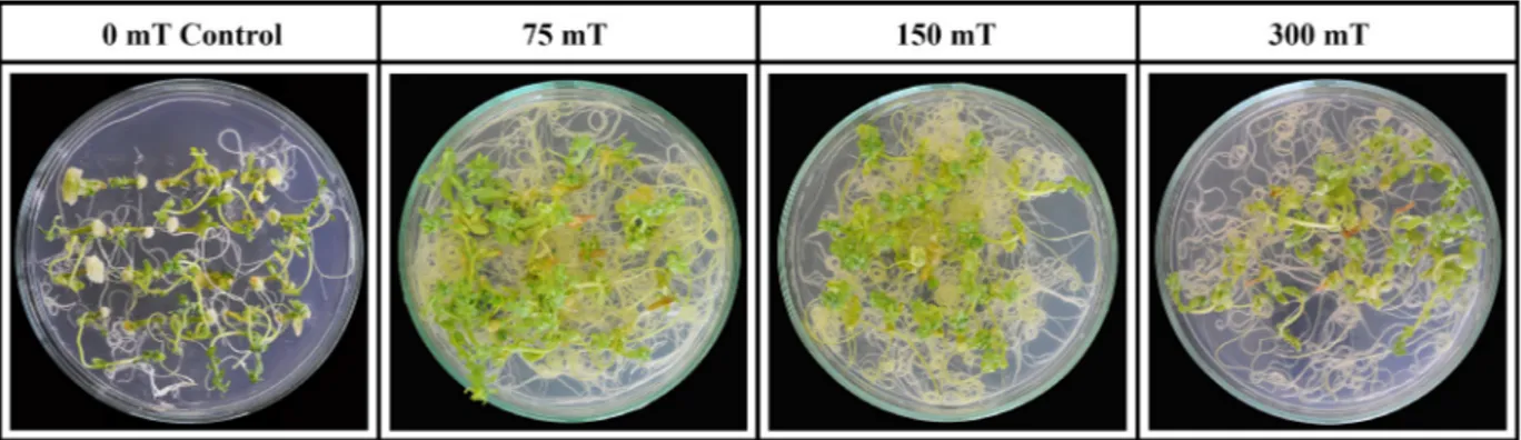
Effect of magnetic field strength on shoot re-generation capacity of flax hypocotyl explants. According to the results, there were statistically significant differences between control (0 mT) and different MF treatments (Table 1). The highest regen-eration percentages were obtained at 75 and 150 mT MF strengths as 84.00% and 85.00% of hypocotyl explant regenerated shoots, respectively (Table 1 and Figure 1). The highest results in the number of shoots per explant, the highest shoot length per explant, total shoot number per Petri dish, the number of rooted explants transferred to the soil and the number of plantlets growing in soil were recorded at 75 mT
Ta
ble 1. The eff
ec t of diff er en t mag ne tic fie ld str eng ths on s ho ot r egenera tion f rom h yp oc otyl e xpl an ts of fl ax Mag ne tic fie ld str eng th (m T ) Re genera tion (%) Sho ot No. per e xpl an t The hig he st s ho ot leng th per e xpl an t ( cm) Tot al s ho ot No. per P etr i di sh The No. of r oot ed e xpl an t transf er re d t o s oil The No. of pl an tle t gr ow ing in s oil Leng th of pl an tle t (c m ) 0 ( con tr ol) 62.00 b 1.54 b 2.62 b 19.20 c 11.80 b 3.40 b 6.00 c 75 84.00 a 2.24 a 3.36 a 37.63 a 26.80 a 21.20 a 7.20 b 150 85.00 a 2.00 a 3.08 a 34.20 b 26.00 a 20.60 a 8.70 a 300 82.00 a 2.00 a 2.96 a 33.00 b 26.40 a 20.10 a 7.80 b Value s w ithin a c olumn f ollowe d by diff er en t le tters ar e sig nific an tly diff er en t a t t he 0.01 le ve l; e ac h v alue i s t he me an of 3 r eplic ations c on taining 20 e xpl an ts p er r eplic ation; m T− millit es la Ta
ble 2. The eff
ec t of diff er en t mag ne tic fie ld str eng ths on Ag ro bacter iu m tu mef acien s-me di at ed gene transf er in fl ax Mag ne tic fie ld str eng th (m T ) Re genera tion (%) Sho ot No. per e xpl an t The hig he st sho ot leng th per e xpl an t (c m ) Tot al s ho ot No. per P etr i di sh The No. of r oot ed ex pl an ts trans -fer re d t o s oil The No. of pl an ts g row ing in s oil 1 The No. of PCR p ositive pl an ts 2 Transf or ma tion effic ienc y (%) (2/1) × 100 Inc re as e in transf or ma tion effic ienc y ac cor ding to c on tr ol (%) 0 ( con tr ol) 41.25 b 1.17 c 1.24 c 9.53 d 8.70 b 1.86 d 0.00 d 0.00 63.00 75 82.00 a 2.40 a 3.40 a 39.40 a 16.40 a 12.60 a 8.00 a 63.00 150 78.00 a 2.36 a 2.76 b 36.80 b 15.60 a 8.40 b 5.00 b 59.00 300 46.00 b 1.70 b 2.54 b 15.60 c 9.20 b 5.50 c 3.00 c 54.00 Value s w ithin a c olumn f ollowe d by diff er en t le tters ar e sig nific an tly diff er en t a t t he 0.01 le ve l; e ac h v alue i s t he me an of 3 r eplic ations c on taining 20 e xpl an ts p er r eplic ation; m T− millit es la
MF strength as 2.24, 3.36, 37.63, 26.80, and 21.20, respectively. The lowest values in all characters were obtained from control (0 mT) where no MF strength was used. From the results, it could be concluded that 75 mT MF strength gave rise to the highest values. Shoot number per explant and total shoot number per Petri dish are the best indicators showing the tissue culture response. In the present study, the number of shoots per explant and total shoot number per Petri dish were recorded as 1.54 and 19.20 in control (0 mT) treatment while they were 2.24 and 37.63 in the above-mentioned treatment, respectively (Table 1). It is well known that regeneration capacity of the tissue decreases significantly in gene transformation studies using A. tumefaciens due to the plant defence mechanism against a pathogenic attack. That is why a higher shoot regeneration frequency is a prerequisite for the success in transformation studies. An increase in the shoot regeneration frequency at 75 mT MF strength could be attributed to the higher mobiliza-tion of nutrients and growth regulators in the tissue. Higher results at 75 mT MF strength could be due to the higher hormone levels of the tissue as reported by Okubo et al. (1991).
Effect of magnetic field strength on Agrobac-terium tumefaciens-mediated transformation of flax. A. tumefaciens, a plant pathogen, is commonly used as a vector for gene transfer to plants (Joubert
et al. 2002). The success of genetic transformation via A. tumefaciens is limited in most of the plant species due to the fact that the plant defence mechanism will be active when the pathogen attacks. For this reason, manipulations of the plant, bacteria, and physical con-ditions have been applied to increase the virulence of bacteria and to increase the transformation efficiency (Chakrabarty et al. 2002). Pre-culturing explants before inoculation (Chakrabarty et al. 2002), modi-fication of temperature (Chakrabarty et al. 2002; De Clercq et al. 2002) and medium pH (De Clercq et al. 2002), addition of chemicals to inoculation medium such as acetosyringone (Chakrabarty et al. 2002; De Clercq et al. 2002; Lopez et al. 2004), changing bacterial density and co-cultivation period (Lopez et al. 2004) and vacuum infiltration (Mahmoudian et al. 2002; Spokevicius et al. 2005) have been reported to increase transformation.
The highest shoot regeneration percentage on a selection medium containing 100 mg/l kanamycin and 500 mg/l Duocid was recorded 82.00% as the highest from 75 mT MF strength. It was 41.25% in control treatment in which no MF was used. Shoot number per explant was obtained from 75 mT MF strength as 2.40, while it was 1.17 in control treatment. The highest shoot length, total shoot number per Petri dish, the number of rooted explants transferred to the soil, the number of plants growing in soil and
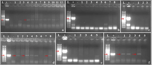
Figure 2. The effects of different magnetic field strengths on gene transfer efficiency in flax: PCR analysis to detect the
nptII gene in putative transgenic plants exposed to 75 mT magnetic field strength (a), PCR analysis to detect the chv gene
in putative transgenic plants exposed to 75 mT magnetic field strength (b), PCR analysis to detect the chv gene in puta-tive transgenic plants exposed to 300 mT magnetic field strength (c), PCR analysis to detect the nptII gene in putaputa-tive transgenic plants exposed to 150 mT magnetic field strength (d), PCR analysis to detect the chv gene in putative trans-genic plants exposed to 150 mT magnetic field strength (e), PCR analysis to detect the nptII gene in putative transtrans-genic plants exposed to 300 mT magnetic field strength (f)
the number of PCR positive plants were recorded as 39.40, 16.40, 12.60, and 8.00 from 75 mT MF strength, respectively. In control treatment, the lowest results were obtained for the highest shoot length per explant, total shoot number per Petri dish, the number of rooted explants transferred to soil, the number of plants growing in soil and the number of PCR positive plants as 1.24, 9.53, 8.70, 1.86, and 0.00, respectively (Table 2).
After 4-week cultivation on a selection medium, rooted explants were directly transferred to the soil by skipping an in vitro rooting stage. From 75 mT MF strength, on average 16.40 rooted explants were transferred to the soil and finally, 12.60 transgenic plants were grown in soil, reached maturity and all were morphologically normal. On the other hand, only 1.86 plants were grown from the control treatment. Out of 8.70 rooted explants transferred to the soil, only 1.86 putative transgenic plants were grown in soil in the control treatment where no MF was used (Table 2).
After PCR analysis, out of 12.60 plants, 8.00 plants were confirmed to be transgenic at 75 mT MF strength (transformation efficiency 63.00%) while none of the plants out of 1.86 was found transgenic in the control treatment. This meant 63.00% increase in transformation efficiency according to control (Ta-ble 2, Figure 2).
Results showed positive effects of magnetic field strength on regeneration and transformation at 75 mT MF strength treatment as compared to control treat-ment. At MF strengths over 75 mT, shoot regenera-tion and transformaregenera-tion were hindered significantly.
The results presented in this study clearly indicate that exposing explants to MF strength before culture improved the regeneration capacity and transforma-tion frequency of flax significantly.
Acknowledgements. This investigation was supported by
Scientific and Technological Research Council of Turkey (TUBİTAK); Grant No. 113O280 to Prof. Dr. M. Yildiz
References
Atak C., Emiroglu O., Alikamanoglu S., Rzakoulive A. (2003): Stimulation of regeneration by magnetic field in soybean (Glycine max L. Merrill) tissue cultures. Journal of Cell and Molecular Biology, 2: 113–119.
Belyavskaya N.A., Fomicheva V.M., Govorun R.D., Danilov V.I. (1992): Structural-functional organization of the mer-istem cells of pea, lentin and flax roots in conditions of screening the geomagnetic field. Biophysics, 37: 657–666.
Carbonell M.V., Martinez E., Florez M., Maqueda R., Lopez-Pintor A., Amaya J.M. (2008): Magnetic field treatments improve germination and seedling growth in Festuca arundinacea Schreb. and Lolium perenne L. Seed Science and Technology, 36: 31–37.
Chakrabarty R., Viswakarma N., Bhat S.R., Kirti P.B., Singh B.D., Chopra V.L. (2002): Agrobacterium-mediated trans-formation of cauliflower: optimization of protocol and development of Bt-transgenic cauliflower. Journal of Biosciences, 27: 495–502.
Dayal S., Shing R.P. (1986): Effect of seed exposure to mag-netic field on the height of tomato plants. Indian Journal of Agricultural Science, 56: 483–486.
De Clercq J., Zambre M., Van Montagu M., Dillen W., An-genon G. (2002): An optimized Agrobacterium-mediated transformation procedure for Phaseolus acutifolius A. Gray. Plant Cell Reports, 21: 333–340.
Florez M., Carbonell M.V., Martínez E. (2004): Early sprout-ing and first stages of growth of rice seeds exposed to a magnetic field. Electromagnetics, 23: 157–166.
Florez M., Carbonell M.V., Martínez E. (2007): Exposure of maize seeds to stationary magnetic fields: effects on germination and early growth. Environmental and Ex-perimental Botany, 59: 68–75.
Funk R.H.W., Monsees T., Ozkucur N. (2009): Electromag-netic effects – from cell biology to medicine. Progress in Histochemistry and Cytochemistry, 43: 177–264. Galland P., Pazur A. (2005): Magnetoreception in plants.
Journal of Plant Research, 118: 371–389.
Goodman E.M., Greenebaum B., Marron M.T. (1995): Effects of electromagnetic fields on molecules and cells. Interna-tional Review of Cytology, 158: 279–338.
Harris S.R., Henbest K.B., Maeda K., Pannell J.R., Timmel C.R., Hore P.J., Okamoto H. (2009): Effect of magnetic fields on cryptochrome-dependent responses in Arabi-dopsis thaliana. Journal of the Royal Society Interface, 6: 1193–1205.
Heberle-Bors E.F., Moreno R.M.B., Alwen A., Stoger E., Vicente O. (1990): Transformation of pollen. In: Nijkamp H.J.J., Van Der Plas L.H.W., Van Aartrijk J. (eds.): Prog-ress in Plant Cellular and Molecular Biology. Kluwer Academic Publications: 244–251.
Hooykaas P.J.J., Schilperoort R.A. (1992): Agrobacterium and plant genetic engineering. Plant Molecular Biology, 19: 15–38.
Jordan M.C., Mc Hughen A. (1988): Glyphosate tolerant flax plants from Agrobacterium-mediated gene transfer. Plant Cell Reports, 7: 281–284.
Joubert P., Beaupère D., Lelièvre P., Wadouachi A., Sangwan R.S., Sangwan-Norreel B.S. (2002): Effects of phenolic compounds on Agrobacterium vir genes and gene transfer
induction. A plausible molecular mechanism of phenol binding protein activation. Plant Science, 162: 733–743. Lopez S.J., Kumar R.R., Pius P.K., Muraleedharan N. (2004):
Agrobacterium tumefaciens mediated genetic transfor-mation in tea (Camellia sinensis (L.) O. Kuntze). Plant Molecular Biology Reporter, 22: 201–202.
Mahmoudian M., Yucel M., Öktem H.A. (2002): Transfor-mation of lentil (Lens culinaris M.) cotyledonary nodes by vacuum infiltration of Agrobacterium tumefaciens. Plant Molecular Biology Reporter, 20: 251–257.
Murashige T., Skoog F. (1962): A revised medium for rapid growth and bioassays with tobacco tissue culture. Phys-iologia Plantarum, 15: 473–479.
Negishi Y., Hashimoto A., Tsushima M., Dobrota C., Ya-mashita M., Nakamura T. (1999): Growth of pea epicotyl in low magnetic field implication for space research. Advances in Space Research, 23: 2029–2032.
Nester E.W., Amasino R., Akiyoshi D., Klee H., Montoya A., Gordon M.P. (1984): The molecular basis of plant cell transformation by Agrobacterium tumefaciens. Basic Life Science, 30: 815–822.
Okubo H., Wada K., Uemoto S. (1991): In vitro morpho-genetic response and distribution of endogenous plant hormones in hypocotyl segments of snapdragon (Anti-rrhinum majus L.). Plant Cell Reports, 10: 501–504. Podlesny J., Misiak L., Podlesna A. (2004): Concentration of
free radicals in pea seeds after pre-sowing treatment with magnetic field. International Agrophysics, 18: 261–267. Reina F.G., Pascual L.A. (2001): Influence of a stationary
magnetic field on water relations in lettuce seeds. Part I Theoretical considerations. Bioelectromagnetics, 22: 589–595.
Savostin P.W. (1930): Magnetic growth relations in plant. Planta, 12: 327.
Simkó M. (2004): Induction of cell activation processes by low frequency electromagnetic fields. Scientific World Journal, 4 (S2): 4–22.
Snedecor G.W., Cochran W.G. (1989): Statistical Methods. 8th Ed., Ames, Iowa State University Press: 217–235.
Soltani F., Kashi A., Arghavani M. (2006): Effect of mag-netic field on Asparagus officinalis L. seed germination and seedling growth. Seed Science and Technology, 34: 349–353.
Spokevicius A.V., Van Beveren K.S., Bossinger G. (2005): Agrobacterium-mediated in vitro transformation of wood-producing stem segments in eucalypts. Plant Cell Reports, 23: 617–624.
Welbaum G.E., Bradford K.J., Yim K., Booth D.T., Oluoch M.O. (1998): Biophysical, physiological and biochemi-cal processes regulating seed germination. Seed Science Research, 8: 161–172.
Yang L., Wang C., Wang L., Xu C., Chen K. (2013): An effi-cient multiplex PCR assay for early detection of Agrobac-terium tumefaciens in transgenic plant material. Turkish Journal of Agriculture and Forestry, 37: 157–162. Yinan L., Yuan L., Yongquing Y., Chunyang L. (2005): Effect
of seed pretreatment by magnetic field on the sensitivity of cucumber (Cucumis sativus) seedlings to ultraviolet-B radiation. Journal Environmental and Experimental Botany, 54: 286–294.
Yildiz M., Er C. (2002): The effect of sodium hypochlorite solutions on in vitro seedling growth and shoot regenera-tion of flax (Linum usitatissimum). Naturwissenschaften, 89: 259–261.
Yildiz M., Özcan S., Er C. (2002): The effect of different explant sources on adventitious shoot regeneration in flax (Linum usitatissimum L.) Turkish Journal of Biol-ogy, 26: 37–40.
Received for publication December 12, 2017 Accepted after corrections May 9, 2018 Publish online June 13, 2018