See discussions, stats, and author profiles for this publication at: https://www.researchgate.net/publication/261447185
Flexible organic-inorganic core-shell nanofibers
by electrospinning and atomic layer deposition
Conference Paper · June 2012 CITATIONS0
READS206
5 authors, including: Some of the authors of this publication are also working on these related projects: RF-Sputtering of Doped Zinc Oxides Thin Films, The Effect of Low Substrate Heating depositionView project Virtual Project on the History of ALD (VPHA) View project Cagla Ozgit-Akgun ASELSAN Inc. 58 PUBLICATIONS 457 CITATIONS SEE PROFILE Inci Donmez Barcelona Microelectronics Institute 36 PUBLICATIONS 385 CITATIONS SEE PROFILE Necmi Biyikli Bilkent University 164 PUBLICATIONS 1,687 CITATIONS SEE PROFILE Tamer Uyar Bilkent University 201 PUBLICATIONS 2,992 CITATIONS SEE PROFILE
All content following this page was uploaded by Cagla Ozgit-Akgun on 09 September 2014. The user has requested enhancement of the downloaded file.
Flexible Organic-Inorganic Core-Shell Nanofibers by Electrospinning and Atomic
Layer Deposition
Fatma Kayaci, Cagla Ozgit-Akgun, Inci Donmez, Necmi Biyikli and Tamer Uyar
**
UNAM – Institute of Materials Science and Nanotechnology, Ihsan Dogramaci Bilkent University,
06800 Ankara, Turkey, [email protected]
ABSTRACT
Organic-inorganic core-shell nanofibers were fabricated by combining electrospinning and atomic layer deposition (ALD). In the first step, nylon66 (polymeric organic core) nanofibers having different average fiber diameters (~100 nm, ~250 nm and ~650 nm) were electrospun by using different solvent systems and polymer concentrations. In the second step, uniform and conformal layer of zinc oxide (ZnO) (inorganic shell) with precise thickness (~90 nm) and composition on the round surface of the nylon nanofibers were deposited by ALD. The core-shell nylon66-ZnO nanofibers have shown unique properties such as structural flexibility due to the polymeric core and photocatalytic activity due to the ZnO shell layer.
Keywords: core-shell nanofiber, electrospinning, atomic layer deposition, photocatalytic activity, zinc oxide
1 INTRODUCTION
Electrospinning has gained growing attention in the past decade since this technique is quite versatile and cost-effective for fabricating functional nanofibers from variety of materials including polymers, polymer blends, metal oxides and composite structures, etc [1-7]. Since the fiber diameter ranging from micron down to a few tens of nanometers, electrospun nanofibers and their nanowebs have several unique characteristics including a very high surface area to volume ratio and pore sizes within the nanoscale [1-10].
The core-shell types of electrospun nanofibers are also very attractive for developing multi-functional nanowebs. For instance, organic-inorganic core-shell composite nanofibers would be quite interesting which combine the advantages of the polymers such as flexibility and light weight along with the properties of the inorganic materials such as high mechanical strength, high thermal stability and excellent electrical, magnetic, optical, catalytical properties, etc [11,12]. Yet, the fabrication of organic-inorganic core-shell nanofibers is somewhat challenging since the deposition of inorganic shell layer requires a high temperature process which can easily deform the polymeric core structure.
Recently, atomic layer deposition (ALD) technique has been explored to produce conformal and very thin inorganic coatings on fibrous systems such as
cellulose-based filter paper, nonwovens, synthetic and natural fibers [13,14]. ALD is a special type of low-temperature chemical vapor deposition, in which the substrate is exposed to sequential pulses of two or more precursors separated by purging periods [15]. Unless decomposition of the precursor occurs; each pulse leads to surface reactions that terminate after the adsorption of a single monolayer. Film growth mechanism of ALD is therefore self-limiting, which gives rise to unique properties such as high uniformity and conformality, as well as sub-nanometer thickness control. ALD process is very flexible so that very thin layers of metals, metal oxides or metal nitrides can be fabricated onto different types of substrates [13-15].
In this study, we have successfully achieved organic -inorganic core-shell nanofibers by combination of electrospinning and ALD in which nylon66 (organic core) nanofibers were obtained by electrospinning, and then, zinc oxide (ZnO) (inorganic shell) were precisely deposited onto electrospun nylon66 nanofibers by ALD technique.
2 EXPERIMENTAL
2.1 Materials
Nylon66 pellets (Mw 262.35 g/mol), formic acid, 1,1,1,3,3,3-hexafluoro-2-propanol (HFIP), rhodamine-B were purchased from Sigma-Aldrich and used without any purification.
2.2 Methods
We produced organic-inorganic core-shell nanofibers by using two processing steps: electrospinning and atomic layer deposition (Figure 1). Firstly, Electrospun nylon66 nanofibers having different average fiber diameters (~100 nm, ~250 nm and ~650 nm) were obtained by varying the solvent type (HFIP and formic acid) and polymer concentrations (5 wt % and 8 wt % nylon66).
In the next processing step, ZnO deposition on the electrospun PA66 nanofibrous membrane was carried out at 200°C in Savannah S100 reactor (Cambridge Nanotech). 800 cycles (~1.4 Å/cycle) of ZnO were deposited using diethyl zinc (Et2Zn or DEZn) and water (H2O) as the zinc and oxygen precursors, respectively. N2 was used as the carrier gas with a flow rate of 20 sccm.
1stprecursor excess precursor molecules
byproduct(s)
(b)
electrospun polymeric nanofiber
ALD cycle (c) ELECTROSPINNING ALD 2ndprecursor excess precursor molecules byproduct(s)
Core-shell organic-inorganic nanofiber
(a)
Nanofibers
Polymer solution high voltage supply
syringe pump 15 kV
Collector
Figure 1: (a, b) Schematic representations of electrospinning and atomic layer deposition (ALD) processes, respectively; (c) schematic representation of the
formation of organic-inorganic core-shell nanofibers.
The morphological, structural and chemical analyses of core-shell nylon66-ZnO nanofiber were performed by using scanning electron microscopy (SEM), transmission electron microscope (TEM) and energy dispersive X-ray (EDX)
The photocatalytic activity of the core-shell nylon66-ZnO nanowebs (weight of nanoweb: 9.3 mg) were tested by monitoring the photocatalytic decomposition of organic dye molecule (rhodamine-B) under UV irradiation at 365 nm wavelength (UV source: 8 W, UVLMS-38 EL). As a control experiment, Rh-B solution without containing nanofibers was also examined under the same UV treatment in order to investigate whether any direct photolysis occurred or not.
3 RESULTS AND DISCUSSION
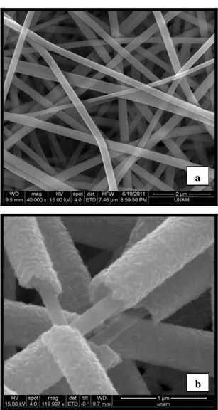
As seen in Figure 2, uniform bead-free nylon66 nanofibers have smooth and uniform surface, however, the surface of the core-shell nylon66-ZnO nanofibers was noticeable rougher due to the grainy structure of the outer ZnO layer [16]. The SEM images also showed that the ALD process yielded uniform coverage of ZnO shell layer over a relatively large surface area of the electrospun nanofibers. In addition the core-shell structure is clearly seen in Figure 2b.
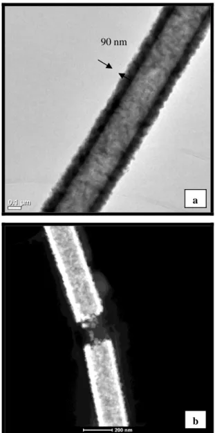
The morphologies of the core-shell nylon66-ZnO nanofibers were further investigated by TEM and HAADF-STEM studies (Figure 3). The TEM and HAADF-HAADF-STEM images also clearly showed that nylon66-ZnO nanofibers have core-shell structure. It is evident that ZnO shell layer was uniformly deposited onto individual nylon66 nanofibers. The surface of the nylon66-ZnO nanofibers was rougher due to nanosized grains of ZnO [17].
The TEM images also revealed that the thickness of the ZnO shell layer was about 90 nm (Figure 3a). The conformal layer-by-layer deposition of ZnO onto the round surfaces of individual electrospun nanofibers is unique to ALD process which resulted in uniform thickness of the ZnO shell layer even the nylon66 nanofibers were randomly distributed in the form of nonwowen and the fiber diameters are very different from each other.
We have shown that these nanowebs can be easily handled and folded as a free standing material (Figure 4).
The energy-dispersive X-ray (EDX) study was also performed for the core-shell nylon66-ZnO nanofibers. The elemental mapping by EDX shown in Figure 5 further confirmed the successful deposition of ZnO shell layer onto nylon66 nanofibers. Zinc (Zn), oxygen (O) and carbon (C) elements were detected in the EDX spectrum, and Zn and O are originated from ZnO shell layer and the C is coming from the organic core structure of nylon66.
a
b
Figure 2: Representative SEM images of (a) nylon 66 nanofibers, (b) nylon66-ZnO core-shell nanofibers.
0.1 µm 0.1 µm
Figure 3: (a) TEM and (b) HAADF-STEM images of nylon66-ZnO core-shell nanofibers.
Figure 4: The photograph of the flexible nylon66-ZnO core-shell nanoweb 0 2 4 6 8 10 Co u nt s ( a .u .) Energy (keV) CO Zn Zn Zn 90 nm a
Figure 5: EDX spectrum of nylon66-ZnO core-shell nanofibers and chemical maps of C, Zn and O.
b
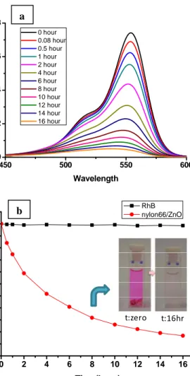
ZnO is a well known excellent nontoxic catalyst owing to its high catalytic activity, high photosensitivity, suitable bandgap and low cost [18-22].The change of the absorption peak of Rh-B at 554 nm in the UV–Vis spectra was monitored as a function of UV irradiation time (Figure 6a) and the absorption peak points were used for calculating the degradation rate of Rh-B defined as C/C0 where C0 and C represent the initial concentration of Rh-B before UV irradiation and after UV irradiation at time t, respectively (Figure 6b). As given in Figure 6, direct photolysis was not detected for the blank Rh-B solution without containing core-shell nylon66-ZnO nanoweb. On the other hand, we observed that the absorbance of dye solution containing this core-shell nanofibers reduced with respect to UV irradiation and therefore pink color of dye solution disappeared in 16h of UV irradiation. Therefore photocatalytic degradation of Rh-B was occurred by core-shell nanofibers successfully.
450 500 550 600 0.0 0.2 0.4 0.6 0.8 Ab sorban ce Wavelength 0 hour 0.08 hour 0.5 hour 1 hour 2 hour 4 hour 6 hour 8 hour 10 hour 12 hour 14 hour 16 hour a
REFERENCES
[1] D. Li, Y. Xia, Advanced Materials, 16, 1151, 2004.
[2] S. Ramakrishna, K. Fujihara, W. Teo, T. Yong, Z. Ma, R. Ramaseshan, Materials Today, 9, 40, 2006. [3] A. Greiner, J. Wendorff, Angewandte
Chemie-International Edition, 46, 5670, 2007.
[4] N. Bhardwaj, S. C. Kundu, Biotechnology Advances, 28, 325, 2010.
[5] Z. Huang, Y. Zhang, M. Kotaki, S. Ramakrishna, Composites Science and Technology, 63, 2223, 2003.
[6] W. E. Teo, S. Ramakrishna, Composites Science and Technology, 69, 1804, 2009.
[7] S. Agarwal, A. Greiner, J. H. Wendorff, Advanced Functional Materials, 19, 2863, 2009. 0 2 4 6 8 10 12 14 16 0.0 0.2 0.4 0.6 0.8 1.0 C/C0 Time(hour) RhB nylon66/ZnO t:zero t:16hr b
[8] S. Ramakrishna, R. Jose, P. Archana, A. Nair, R. Balamurugan, J. Venugopal, W. Teo, Journal of Materials Science, 45, 6283, 2010.
[9] X. Lu, C. Wang, Y. Wei, Small, 2009, 5, 2349. [10] V. Thavasi, G. Singh, S. Ramakrishna, Energy
Environmental Science, 1, 205, 2008.
[11] J. Kong, H. R. Tan, S. Y. Tan, F. Li, S. Y. Wong, X. Li, X. Lu, Chemical Communications, 46, 8773, 2010.
[12] J. M. F. Jabal, L. McGarry, A. Sobczyk, D. E. Aston, Langmuir, 2010.
[13] G.K. Hyde, K.J. Park, S.M. Stewart, J.P. Hinestroza, G.N. Parsons, Langmuir, 23, 9844, 2007.
[14] Kemell, M., Pore, V., Ritala, M., Leskelä, M., Lindén, M., Journal of the American Chemical Society, 127, 14178, 2005.
Figure 6: The UV–vis spectrum of the Rh-B solution containing nylon66-ZnO core-shell nanofibers (a) as a function of the UV irradiation time, (b) the rate (C/C0) of
Rh-B degradation and the colour change of the RhB solution after 16 hr.
[15] G. M. Kim, S. M. Lee, G. Michler, H. Roggendorf, U. Gosele, M. Knez, Chemistry of Materials, 20, 3085, 2008.
[16] Z. Zhang, X. Li, C. Wang, L. Wei, Y. Liu, C. Shao, The Journal of Physical Chemistry C, 113, 19397, 2009.
4 CONCLUSION
In this study we have presented the fabrication of core-shell nylon66-ZnO nanofibers by combination of electrospinning and ALD techniques.
Here, we have shown that the resulting core-shell PA66-ZnO nanowebs can be a very good candidate as a filtering material because of the flexible polymeric core and the photocatalytic activity of the ZnO shell layer.
[17] Y. Wang, W. Jia, T. Strout, A. Schempf, H. Zhang, B. Li, J. Cui, Y. Lei, Electroanalysis, 21, 1432, 2009.
[18] A. A. Aal, S. A. Mahmoud, A. K. Aboul-Gheit, Materials Science and Engineering: C, 29, 831, 2009.
[19] M. A. Kanjwal, N. A. M. Barakat, F. A. Sheikh, S. J. Park, H. Y. Kim, Macromolecular Research, 18, 233, 2010.
ACKNOWLEDGEMENT
[20] H. Liu, J. Yang, J. Liang, Y. Huang, C. Tang, Journal of the American Ceramic Society, 91, 1287, 2008.
State Planning Organization (DPT) of Turkey is acknowledged for the support of UNAM-Institute of Materials Science and Nanotechnology. Dr. Uyar and Dr. Biyikli acknowledge Marie Curie International Reintegration Grant (IRG) for funding NANOWEB (PIRG06-GA-2009-256428) and NEMSmart (PIRG05-GA-2009-249196) projects. F. Kayaci and C. Ozgit-Akgun thank to TUBITAK-BIDEB for their national PhD study scholarships.
[21] Z. Zhang, C. Shao, X. Li, C. Wang, M. Zhang, Y. Liu, ACS Applied Materials & Interfaces, 2, 2915, 2010.
[22] M. A. Kanjwal, F. A. Sheikh, N. A. M. Barakat, I. S. Chronakis, H. Y. Kim, Applied Surface Science, 257, 7975, 2011.
View publication stats View publication stats