344 Turk Gogus Kalp Dama 2012;20(2):344-347
Türk Göğüs Kalp Damar Cerrahisi Dergisi Turkish Journal of Thoracic and Cardiovascular Surgery
doi: 10.5606/tgkdc.dergisi.2012.064
Peripheral bypass grafting in a patient with essential thrombocytosis:
What should a vascular surgeon know?
Esansiyel trombositozu olan hastada periferik baypas greft cerrahisi:
Damar cerrahı neleri bilmeli?
Burak Onan,1 Kürşad Öz,1 İsmihan Selen Onan2
1Department of Cardiovascular Surgery, Dr. Lütfi Kırdar Kartal Training and Research Hospital, İstanbul, Turkey; 2Department of Cardiovascular Surgery, İstanbul Bilim University, Florence Nightingale Hospital, İstanbul, Turkey
Hiperkoagülasyon durumları nativ arter ve arteriyel greft trombozuna neden olabilir. Doğuştan protein eksiklikleri dışında, miyeloproliferatif hastalıklar da trombotik vas-küler olaylara yol açabilir. Esansiyel trombositoz (ET), periferik arter trombozu ve buna bağlı bacak iskemisinin nadir bir nedenidir. Bu yazıda, istirahat ağrısı ve sol ayak başparmağında iyileşmeyen iskemik ülser ile başvuran ve ET’ye sekonder tromboz ve buna bağlı şiddetli yüzeyel femoral arter stenozu tanısı konan 57 yaşında bir erkek olgu sunuldu. Ayrıca bu yazıda, bacak iskemisine neden olan ET literatür bilgileri ışığında gözden geçirildi ve bu ender patolojinin klinik tedavisi cerrahi bakış açısıyla sunuldu.
Anah tar söz cük ler: Esansiyel trombositoz; bacak iskemisi;
peri-ferik arter hastalığı.
Hypercoagulable states can be associated with native arterial and arterial graft thrombosis. Other than congenital protein deficiencies, myeloproliferative disorders may also lead to thrombotic vascular events. Essential thrombocytosis (ET) is a rare cause for peripheral arterial thrombosis and associated extremity ischemia. In this article, we present a 57-year-old male who was admitted with rest pain and non-healing ulcers on the left toe and diagnosed with an isolated severe stenosis of superficial femoral artery due to ET. This report also reviews the literature on ET which may cause extremity ischemia, and clinical management of this rare pathology from the surgical point of view.
Key words: Essential thrombocytosis; extremity ischemia;
peripheral artery disease.
Received: November 23, 2009 Accepted: January 20, 2010
Correspondence: Burak Onan, M.D. Dr. Lütfi Kırdar Kartal Eğitim ve Araştırma Hastanesi, Kalp ve Damar Cerrahisi Kliniği, 34890 Cevizli, Kartal, İstanbul, Turkey. Tel: +90 553 - 622 38 78 e-mail: [email protected]
Myeloproliferative disorders (MPDs) may present with hemorrhagic and thrombotic complications related to the increased number and dysfunction of platelets. Essential thrombocytosis (ET) is defined by the exclusion of other MPDs or secondary causes of reactive thrombocytosis as well as the demonstration of autonomous platelet production.[1] Although microvascular arterial thrombosis is predominant in this disorder, lower extremity arterial thrombosis can be evident. In this report, we present the case of a patient diagnosed with ET who had critical extremity ischemia from an isolated superficial femoral artery (SFA) thrombosis.
CASE REPORT
A 57-year-old man with right hemiparesis presented with a four-week history of painful left toe ulcers. The patient was also suffering from intermittent
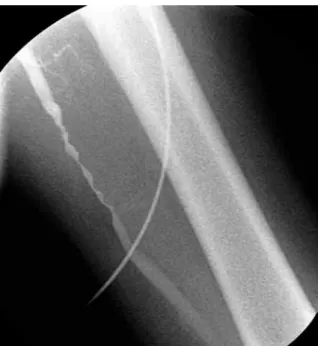
claudication that had gradually increased over the previous six weeks. An angiogram documented an isolated critical stenosis on the left SFA without sufficient collateral flow (Figure 1). The patient’s medical history included cigarette use for almost 30 years, and a cerebrovascular transient ischemic event of unknown origin that had caused right hemiparesis four years previously. No other risk factors for vascular complications, such as markers of hypercoagulability, for example thrombophilia, factor V leiden, and antiphospoholipid antibodies, or, general cardiovascular risk factors, such as diabetes mellitus, hypertension, hypercholesterolemia, and obesity, were evident in this patient. In the past, no biochemical abnormality had been detected, nor history of major trauma to the left extremity or symptoms suggestive of peripheral ischemia had been found.
Onan et al. Peripheral bypass in essential thrombocytosis
345 The patient presented with sinus rhythm. On
physical examination, arterial pulses below the knee were nonpalpable on the left, and there were painful ulcerative lesions with regular margins on the first and second toe. The ankle-brachial index (ABI) was 0.35. In addition, abdominal palpation revealed splenomegaly.
Recent laboratory values showed the platelet count being remarkably elevated to 1.940x10⁹/L of blood. Although there was no abnormality on the blood coagulation profiles, a functional test of thrombocytes revealed an increased aggregation response to epinephrine. A microscopic evaluation of blood cells revealed multiple enlarged thrombocytes with lots of aggregates (Figure 2, Left panel). Bone marrow aspiration demonstrated an increase and clustering of enlarged mature megakaryocytes and diagnosed ET (Figure 2, Right panel) confirming an underlying myelodysplastic disorder.
A cardiac echocardiogram did not note an intracardiac thrombus, which can be a thrombotic source for arterial embolization. Left leg arterial duplex ultrasound (US) imaging showed velocities >400 cm/s at the lower SFA level. Magnetic resonance (MR) angiography of the chest and abdomen was unremarkable for an intravascular pathology while thoracic and abdominal aorta, iliac arteries, and peripheral arteries, other than SFA, were free from an underlying atherosclerotic disease process or calcification of the vessel wall. These findings clarified the pathophysiology of critical extremity ischemia from a likely SFA thrombosis, and medical treatment was then recommended.
In the preoperative period, the patient was anticoagulated with enoxaparin sodium, and a dual antiplatelet treatment with hydroxyurea of 15-20 mg/kg and aspirin of 300 mg daily was started. The platelet count was carefully titrated below 400x109/L with hydroxyurea without allowing neutropenia. However, medical therapy failed to relieve the symptoms, and
Figure 1. Peripheral angiography image demonstrates a critical long segment stenosis of the superficial femoral artery with an irregular border. Proximal and distal segments of the superficial femoral artery were free from a disease process such as atherosclerosis or calcification.
Figure 2. Microscopic views demonstrate a peripheral blood smear of the patient with essential throm-bocytosis and reveal an increased number and aggregates of large dysmorphic thrombocytes (A, left panel). The bone marrow aspirate demonstrates microscopically megakaryocytic hyperplasia with a clustering of enlarged megakaryocytes (B, right panel). (Wright’s stain, x 400).
Turk Gogus Kalp Dama
346
arterial duplex US imaging did not reveal a decrease in the flow velocities. Arterial thrombectomy was not considered because an increased number of dysfunctional thrombocytes might lead to secondary thrombosis after the procedure. Therefore, an elective surgical intervention was scheduled. The patient underwent an uneventful superficial femoral to above-knee popliteal artery bypass grafting by using a reversed saphenous vein. Perioperatively, proximal and distal segments of the popliteal artery were free from a disease process.
In the postoperative period, antiplatelet therapy with hydroxyurea to keep the platelet count below 400.000/mm3 was maintained. Aspirin treatment was started 300 mg once a day to inhibit postoperative collagen-induced thrombocyte aggregation. During the hospital stay, enoxaparin sodium was also delivered until international normalized ratio (INR) reached a satisfactory level with the use of warfarin.
At the one-month follow-up, the ABI was 1.0 on the left, and a repeat left femoral arterial duplex US showed velocities of 138 cm/s. The patient is currently doing well one-year postoperatively with a patent graft as seen on angiography and healed ulcers (Figure 3).
DISCUSSION
Essential thrombocytosis is a chronic myeloproliferative disorder characterized by persistent thrombocytosis greater than 400.000/mm3 and confirmed by a bone marrow biopsy demonstrating megakaryocytic hyperplasia. The diagnosis is established by excluding other myeloproliferative disorders and secondary causes of thrombocytosis such as splenectomy, malignancy, chronic inflammatory diseases, iron-deficiency anemia, and acute blood loss. Bone marrow, blood smear, and functional studies of platelets help the diagnosis.[2] Despite the recent identification of some mutations such as the JAK2V617F, the detailed pathogenetic mechanism is still a matter of discussion.[3]
Essential thrombocytosis is rarely recognized as an isolated predisposing factor in patients with thromboembolic events. In the presence of thrombogenic lesions such as arterial aneurysm, atherosclerotic plaque formation, and/or dissection, ET increases the risk of associated thrombosis and, therefore, should be excluded to confirm the pathogenesis of arterial thrombosis. Clinically, 20% to 50% of patients with ET may have some manifestations when a markedly elevated platelet count is discovered.[2] An episode of arterial thrombosis may complicate the course of disease in 30-40% of cases.[4] Petrides and Siegel[5] reported that thrombosis clinically presents with an incidence of 6.6% per patient
in a year. Moreover, a significant increase in the risk of thromobosis and mortality occurs with patients who have a history of previous thrombotic events and who are older than 60 years of age.[6]
Ischemic events in ET usually occur at the microvascular level and include mostly cerebral vessels with relapsing episodes, as had been diagnosed in the history of our patient.[7] The patients are generally asymptomatic between attacks unless a permanent deficit occurs. Larger arteries are involved in less than 1% of patients, but the incidence of extremity ischemia related to thrombosis on medium-sized arteries, including SFA, is not clear.[4]
In our literature review, few clinical reports have been published describing the clinical onset of critical extremity ischemia in ET.[8-13] In those reports, extremity ischemia was mostly observed after arterial thrombosis and was due to paradoxical embolization from a venous thrombosis in only one case.[13] On the other hand, patients mostly presented with thrombosis on native vessels. Nevertheless, Sugimoto[11] noted that ET complicated the course of an underlying
Figure 3. A postoperative magnetic resonance angiogram shows the patency of the reversed saphenous vein graft, which was anastomosed between the proximal segment of the superficial femoral artery and the above-knee popliteal artery.
Onan et al. Peripheral bypass in essential thrombocytosis
347 atherosclerosis. To our knowledge, ours is the first
reported case of isolated thrombosis of SFA causing critical extremity ischemia in a patient with ET. Sadahiro et al.[12] previously noted an isolated arterial thrombosis, but it was discovered on the common iliac artery. Although it would be more acceptable to document vascular thrombosis pathologically with an intraoperative biopsy from the stenotic segment of the artery to exclude an atherosclerotic process, clinical and radiological findings led us to confirm ET. The absence of any stenotic lesion at the bifurcation of the peripheral arteries in preoperative MR imaging excluded atherosclerosis. In addition, the vessel wall did not show a thickening or fibrotic degeneration intraoperatively.
There is no consensus on the ideal medical therapy in the perioperative management of ET. However, maintaining the platelet count below 400x109/L seems to be of primary importance to avoid a reactive thrombosis after surgery. Most patients benefit from aspirin and antiplatelet agents, such as hydroxyurea, as primary or secondary preventive therapy in ET.[14] Aspirin alone is sufficient in only young patients due to a lack of risks for that age group. Hydroxyurea, the current gold-standard drug, is a potent cytoreductive agent in high-risk patients and should be titrated according to the platelet count of the patient.[15] Cortelazzo et al.[15] reported that a dual treatment decreases the risk of arterial thrombosis by more than 80%. In addition to dual antiplatelet therapy, postoperative use of warfarin sodium is also beneficial in high-risk patients. Thrombolytic therapy can be beneficial in the early period of extremity ischemia.
In summary, isolated thrombosis of SFA as a cause for extremity ischemia is an atypical presentation of ET. We believe that an underlying hematological disorder should be evaluated in patients presenting with extremity ischemia and elevated platelet counts. In particular, ET requires an aggressive antiplatelet therapy before surgical intervention as well as in the postoperative period.
Declaration of conflicting interests
The authors declared no conflicts of interest with respect to the authorship and/or publication of this article.
Funding
The authors received no financial support for the research and/or authorship of this article.
REFERENCES
1. Vardiman JW, Harris NL, Brunning RD. The World Health Organization (WHO) classification of the myeloid neoplasms. Blood 2002;100:2292-302.
2. Harrison CN, Green AR. Essential thrombocythemia. Hematol Oncol Clin North Am 2003;17:1175-90.
3. Michiels JJ, Bernema Z, Van Bockstaele D, De Raeve H, Schroyens W. Current diagnostic criteria for the chronic myeloproliferative disorders (MPD) essential thrombocythemia (ET), polycythemia vera (PV) and chronic idiopathic myelofibrosis (CIMF). Pathol Biol (Paris) 2007;55:92-104.
4. Bellucci S. Vascular complications of essential thrombocythemia. Bull Acad Natl Med 2007;191:519-30. [Abstract]
5. Petrides PE, Siegel F. Thrombotic complications in essential thrombocythemia (ET): clinical facts and biochemical riddles. Blood Cells Mol Dis 2006;36:379-84.
6. Passamonti F, Rumi E, Pungolino E, Malabarba L, Bertazzoni P, Valentini M, et al. Life expectancy and prognostic factors for survival in patients with polycythemia vera and essential thrombocythemia. Am J Med 2004;117:755-61.
7. Vemmos KN, Spengos K, Tsivgoulis G, Manios E. Progressive stroke due to essential thrombocythemia. Eur J Intern Med 2004;15:390-392.
8. Sohn V, Arthurs Z, Andersen C, Starnes B. Aortic thrombus due to essential thrombocytosis: strategies for medical and surgical management. Ann Vasc Surg 2008;22:676-80. 9. Hon JK, Chow A, Abdalla S, Wolfe JH. Myeloproliferative
disorder as the cause of peripheral ischemia in a young patient. Ann Vasc Surg 2008;22:456-8.
10. Johnson M, Gernsheimer T, Johansen K. Essential thrombocytosis: underemphasized cause of large-vessel thrombosis. J Vasc Surg 1995;22:443-7.
11. Sugimoto T, Kitade T, Mimura T, Koyama T, Hatta T, Kurisu S, et al. Infrainguinal bypass surgery for chronic arterial occlusive disease associated with essential thrombocythemia: report of a case. Surg Today 2004;34:632-5.
12. Sadahiro M, Hino H, Senoo S. A case report: abdominal aorta-femoral artery bypass grafting in patient with essential thrombocythemia. Nihon Geka Gakkai Zasshi 1995;96:198-201. [Abstract]
13. Ahmed S, Sadiq A, Siddiqui AK, Borgen E, Mattana J. Paradoxical arterial emboli causing acute limb ischemia in a patient with essential thrombocytosis. Am J Med Sci 2003;326:156-8.
14. Schafer AI. Thrombocytosis and thrombocythemia. Blood Rev 2001;15:159-66.
15. Cortelazzo S, Finazzi G, Ruggeri M, Vestri O, Galli M, Rodeghiero F, et al. Hydroxyurea for patients with essential thrombocythemia and a high risk of thrombosis. N Engl J Med 1995;332:1132-6.