KADIR HAS UNIVERSITY
GRADUATE SCHOOL OF SCIENCE AND ENGINEERING
IN SILICO DESIGN AND MODELING OF COUMARIN
DERIVATIVES AS SELECTIVE MONOAMINE OXIDASE A
INHIBITORS
GRADUATE THESIS
DİLARA KARAMAN
2 Dilar a Ka ra man M.S . The sis 201 4 S tudent’ s F ull Na me P h.D. (or M.S . or M.A .) The sis 201 1
IN SILICO DESIGN AND MODELING OF COUMARIN
DERIVATIVES AS SELECTIVE MONOAMINE OXIDASE A
INHIBITORS
DİLARA KARAMAN
Submitted to the Graduate School of Science and Engineering in partial fulfillment of the requirements for the degree of
Master of Science In
COMPUTATIONAL BIOLOGY AND BIOINFORMATICS
KADIR HAS UNIVERSITY May, 2014
ii
KADIR HAS UNIVERSITY
GRADUATE SCHOOL OF SCIENCE AND ENGINEERING
IN SILICO DESIGN AND MODELING OF COUMARIN DERIVATIVES AS SELECTIVE MONOAMINE OXIDASE A
INHIBITORS
DİLARA KARAMAN
APPROVAL DATE: 22/May/2014 AP PE ND IX C APPENDIX B APPENDIX B
i
IN SILICO DESIGN AND MODELING OF COUMARIN
DERIVATIVES AS SELECTIVE MONOAMINE OXIDASE A
INHIBITORS
Abstract
Selective and reversible inhibition of Monoamine Oxidase (MAO) isoenzymes has an important place in treatment of various neurological disorders. Out of the two isoforms of Monoamine Oxidase enzymes, inhibition of MAO-A have been giving positive results in treatment of depression, and inhibition of MAO-B in cure of Parkinson’s disease. The difference in treatment is due to the fact that these two enzymes have different substrate specificities.
In this study, 125 different coumarin derivatives were designed by adding 5 different side groups to 3rd, 5th, 7th positions of coumarin nucleus. These coumarin derivatives were tested in terms of affinity to MAO enzymes by using computational methods in silico. Using to AutoDock4.2 docking software’s results, we have found that most of these derivatives had affinity for both MAO-A and MAO-B enzymes at nanomolar and micromolar levels. At the same time we have seen that the coumarin derivatives had more inhibition properties with MAO-A. Binding properties of each the best five derivatives for MAO-A and MAO-B were examined comprehensively by using Accelrys Discovery Studio software. According to these results, M123 ligand might be the best coumarin derivative in the 125 ligands, because M123 ligand was the best second inhibitor and the most selective inhibitor. Other results from this study showed that, while using phenyl as side group increased the selectivity, using of phenyl and bromine increased the affinity to MAO-B but also decreased the selectivity. This study demonstrates that coumarin derivatives having particular phenyl at 7th position are effective at the MAO-A inhibition and coumarin derivatives that will be improved in this direction may be candidates in treatment of depression.
ii
KUMARİN TÜREVLERİNİN SEÇİCİ VE GERİ DÖNEBİLİR
MONOAMİN OKSİDAZ-A İNHİBİTÖRLERİ OLARAK
TASARLANMASI VE MODELLENMESİ
Özet
Monoamin oksidaz (MAO) izoenzimlerinin seçici ve geri dönebilir inhibisyonu, çeşitli nörolojik hastalıkların tedavisinde önemli bir yere sahiptir. Monoamin oksidaz enzimlerinin iki izoformu içinde MAO-A inhibisyonu depresyonun tedavisinde, MAO-B inhibisyonu ise Parkinson hastalığının tedavisinde olumlu sonuçlar vermektedir. Bu iki enzim farklı substrat özgünlüklerine sahiptir.
Bu çalışmada, kumarin çekirdeğinin 3’üncü, 5’inci ve 7’nci pozisyonlarına 5 farklı yan grup eklenerek 125 farklı kumarin türevi tasarlandı. Bu türevler hesaplamalı yöntemler ile in silico ortamda MAO enzimlerine uygunlukları açısından test edildiler. AutoDock4.2 programının sonuçlarına göre bu türevlerin çoğu hem MAO-A hem de MMAO-AO-B enzimlerine nanomolar ve mikromolar düzeyde afiniteye sahip olduğu gözlemlendi. Aynı zamanda bu kumarin türevlerinin MAO-A ile daha iyi inhibisyon özelliklerine sahip olduğu görüldü. En iyi beşer ligandın MAO-A ve MAO-B ile bağlanma özellikleri Accelrys Discovery Studio programı kullanılarak detaylı şekilde incelendi. Bu sonuçlara göre M123 ligandı bu 125 ligand içinde en iyi kumarin türevi olabilir çünkü M123 ligandı MAO-A için en iyi ikinci inhibitör ve en seçici inhibitördü. Bu çalışmadan çıkan diğer sonuçlar da düşünüldüğünde yan grup olarak fenil kullanılması seçiciliği arttırırken, fenil ve brom kullanılması MAO-B’ye ilgiyi arttırıyor ancak seçicilik de azalıyor. Bu çalışma, kumarin türevlerinin özellikle 7. pozisyonda fenil bulunması durumunda MAO-A inhibisyonunda etkili olduğu ve bu yönde geliştirilecek kumarin türevlerinin depresyon tedavisinde kullanılmaya aday olabileceğini göstermektedir.
iii
Acknowledgement
Firstly, thanks to my golden advisor Prof. Dr. Kemal Yelekçi who inoculated hope into me when I had lost all my belief on occurring of this thesis and he leaded me perfectly. It was a perfect light for me his correctness and assiduity. Apparently he improved this study facility in Kadir Has University to us by supporting materially.
I’m full of gratitude for great helping of Serkan Altuntaş, who is second advisor and my teachers and my friends for their unforgettable helps during lessons.
I’m very thankful to Dr. Hatice Bahar Şahin and Prof. Dr. Safiye Sağ Erdem for their valuable helps and fastidious reviews with a great attention and self-sacrifice.
Thanks to the remarkable person provided us to continue studying in Kadir Has University, Kadir Has University’s esteemed General Secretary Fügen Çamlıdere who helped us immensely in our difficult times, gave tremendous support both in terms of emotional with continual encouragement and as material by giving
fellowship, additionally a computer me to write this thesis and even meal opportunity in the University refectory. I believe that she is the most important supporter for all students in Kadir Has University.
iv
v
Table of Contents
Abstract i Özet ii Acknowledgements iii Dedication iv Table of Contents vList of Tables viii
List of Figures ix
List of Abbreviations xiii
1 Monoamine Oxidase Enzymes 1
1.1 Introduction……….... 1
1.2 Related Disorders………..……….….2
1.3 General Structure of Monoamine Oxidase Enzymes…….…….4
1.4 Structural Properties of MAO Isoenzymes……….……6
1.5 Effects of MAO Isoenzymes on Neurotransmitters……….…...9
1.6 Amine Catalysis Reaction of MAO Isoenzymes…...………10
1.7 Monoamine Oxidase Inhibitors…………...…….……….…….11
2 Properties of Human Monoamine Oxidase Genes 14
2.1 Introduction……….…...14
2.2 MAOA Gene………...15
2.2.1 Positional Properties of MAOA Gene……….…..…15
2.2.2 Expression of MAOA Gene……….……..16
2.2.3 Disorders Related to MAOA Gene………...………...18
2.3 MAOB Gene……….……….……..19
2.3.1 Positional Properties of MAOB Gene…………...………19
2.3.2 Expression of MAOB Gene……….……..21
vi
3 Coumarin Derivatives 24
3.1 The Potential Behind of Vanilla Smell………..….…. 24
3.2 Chemical and Structural Properties of Coumarin ………….…..25
3.3 Toxic Effects of Coumarin………...…26
3.4 Founding of Coumarins in Natural Products and Artificial Synthesis………..…………..………...27
3.5 Classification of Coumarin Derivatives....………...………29
3.6 Effects of Coumarin Derivatives on MAO Enzymes……….…..31
3.7 Other Medical Effects of Coumarin Derivatives…………..……33
3.7.1 Antioxidant Activity……….…...…33
3.7.2 Anti-inflammatory Effects of Coumarins………34
3.7.3 Roles of Coumarin in Treatment of High Protein Edema...34
3.7.4 Anti-tumor Activity……….35
3.7.5 Coumarin in Leukemia………35
3.7.6 Chromone and Coumestan Derivatives 4 Material and Methods 36
4.1 Introduction……….……...36
4.2 Preparation of Ligands………..……..….37
4.3 Preparation of Proteins ………38
4.4 Docking Study of Coumarin Derivatives with MAO-A and MAO-B………39
4.5 Molecular Modeling……….59
4.6 Molecular Mechanics………...59
4.7 The Environment in the Molecular Modeling………..60
4.8 Application Areas……….………...61
4.9 AutoDock 4.2………..61
4.10 Equations……….62
5 Results and Discussion 64
5.1 Introduction………..………64
5.2 Docking Results ………..……….……....……..…..…71
5.3 Evaluation of M029 Ligand with MAO-A Enzyme……...….75
5.4 Evaluation of M029 Binding Properties for MAO-B…..…….…77
5.5 Evaluation of M109 Ligand with MAO-A Enzyme………..…...79
vii
5.7 Evaluation of Binding Properties between Compound M115
and MAO-A Enzyme ……….…….84
5.8 Evaluation of M115 and MAO-B Binding Properties…..……...86
5.9 Evaluation of M118 Ligand and MAO-A Enzyme Binding Properties………...88
5.10 Evaluation of M118 Ligand Position in the MAO-B Enzyme....90
5.11 Evaluation of Ligand M123 and MAO-A Enzyme Binding Properties……….…93
5.12 Evaluation of Compound M123 and MAO-B Binding Properties……….95
5.13 Evaluation of M106 Ligand with MAO-B Enzyme Complex….97 5.14 Evaluation of M061 Ligand with MAO-B Enzyme Active Site..99
5.15 Evaluation of M101 Ligand and MAO-B Enzyme Binding Properties………101
5.16 Evaluation of M122 Ligand and MAO-B Enzyme Binding Properties………...….103 6 Conclusion 106 References 109 Curriculum Vitae 119 AP PE ND IX C APPENDIX B
viii
List of Tables
Table 1.1 MAO inhibitors ... 11-12
Table 2.1 MAOA gene mRNA products ... 17
Table 2.2 MAOB gene mRNA products ... 21
Table 3.1 Common natural coumarin derivatives ... 29
Table 3.2 Examples of coumarin sub-types ... 30
Table 4.1 2-D Structures of 125 coumarin derivatives ... 46-58
Table 5.1 Ki and free binding energy values of 125 ligands for MAO enzymes ... 72-74
ix
List of Figures
Figure 1.1 Monoamine Oxidase A secondary structure ... 4
Figure 1.2 Monoamine Oxidase B secondary structure ... 4
Figure 1.3 Hydrophobicity graphic of MAO-A enzyme’s amino acids ... 5
Figure 1.4 Hydrophobicity graphic of MAO-B enzyme’s amino acids ... 5
Figure 1.5 3-D representation of FAD coenzyme and its interactions with MAO-B ... 6
Figure 1.6 Superimpose of MAO isoenzymes. ... 7
Figure 1.7 Hydrophobic binding sites. ... 8
Figure 1.8 MAO-A enzyme 3-D representation. ... 8
Figure 1.9 MAO-B enzyme 3-D representation ... 9
Figure 1.10 3-D representation of neurotransmitters related to MAO enzymes ... 10
Figure 1.11 Oxidation of amines by MAO-bound FAD ... .11
Figure 1.12 Molecular 3-D structures of inhibitors. ... 13
Figure 2.1 MAOA cytogenetic location on chromosome Xp11.3 ... 15
Figure 2.2 MAOA gene’s exons map. ... 16
Figure 2.3 Transcription factors and their binding sites in MAOA gene promoter. ... 16
Figure 2.4 MAOA estimated expression levels on different tissues and blood ... 17
Figure 2.5 MAOB gene location on P arm of X chromosome ... 19
Figure 2.6 MAOB exons map ... .20
Figure 2.7 Transcription factors and their binding sites in MAOB gene promoter. ... 21
Figure 2.8 MAOB estimated expression levels on different tissues and blood ... 22
x
Figure 3.2 Rings in coumarin (C9H6O2) ... .25
Figure 3.3 Perkin coumarin synthesis reaction. ... 27
Figure 3.4 Penchmann coumarin synthesis ... 28
Figure 3.5 Palladium-catalyzed oxidative cyclocarbonylation reaction of 2-vinylphenols ... 28
Figure 3.6 Scaffold for natural coumarin derivatives ... 29
Figure 3.7 3-D structure of 3,4-Benzo-7-(6-bromoallyloxy)-8-methylcoumarin... .31
Figure 3.8 3-D representation of resveratrol-coumarin hybrid compounds . 32 Figure 3.9 7-Oxycoumarin derivative ... 33
Figure 3.10 3-D structure of an anti-inflammatory coumarin derivative ... 34
Figure 4.1 Coumarin scaffold ... .37
Figure 4.2 Atoms’ positions in isoalloxazine ring of FAD . ... 39
Figure 4.3 Grid box options to adjust grid parameter (left), AutoDock4.2 grid box performing (right). ... 40
Figure 4.4 AutoDock 4.2 molecular graphics... 61
Figure 5.1 Ki values of the best 25 ligands for MAO-A enzyme ... .65
Figure 5.2 Ki values of the best 26-50 ligands for MAO-A enzyme. ... 65
Figure 5.3 pKi values of the best 25 ligands for MAO-A enzyme ... 66
Figure 5.4 Comparison of Ki values of 25 ligands had the best results for MAO-A enzyme ... 67
Figure 5.5 Representation of selectivity index (SI) of first the best 25 ligands for MAOA. ... .68
Figure 5.6 The best 25 ligands’ Ki values for MAO-B enzyme. ... 69
Figure 5.7 The best 26-50 ligands’ Ki values for MAO-B enzyme ... 69
Figure 5.8 pKi values of the best 25 ligands for MAO-B enzyme ... 70
Figure 5.9 2-D representation of the placing of M029 in MAO-A binding site ... .75
Figure 5.10 3-D representation of M029 with MAO-A. ... 76
Figure 5.11 2-D representation of M029 ligand’s position in MAO-B enzyme ... 78
Figure 5.12 3-D representation of M029’ position in MAO-B enzyme ... 79
Figure 5.13 2-D representation of compound M109 and amino acids placing in binding site of MAO-A enzyme ... .80
xi
Figure 5.14 3-D representation of M109 ligand and MAO-A enzyme. ... 81 Figure 5.15 2-D representation of compound M109 and amino acids
placing in binding site of MAO-B enzyme ... 82 Figure 5.16 3-D representation of M109 ligand and amino acids placing active site of MAO-B enzyme... 83 Figure 5.17 2-D representation of M115 ligand and MAO-A enzyme’s amino acids positon in the active site ... .84 Figure 5.18 3-D representation of compound M115 and MAO-A enzyme. 85 Figure 5.19 M115 ligand and MAO-B enzyme binding properties 2-D representation. ... 86 Figure 5.20 3-D representation of M115 ligand and MAO-B enzyme
binding properties... 87 Figure 5.21 2-D representation of ligand M118 and MAO-A enzyme
active site ... .88 Figure 5.22 3-D representation of M118 and MAO-A enzyme. ... 89 Figure 5.23 2-D representation of M118 ligand and MAO-B’s amino
acids in the active site ... 90 Figure 5.24 3-D representation of compound M118 and MAO-B active
site ... 91 Figure 5.25 3-D representations of valence electrons of M118 with
MAO-B active site ... .92 Figure 5.26 2-D representation of M123 ligand and MAO-A enzyme ... 93 Figure 5.27 3-D representation of M123 ligand and MAO-A enzyme’s
active site ... 94 Figure 5.28 2-D representation of M123 ligand and MAO-B enzyme’s related amino acids ... 95 Figure 5.29 3-D representation of M123 ligand and MAO-B enzyme
positions. ... .96 Figure 5.30 2D representation of M106 ligand and MAO-B binding site ... 97 Figure 5.31 3-D representation of compound M106 and MAO-B
enzyme’s binding site amino acids ... 98 Figure 5.32 2-D representation of M061 ligand and MAO-B enzyme ... 99 Figure 5.33 3-D representation of M061 ligand and MAO-B enzyme ... 100 Figure 5.34 2-D representation of compound M101 and MAO-B enzyme.101
xii
Figure 5.35 M101 ligand and MAO-B binding position 3-D
representation ... 102 Figure 5.36 M122 ligand and MAO-B enzyme binding properties 2-D
representation ... 103 Figure 5.37 3-D representation of compound M122 and MAO-B enzyme.104
xiii
List of Abbreviations
MAO Monoamine Oxidase MAOA Monoamine Oxidase A MAOB Monoamine Oxidase B
ADMET Absorption, Distribution, Metabolism, Excretion, Toxicity PD Parkinson ’s Disease
AD Alzheimer’s Disease
FAD Flavin Adenine Dinucleotide VNTR Variable Number Tandem Repeat 3-D Three Dimensional
2-D Two Dimensional 5-HT Serotonin
MPTP 1-methyl-4-phenyl,1,2,3,6-tetrahydropridin NA Norepinephrine
ADHD Attention-Deficit/Hyperactivity Disorder PARK2 Parkin RBR E3 Ubiquitin Protein Ligase ERR Estrogen-Related Receptors
TDI Tolerable Daily Intake LD50 Lethal Dose, 50% PDB Protein Data Bank
1
Chapter 1
Monoamine Oxidase Enzymes
1.1 Introduction
A healthy nervous system is the most valuable property that a person has got. Some disorders characterized with malfunction and damages in the nervous system are depression, Parkinson’s disease (PD) and Alzheimer’s disease [1, 2, 3, 4, 5]. These disorders had been studied extensively, numerous medicines had been improved. Selective and reversible inhibitors of Monoamine Oxidase (MAO) isoenzymes are promising methods in treatment of these disorders.
Since high amount of expenditures are made to synthesize compounds with drug potential and to search their ADMET properties, prediction of binding properties of inhibitors to protein and pre-selection through candidates have an important place in decreasing the expenditures and load of work.
Previous studies providing information about the most available scaffold models can be used for MAO enzymes. Recent studies [6, 7, 8, 9, 10] provides scaffolds of coumarin. In this study 125 different coumarin derivatives were tested with MAO-A and MAO-B enzymes and compared for their inhibitor properties.
2
In the first chapter, general properties of MAO isoenzymes, reaction mechanism of MAO enzyme and some MAO inhibitors’ chemical structures and their effects on MAO enzymes were presented.
In the second chapter, genetic structures and expression properties of MAO isoenzymes were summarized.
In the third chapter, an overview of coumarin’s properties, synthesis reactions and known properties of effects on MAO isoenzymes of coumarin derivatives and inhibition effects were given.
In the fourth chapter, material and method used in the design of coumarin derivatives, and calculation methods were presented.
In the fifth chapter, binding properties obtained from the result of docking studies, comparison of energy values and Ki values, and representation of interactions of the best ligands with MAO-A and MAO-B enzymes were outlined.
1.2 Related Disorders
Monoamine Oxidase is one of known targets for many neurological disorders [4]. Abnormal activation or level of MAO enzymes in humans leads to depression, hyperactivity, schizophrenia, irregular sexual maturation and other diseases [5].
3
striatal-projecting dopaminergic neurons in the substantia nigra leads to the characteristic symptoms [2]. There are several mechanism causing PD.
Alzheimer is also age-related but caused by misfolding of beta amyloid proteins forming senile plaques in the brain [3], however as PD, Alzheimer may be result of multifactor. Consequently in Alzheimer disease brain structure and functions are affected negatively.
Another related disease to MAO enzymes is Schizophrenia is direct cause to psychiatric disorder. Although it cannot be said that polymorphism to schizophrenia, some polymorphism studies showed that there can be a correlation between MAOA and MAOB genes polymorphism (uVNTR and rs1137070 on MAOA and rs1799836 on MAOB genes) and schizophrenia. But more comprehensive studies on this topic are needed [11].
Depression is a neurological disorder might depend on environmental stress factors and hormonal modulator for instance reducing level of serotonin (5-HT) in neural system cause lower brain function. Since MAO-A enzyme catalyzes the serotonin, high MAO-A activity or increased number of MAO-A enzyme might involve in depression disorder [12].
4
1.3 General Structure of Monoamine Oxidase Enzymes
MAO (Monoamine oxidase) is a flavoenzyme which exists in all mammalian tissues, placed at the outer membrane of mitochondria as an integral protein. MAO isoenzymes have two isoform: MAO-A and MAO-B [1].
MAO-A consists of 527 amino acids, MAO-B consists of 520 amino acids [13]. Considering their secondary structures, they are very similar in domain structure. Figure 1.1 and Figure 1.2 shows the secondary structures. As seen in the figures, particularly first 50 residues are very similar, and they have helix structures between 120-225 residues and β-strand structures between 60- 110 residues and 265-400 residues intensively.
Figure 1.1: Monoamine Oxidase A secondary structure [14]. Blue lines point to helix, green lines to strand, pink lines to turn. Structure is taken from Uniprot website.
Scale is generated via Paint.
Figure 1.2: Monoamine Oxidase B secondary structure [15]. Blue lines point to helix, green lines to strand, pink lines to turn. Structure is taken from Uniprot website
5
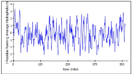
The hydrophobicity is a property related to the solubility of an amino acid in water. Hydrophobic amino acids are localized in the interior of protein, and hydrophilic amino acids interact with the aqueous environment [16]. Figure 1.3 and Figure 1.4 show more hydrophobic regions of MAO isoenzymes.
Figure 1.3: Hydrophobicity graphic of MAO-A enzyme’s amino acids. Figure was drawn via Discovery Studio 3.5 Accelrys.
Figure 1.4: Hydrophobicity graphic of MAO-B enzyme’s amino acids. Figure was drawn via Discovery Studio 3.5 Accelrys.
6
Both isoenzymes hold FAD (flavin adenine dinucleotide) coenzyme in substrate binding site (Figure 1.5) and FAD is bound to Cysteine amino acid (Cys406 of MAO-A and Cys497 of MAO-B) covalently making thioether linkage with 8-α-methyl group of FAD [13, 17].
Figure 1.5: 3-D representation of FAD coenzyme and its interactions with MAO-B. Purple residue balls represent electrostatic interactions; green residue balls represent
van der Waals interactions with FAD coenzyme. Figure generated via Accelrys.
1.4 Structural Properties of MAO Isoenzymes
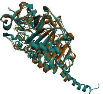
MAO-A works as monomers whereas MAO-B dimers. Relatively minor differences in the architectures of their respective active sites are thought to determine the observed differences in specificities of MAO-A and MAO-B [18]. In order to see structural comparison of three dimensional structures of MAO isoenzymes,
7
arrives to substrate-binding cavity. The volume of the MAO-A active site is ~400 Å3 while the MAO-B active has ~ 700 Å3 volume [18, 19].
Figure 1.6: Superimpose of MAO isoenzymes. Blue structure represents MAO-A and orange structure represents MAO-B enzyme. Superimpose generated via Accelrys.
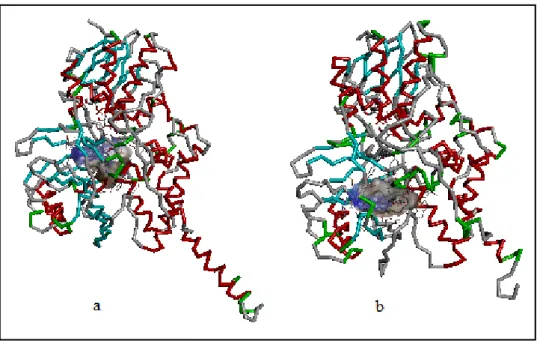
As seen in Figure 1.7, both isoenzymes have hydrophobic binding sites. While the MAO-A active site is a single cavity, the MAO-B active site consists of an entrance cavity (300 Å3) which leads, from the surface of the enzyme, to the substrate cavity (~ 400 Å3
) [18, 3]. Since MAO-A does not have an entrance cavity; substrate reaches directly to one cavity. Specifically their active regions show similar amino acid sequences [13]. The most important difference is in the cavity shaping loop of MAO-A between the residues 210-216. In MAO-B they are located between the residues 201-207 which shows that Cα movement up to 6 Å. Therefore MAO-A has wider aromatic cage than that of MAO-B taking into bulkier aromatic groups [20].
8
Figure 1.7: Hydrophobic binding sites. a) MAO-A , b) MAO-B enzymes. Blue surface represents lower and brown surface represents higher hydrophobicity.
Figure 1.8 shows 3-D shape of MAO-A enzyme and Figure 1.9 shows MAO-B enzyme.
9
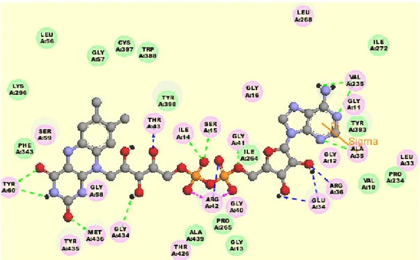
Substrate cavity involves Ile180, Asn181, Phe208 and Ile335 residues in MAO-A and Leu171, Cys172, Ile199, Tyr325 in MAO-B. FAD coenzyme is surrounded by
Tyr407, Tyr444 in MAO-A and Tyr398, Tyr435 in MAO-B [13, 21, 22, 23, 24].
Figure 1.9: MAO-B enzyme 3-D representation
The essential amino acids for ligand in MAO-A are 161st and 375th amino acids, as for MAO-B they are 152nd and 366th amino acids [13, 21, 22, 23, 24].
1.5 Effects of MAO Isoenzymes on Neurotransmitters
The neurotransmitters oxidized by MAO enzymes are dopamine, epinephrine, norepinephrine (NA), 1-methyl-4-phenyl,1,2,3,6-tetrahydropridin (MPTP) and serotonin (5-HT). All these monoamines are responsible for changing myocardial function. Since heart tissues are affected by free radicals in the neural and hormonal system, the decrease of monoamines increases MAO-derived H2O2 production in
10
In the nervous system norepinephrine and serotonin oxidation is catalyzed with MAO-A enzyme, phenyl ethylamine and benzyl amine oxidation catalyzed with B. Dopamine, tyramine and tryptamine are non-selective substrates for MAO-A and MMAO-AO-B [26]. MMAO-AO-MAO-A is primarily responsible for the oxidation of tyramine (Figure 1.10). Therefore MAO-A’s peripheral inhibition has been associated with the risk for an acute hypertensive syndrome known as the ―cheese reaction‖ [27, 28].
Figure 1.10: 3-D representation of neurotransmitters related to MAO enzymes
1.6 Amine Catalysis Reactions of MAO Isoenzymes
FAD (flavin adenine dinucleotide) is a redox cofactor in the reactions catalyzed by MAO (Figure 1.11) [29]. Monoamines are changed into related aldehydes with this reaction. The starting point is the breaking of Cα-H bond. FAD reduced to FADH2
and meanwhile amine is transformed to imine. In MAO reaction at the after step; when hydrogen peroxide is generated, FADH2 is oxidized. Meanwhile imine
11
Figure 1.11: Oxidation of amines by MAO-bound FAD [29]
1.7 Monoamine Oxidase Inhibitors
MAO inhibitors are divided into two types as reversible or irreversible inhibitors. The first produced MAO inhibitors inhibit based on mechanism and they bind to proteins covalently [13]. These irreversible inhibitors have serious side effects like hallucination, schizophrenia and hypertension. Table 1.1 shows general MAO inhibitors’ 2-D structures and their known properties against MAOs.
Table 1.1: MAO inhibitors
Structure of MAO Inhibitors Medical Properties Binding Properties
Moclobemide
Anti-depression
Reversible
Selective MAO-A Inhibitor
12 Selegiline Anti-Parkinson Irreversible Selective MAO-B Inhibitor Pargyline Anti-depression Irreversible Selective MAO-B Inhibitor Rasagiline Anti-depression Irreversible Selective MAO-B Inhibitor Tranylcypromine Anti-depression Irreversible Nonselective MAO-A Inhibitor Clorgyline Anti-depression Irreversible Selective MAO-A Inhibitor Pirlindole Anti-depression Reversible Selective MAO-A Inhibitor Continuation of Table 1.1
13
Irreversible inhibitors have different mechanism than that of reversible inhibitors. Unknown selectivity of MAOIs causes lots of side effects as like hallucination, hypertension and hepatotoxicity [13].
As a new irreversible and selective MAO-B inhibitor Rasagiline, as a selective and reversible MAO-A inhibitor Harmine (Figure 1.12 (b)), as a reversible and selective MAO-B inhibitor Safinamide (Figure 1.12 (a)) [34, 35] are used.
(a) (b)
Figure 1.12: Molecular 3-D structures of inhibitors. (a) Safinamide, (b) Harmine. Turquoise ball represents Fluorine, red ball is Oxygen, navy blue ball is Nitrogen,
14
Chapter 2
Properties of Human Monoamine Oxidase Genes
2.1 Introduction
MAO genes, that have common ancestral gene [3], lay side by side on the X
chromosome. Males are affected directly because of MAO genes’ inheritance condition, if they have mother carrying a MAO gene mutation.
Recent studies on MAO genes deficient mice show that both isoenzymes involve in a large spectrum of mental disorders, some of them are autism, anxiety, impulse-control disorders and ADHD [36].
Recent data shows that MAOA gene is related to 107 disorders and to MAOB gene is related to 59 disorders [37, 38]. When the excess of disorders is pointed out, it can be said that also MAOA and MAOB genes and protein function must be studied comprehensively.
Inheritance pattern of this gene creates an advantage for polymorphism studies on
15 2.2 MAOA Gene
MAOA gene is a member of a MAOs gene family being responsible for production
Monoamine oxidase A enzyme which catalyzed monoamines. A mutation in this gene causes Brunner Syndrome that is characterized by non-dysmorphic mild mental retardation including disturbed regulation of impulsive aggression. Male patients are affected by borderline mental retardation and exhibit abnormal behavior, but female carriers have normal intelligence and behavior [39]. This gene has multiple transcript variants according to splicing process [40].
2.2.1 Positional Properties of MAOA Gene
MAO genes are placed in the p arm of X chromosome (Figure 2.1), so males inherit
only a single maternal copy.
Figure 2.1: MAOA cytogenetic location on chromosome Xp11.3 [41]. Figure was taken from Genetics Human References website.
MAOA gene is located on the short arm of the X chromosome [42] at position p11.3.
16
43,654,906 to base pair 43,746,823 of X chromosome [42].
Figure 2.2: MAOA gene’s exons map [43]. Figure was taken from Genatlas website.
2.2.2 Expression of MAOA Gene
MAOA gene is expressed with over 200 transcription factors [44]. Eight of them are
shown in Figure 2.3 corresponding to their binding sites in this gene promoter.
Figure 2.3: Transcription factors and binding sites in MAOA gene promoter [44]. Figure was taken from Sabiosciences website.
17
This gene has 4 transcripts (Table 2.1) [40]. MAOA-001 is expressed at different levels on different tissues. Expression levels are shown in Figure 2.4.
Table 2.1: MAOA gene mRNA products [40] Length of nucleotide sequence
(bp) Length of protein MAOA-001 4015 527 Protein coding MAOA-201 2383 394 Protein coding MAOA-002 935 - Processed transcript MAOA-003 557 - Processed transcript
Figure 2.4: MAOA estimated expression levels on different tissues and blood [45]. Figure was taken from GeneCards website.
18 2.2.3 Disorders Related to MAOA Gene
MAOs play roles in several psychiatric conditions, including chronic stress, major depressive disorder and alcohol dependence [46]. 10 disorders from 107 disorders related to MAOA according to MalaCards [47] are; social phobia, substance abuse, paranoid schizophrenia, serotonin syndrome, borderline personality disorder, novelty seeking personality, hepatic encephalopathy, anxiety disorder, post-traumatic stress disorder, specific phobia.
Novel roles of MAOA, MAOB and serotonin are regulating of intermediate progenitor cells proliferation in the developing brain [48].
The over expression of protein in female causes panic disorders. The protein under expression causes lower platelet MAOA activity in children by ADHD (Attention-deficit/hyperactivity disorder), inattentive, and hyperactive changes than control children [49].
The MAOA gene was called as ―warrior gene‖ after a study was done with Rhesus macaque monkeys [50]. When Rhesus monkeys had five and six repeat in the MAOA gene’s VNTR upstream, they had 1.3 fold overall activity than 7-repeat or more repeats [50]. On the other hand, different from human MAOA gene, if there are lower repeats in this gene, MAO-A enzyme is expressed at higher levels [50]. In fact that
MAOA gene itself is not responsible for aggression. Therefore it is not a particular
gene for psychiatric disorders had a direct relation with violence propensity [51].
19
was related to transcriptional regulation had a 30 bp repeat polymorphism (MAO-A30bp-rpt) [52].
Looking into MAO-A30bp-rpt allele frequencies, it can be seen vary between ethnic groups worldwide [53].
Most of epidemiological studies on variants of MAO-A30bp-rpt revealed a correlation between MAO-A30bp-rpt and some disorders as like anxiety, depression, and addiction (for example alcoholism). Some studies related to the 3-repeat allele of MAO-A30bp-rpt, alleged without being based on clear data that lower MAO-A activity and higher dopamine levels are related with risk-taking [53, 54]. Hence, Gibbons supposed it as a ―warrior gene‖ in 2004 [55].
2.3 MAOB Gene
2.3.1 Positional Properties of MAOB Gene
MAOB gene is placed in p11.3 of X chromosome, between 43,625,858-43,741,693
nucleotides of reverse strand (Figure 2.5) [56].
Figure 2.5: MAOB gene location on P arm of X chromosome (bands according to Ensembl, locations according to GeneLoc) [56]
20 DNA size: 115.81 Kb
mRNA size: 2537 bp 15 exons (Figure 2.6)
Figure 2.6: MAOB exons map [57]. Figure was taken from Genatlas website.
Human MAOB gene consist of 15 exons, exon 12 codes FAD-binding site of MAO gene as the most conserved region and this region shows 93.9 % similarity in MAOA and MAOB genes. When high similarity is considered, it can be said that these two genes might have a common ancestral gene that had been duplicated afterwards. The high structural and functional similarity of these two MAO enzymes based on their genetic relationship [58].
The promoter region of MAOB gene to base -1,369 from ATG (starting point of mRNA translation) was also sequenced to identify variants with potential functional effects on gene transcription [59]. Recent studies demonstrated that Transforming growth factor –β-inducible early gene (TIEG)2 shows dual functions as a repressor at
CACCC element and as an activator at the distal Sp1 sites of MAOB promoter.
TIEG2 has an effect on MAOB expression and mRNA level [60, 61]
21 2.3.2 Expression of MAOB Gene
Figure 2.7 displays the most relevant transcription factor binding sites in this gene promoter [62].
Figure 2.7: Transcription factors and binding sites in MAOB gene promoter [62] Figure was taken from Sabiosciences website.
This gene has 5 transcripts (Table 2.2) [63]:
Table 2.2: MAOB gene mRNA products
Nucleotide sequence length (bp)
Protein length (a.a)
MAOB-001 2566 520 Protein coding
MAOB-201 1862 504 Protein coding
MAOB-202 1660 411 Protein coding
MAOB-002 857 - Processed transcript
22
MAOB expression levels differ tissue by tissue. PARK2 suppresses the transcription
of MAOA and MAOB to control oxidative stress induced by dopamine oxidation [64]. In the adrenal gland, cranial nerve, uterus and platelet it is expressed highly for human. Expression levels in other tissues and blood is shown on Figure 2.8.
Figure 2.8: MAOB estimated expression levels on different tissues and blood [56]. Figure was taken from GeneCards website
Three types (α, β, γ,) Estrogen-related receptors (ERR) increased the transcription of
MAOs A and B, the effects were abolished by parkin, but not by its PD-linked
mutants [64].
Mutagenesis studies show that on the 156th amino acid C/S change [65], 345th C/S change [65], 389th C/A change [66, 65], 397th C/S change [65] cause complete loss of activity of protein product of MAOB gene. On 158th amino acid T/A change causes
23
dramatic loss of activity [66], on 382nd H/R change causes significant loss of activity [66].
2.3.3 MAOB Gene Related Disorders
In line with hypothesis of lower platelet MAO activity in different types of psychopathology, children with different subtypes of ADHD (Attention-deficit/hyperactivity disorder) had significantly lower platelet MAO-B activity than control children [49].
The increase of MAO-B protein may be accepted as a signal for Parkinson disease and also MAO-B platelet protein level may serve as a biomarker for age-related dementia, especially AD [67]. Although it has been reported that over MAOB mRNA and enzymatic activity in platelet related to Alzheimer’s and Parkinson’s diseases, it is not clear what is the cause of enhanced MAOB mRNA level causing these diseases in the brain [68, 59].
24
Chapter 3
Coumarin Derivatives
3.1 The Potential Behind of Vanilla Smell
If it could be gone into deeps of luscious vanilla’s secretion around the seed, it would be encountered with molecule at Figure 3.1. The gallant of the odoriferous is one of the main characters of our thesis at the same time. Actually, Guibourt had been encountered with this molecule by using his chemist abilities in 1820. The name derives from ―cumaru‖, an Amazonian dialect name for the Tonka been [69] which had been first isolated plant.
25
Coumarins comprise of a large family compounds. Some of which have natural and synthetic origin may have several pharmacologic activities. MAO inhibition modifies coumarins too. Coumarin can be obtained from plants and used in medicinal chemistry [70]. The usage of coumarin in the medical reach areas dates back many years. The reason why they are used based on the fact that they have lots of derivatives. Approximately; 1300 coumarin derivatives are obtained from plants, bacteria and fungi [71].
3.2 Chemical and Structural Properties of Coumarin
Coumarin with other name 2-H-Chromen-2-one is a white lactone consisting of a benzene ring and α-pyrone ring (Figure 3.2). The coumarin nucleus (benzo-2-pyrone) is derived from cinnamic acid (phenyl acrylic skeleton) via bio-synthesis. The hydroxyl group at 7th position of coumarin nucleus has an important place in biosynthesis. Since coumarin family has variable structures, their structural variations can change their biological activity [72].
26 Molecular weight: 146
Crystal structure: Orthorhombic
Solubility: Very soluble in alcohol and ether, not soluble in water Organoleptic Properties: Vanilla smell
Melting Point: 69 0C, Boiling Point: 290 0C [72]
The special nice smell just like newly-mown hay of coumarin is one of the main reasons for the industrial synthesis [73]. On the other hand, coumarins possess a number of biological activities like anticoagulant, antimicrobial, anti-inflammatory, analgesic, antioxidant, anticancer, antiviral, antimalarial [74], anti-osteoporosis, antiseptic, anti-HIV, anti-hypertension, anti-arrhythmia, antifungal [75], antinociceptive [76]. Coumarin itself and 7-hydroxycoumarin have been reported to inhibit the proliferation of a number of human malignant cell lines in vitro and have demonstrated activity against several types of animal tumors [77]. Although coumarin is a secondary phytochemical with hepatotoxic and carcinogenic properties, according to clinical data related to hepatotoxicity effect of coumarin, this compound is rather useful for using as a medicine [78].
3.3 Toxic Effects of Coumarin
It should be noted that coumarin is moderately toxic with a LD50 of 275 mg/kg [73].Using the human data, a tolerable daily intake (TDI) of 0.1 mg/kg body weight was derived, confirming that of European Food Safety Authority. Nutritional exposure may be considerably, and is mainly due to use of cassia cinnamon, which is a popular spice [78].
27
3.4 Founding of Coumarins in Natural Products and Artificial Synthesis
Being fragrant chemical compound, it can be found in natural products, such as in vanilla grass (Anthoxanthum odoratum), sweet woodruff (Galium odoratum), sweet clover (Meliotus L.), sweet grass (Hierochloe odorata) [69], tonka been tree (Dipteryx odorata), cassia cinnamon (Cinnamomum aromaticum), fraxinus bark (Cortex fraxini) [79].
On the other hand, the most accessible route to coumarin is the one originally presented by Sir William Perkin. Coumarin can be obtained with following reaction as shown Figure 3.3,
1) Phenol is converted to salicylaldehyde with Reimer-Tiemann reaction.
2) Then salicylaldehyde is exposed to Perkin reaction with acetic acid and sodium acetate to occur an unsaturated acid.
3) In result of intramolecular esterification of the last product coumarin is formed [73].
28
Another coumarin synthesis pathway (Figure 3.4) is Penchmann Coumarin Synthesis [80]:
Figure3.4:Penchmann coumarin synthesis [80]
For a direct synthesis of various coumarins, the following palladium-catalyzed oxidative cyclocarbonylation reaction is useful (Figure 3.5).
Figure 3.5: Palladium-catalyzed oxidative cyclocarbonylation reaction of 2-vinylphenols [81]
This reaction begins with 2-vinylphenols for the synthesis of various coumarins directly in good yields in the presence of low pressures of CO, and air or 1,4-benzoquinone as the oxidant. The reaction conditions are attractive in terms of environmental considerations and operational simplicity [81].
29 3.5 Classification of Coumarin Derivatives
Coumarin family has numerous kinds of structures, since their basic structures and lots of types substitutions allows this kindness. The most common coumarin derivatives in nature are Umbelliferone (7-hydroxycoumarin), Esculetin (6,7-Dihidroxycoumarin) and Scopoletin (7-hydroxy-6-methoxycoumarin) [72]. Table 3.1 shows most often common coumarin derivatives in nature with specific names. Figure 3.6 shows coumarin scaffold’s 4th, 6th and 7th positions which can have side groups shown with Table 3.1.
Figure 3.6:Scaffold for natural coumarin derivatives
Table 3.1: Common natural coumarin derivatives [72]
Type of derivative R1 R2 R3 Coumarin H H H Herniarin H H OCH3 Esculetin H OH OH Umbelliferone H H OH Scopoletin H OCH3 OH Methyl-umbelliferone CH3 H OH
30
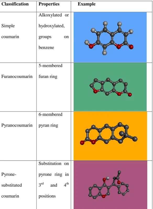
Also four main coumarin subtypes as can be seen in Table 3.2 are the simple coumarins, furanocoumarins, pyranocoumarins and the pyrone-substituted coumarins [82]. Their 3-D structures are drawn by Discovery Studio Accelrys program.
Table 3.2: Examples of coumarin sub-types. Grey ball represents Carbon, red ball represents Oxygen, white ball represents Hydrogen.
Classification Properties Example
Simple coumarin Alkoxylated or hydroxylated, groups on benzene Furanocoumarin 5-membered furan ring Pyranocoumarin 6-membered pyran ring Pyrone-substituted coumarin Substitution on pyrone ring in 3rd and 4th positions
31
3.6 Effects of Coumarin Derivatives on MAO Enzymes
The interpretation of the chemical binding structure of models allows derivative some chemical features, which can be considered important in the hMAO-B selectivity. For instance, the furan ring or the =CRX fragment (R: any group linked though carbon; X: electronegative atom and =: double bonds) in the ligand structure decreases the selectivity [27].
Compounds that have acetonyl/bromoallyloxy groups in the 7th position of coumarin have good inhibitory activity against MAO-A and MAO-B (Figure 3.7). Bulky groups such as cyclohexyl or phenyl in the 3,4-positions of the 7-acetonyl coumarin derivatives increase inhibitory activities of them with both MAO-A and MAO-B but decrease the selectivity. On the other hand replacement of the acetonyl substituent at the 7-position with the bromoallyloxy group shows very high MAO-B inhibitory activity (IC50 of about 1.2 nM and 1.5 nM) and MAO-B selectivity (nearly 100-fold
and 1600-fold) with respect to MAO-A isoform [7].
Figure 3.7: 3-D structure of 3,4-Benzo-7-(6-bromoallyloxy)-8-methylcoumarin
Lipophilicity is an important property for MAO-B inhibition potency of 7-substituent coumarins, correlation of pIC50 values with log P values demonstrates it [83].
32
It is unclear why the inhibitory potency against MAO-B is affected by the length of the side chain [84]. Coumarin analogs interact with non-covalent bonds to human MAO-B complexes [85].
When an anaryl group is in the 3rd position in coumarin nucleus, substituent which is in the 7th position does not increase the potency against MAO-B inhibition. Actually the important thing in terms of QSAR approach is the moiety which is in the pyrone ring for a 7-substituted coumarin derivative by the time antiparkinson activity of coumarin is considered. [10].
The resveratrol-coumarin hybrid compounds ((a), (b), (c) in Figure 3.8), shows high selectivity for the MAO-B isoenzyme and inhibitory activity in the nano to picomolar range [8].
Figure 3.8: 3-D representation of resveratrol-coumarin hybrid compounds.
In addition, Santana et al. also found that coumarins with electronegative groups substituted at the position 3 of γ-pyrone nucleus decrease the hMAO-B selectivity [27, 9].
According to recent study of Abdelhafez (2013) the main atoms and groups of the 7-Oxycoumarin derivatives (Figure 3.9) interacts with hydrogen bonds to MAO-A
33
active site. In the result of this study, correlation in coumarin derivatives and % inhibition ratio of MAO-A was found at pM levels and of MAO-B at µM levels with AutoDock binding affinities. 7-Oxycoumarin derivatives (4-methyl and/or 3,4-dimethylumbelliferone with acyclic acetohydrazide moiety) in vitro shows % inhibition against MAO-A of 5.01 pM and in vivo ED 50 % of 0,009143 µM. This compound was tightly bound into MAO-A through four hydrogen bonds via its 2-C=O of pyrone and 2-C=O of pyrrolidine or pyrazolidine. The interacted amino acids of MAO-A receptor are NH of Asn181, OH of Tyr444 and with a lower extent NH of Gln215 and C=O of Ala111 [6].
Figure 3.9: 7-Oxycoumarin derivative a) 3-D image (4-methyl and/or 3,4-dimethylumbelliferone with acyclic acetohydrazide moiety) b) Its 2-D image (drawn
with Accelrys)
3.7 Other Medical Effects of Coumarin Derivatives
3.7.1 Antioxidant Activity
The free radical scavenging activities of coumarin derivatives are related to the number and position of the hydroxyl group on the benzenoid ring of the coumarin. In
34
hydroxylated coumarin, the substituent at C-2, C-4, C-7 positions is reported to play a key role in enhancing the activity [86, 87]. To show antioxidant activity, a coumarin derivative has to posses at least one hydroxyl group [88].
3.7.2 Anti-inflammatory Effects of Coumarins
The compound (3-chloro-7-methyl-9H-pyrano [2,3-e] benzo-1,4-oxazine-2,9-dione) in the Figure 3.10 shows maximum anti-inflammatory effect on time dependant study and this may due to the presence of chlorine at 3rd positions, methyl at 7th positions on the aromatic ring of the coumarin respectively [74].
Figure 3.10: 3-D structure of an anti-inflammatory coumarin derivative
3.7.3 Roles of Coumarin in Treatment of High Protein Edema
Coumarin and numerous other benzopyrones have been tested in high protein edema, and all have been shown to successfully reduce the swelling [69]. Coumarin (particularly Esculetin) is ideal natural reducer of edema in legs. If coumarins are used with compression stacking, they give positive results in reducing swelling in legs. Therefore in case venous are insufficient, coumarin cure is recommended [89].
35 3.7.4 Anti-tumor Activity
Specifically it is expected to increase biological activity in case fluorine and sulfonamide groups are inserted at the coumarin nucleus, because they have known antibacterial and antitumor activity when fluorine and heterocyclic groups are together in coumarin derivative [90].
3.7.5 Coumarin in Leukemia
8-nitro-7-hydrocoumarin displays cytotoxic properties, inducing cell death by apoptosis. Overall esculetin exhibits the strongest antiproliferative effect on the carcinoma cell lines tested. Esculetin (6,7-Dihydroxycoumarin) and
7-hydroxycoumarin inhibits tyrosine phosphorylation in EGF-stimulated tumour cells in a time – and dose- dependent manner [82].
3.7.6 Chromone and Coumestan Derivatives
Chromone and coumestan scaffolds are structurally similar to coumarin. In this concept when the properties of chromone and coumestan derivatives were considered C6-substituted chromones are more potent MAO-B inhibitors than the C7-substituted chromones, because there is a potential hydrogen bond among these with Tyr398 which is anticipated to occur with the C6-substituted chromones, but not the C7-substituted chromones [18]. As for coumestan derivatives, in these derivatives, methoxy and allyloxy coumestan derivatives are more active than methylenedioxy and ethylenedioxy coumestan derivatives. Methoxy derivatives showed better activity than allyloxy derivatives [91].
36
Chapter 4
Material and Methods
4.1 Introduction
The first step was to obtain available coordinate files of ligands and protein molecules. The second step was to perform grid calculations for every ligand molecule. At the third step, protein and ligand was docked.
During this study various computational molecular modeling tools were used. The drawing was done with Discovery Studio Accelrys software. Docking was performed with AutoDock 4.2. The MAO-A and MAO-B proteins had been taken from PDB (Protein Data Bank). The results of docking were read from *.dlg files. The best 50 results of MAO-A, the best 50 results of MAO-B, the best 25 results for correlation of Ki values between MAO-A and MAO-B according to selectiveness properties being corresponding differences of affinities, were shown. At the light of these results, the best five results for each one were illustrated to cast light on the interactions between the ligand and the binding site of MAO protein cavity.
37 4.2 Preparation of Ligands
Our aim is to perform 125 different ligand structures using 5 different side groups on coumarin scaffold. Previous studies [76, 92, 10, 8] shows that 3rd, 4th and 7th positions on coumarin are specifically important for MAO inhibition potency. Hence in this study 3rd, 5th and 7th positions were selected to add side groups to observe changes in the activity (Figure 4.1).
The selected five side groups are;
Methoxy (-OCH3), Fluorine (-F), Bromine (-Br), Amide (-C(O)NH2) and Phenyl (-C6H5).
Especially Fluorine and bulky groups affect the potency, additionally hydroxy group in 7th position increase the affinity to MAO-B enzyme. Previous studies demonstrate the Bromine, amide and phenyl have the most potent inhibitory activity against MAO-B enzyme [7].
Figure 4.1: Coumarin Scaffold
The drawing was performed first on the paper. These 2D pictures were drawn in 3D using Discovery Studio 3.5. After performing 3D structure of the ligand on a new
38
window, 2D image was taken applying ―Show 2D Structure‖ by changing ―Display Style‖ at the end of adjusting 2D images were saved as *.png format and 3D files were saved as *.mol2 and *.pdb formats.
These 125 *.pdb files firstly were tried to minimize with pdb2pqr web tool but files could not be transformed pqr format. Hence another web based minimizer were tried, but also YASARA’s transformed files were not reliable for pdb format, at the last, ―clean geometry‖ tool of Discovery Studio was used to optimize these 125 ligands’ 3D shapes. At the end of the optimization, these files were saved as both pdb and mol2 formats. Because *.mol2 files could not be opened in AutoDock4.2 on Windows7, *.pdb files were used to obtain *.pdbqt files for each ligands. At the stage of drawing, all ligands were drawn together all hydrogen atoms in Discovery Studio. The redundant hydrogen atom on the phenyl ring being side group that had been added by program was removed meticulously by rotating the molecule. Pdbqt files are necessary file formats for AutoDock 4.2. Because the polar hydrogens and partial charges are added to molecule and AutoDock 4 atom types are in the pdbqt file.
4.3 Preparation of Proteins
Pdbqt files of MAO-A and MAO-B were obtained directly from *.pdb files that had been prepared at previous study [13]. MAO isoenzymes’ cocrystalized structures had been reached from PDB (Protein Data Bank); human MAO-A (2Z5X) with harmine (resolution 2.2 Å) and human MAO-B (2Z5V) with safinamide (resolution 1,6 Å). These pdb files had been opened with Discovery Studio 3.5 and first, ligand (harmine
39
for MAO-A and safinamide for MAO-B) had been cleaned. Lost residues had been completed, then atoms which are in the active site had been minimized. Waters which are around the protein had been removed. pH value had been adjusted 7.2. At the last step *.pdb files had been converted to *.pdbqt files using AutoDock 4.2
4.4 Docking Study of Coumarin Derivatives with MAO-A and MAO-B
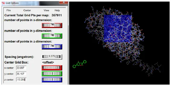
The prepared *.pdbqt files for coumarin derivatives and MAO-A and MAO-B were used to perform *.gpf files for each docking.
Grid option was used to perform *.gpf (Grid Parameter File) files. For Grid Calculation, pdbqt file which belong to MAO-A was selected as ―Macromolecule‖. Ligand’s pdbqt file was opened as ―Set Map Types‖. In the grid box, ―spacing longs‖ was taken as 0.375, grid box size was kept 70x70x70. X, Y, Z values in the ―center grid box‖ is the N5 atom of the FAD (Figure 4.2).
Figure 4.2: Atoms’ positions in isoalloxazine ring of FAD [93].
Therefore firstly MAO-A *.pdb file was viewed in the VMD. Molecule was imaged as ball and stick format, after confirming the FAD and its N5 atom in the MAO-A,
40
atom names were shown by using VMD tools and it was clear the N5 atom’s name is FAD 600:NY1. Then MAO-A pdb file was opened with WordPad and in the Heteroatoms NY1 of FAD was found. Its coordinates are: 33.897 35.157 -13.250 Also MAO-B pdb file was opened, NY1’s coordinates for MAO-B are: 55.724 151.605 21.259. Before closing this grid box window ―close saving current‖ was clicked in the ―File‖ option (Figure 4.3).
Figure 4.3: Grid box options to adjust grid parameter. AutoDock4.2 grid box (left), while performing (right).
In the output it was saved with each ligand’s name as *.gpf file. After 125 ligand’s grid calculations were done with MAO-A, also it was done with MAO-B but at this time center grid box values were changed according to MAO-B NY1 of FAD’s X, Y, Z coordinates.
Map files’ names which will be performed are written in the *.gpf file. Numbers of map files are equal to four more of number of atom types of ligand. *.glg file and map files are performed by autogrid4 writing on the terminal window;
41 …/autogrid4 –p M001.gpf –l M001.glg
A sample gpf file is below.
npts 70 70 70 # num.grid points in xyz gridfld maoA.maps.fld # grid_data_file spacing 0.375 # spacing(A)
receptor_types A C HD N NA OA P SA # receptor atom types ligand_types A C HD OA N # ligand atom types
receptor maoA.pdbqt # macromolecule
gridcenter 33.897 35.157 -13.25 # xyz-coordinates or auto smooth 0.5 # store minimum energy w/in rad(A) map maoA.A.map # atom-specific affinity map map maoA.C.map # atom-specific affinity map map maoA.HD.map # atom-specific affinity map map maoA.OA.map # atom-specific affinity map map maoA.N.map # atom-specific affinity map elecmap maoA.e.map # electrostatic potential map dsolvmap maoA.d.map # desolvation potential map
dielectric -0.1465 # <0, AD4 distance-dep.diel;>0, constant
After two minutes, necessary map files and *.glg file are arisen. During these operations, apparent directories were formed for each ligands. At the beginning in each directory only one ligand’s pdbqt, one protein pdbqt and related *.gpf file must exist. Otherwise, if it was more than one *.gpf files in the same directory, it would be
42
fault in performing new map files as from second ligand. Although all *.glg files are obtained, docking results are different when they were compared with apparent directories.
After performing *.gpf files, *.dpf files which are main docking parameter files were prepared by using AutoDock 4.2 docking algorithm. For this purpose, number of runs is taken as 10, number of generation = 27.000, number of evolutions = 5.000.000 since torsion numbers of ligands are smaller than 10.
Each *.dpf file that is belong to separate ligand was saved in the directory that is belong to one ligand and contain its *.gpf file additionally its ligand *.pdbqt and macromolecule *.pdbqt files.
A sample *.dpf file is below.
AutoDock_parameter_version 4.2 # used by AutoDock to validate parameter set outlev 1 # diagnostic output level
intelec # calculate internal electrostatics seed pid time # seeds for random generator ligand_types A C HD OA N # atoms types in ligand fld maoB.maps.fld # grid_data_file
map maoB.A.map # atom-specific affinity map map maoB.C.map # atom-specific affinity map map maoB.HD.map # atom-specific affinity map map maoB.OA.map # atom-specific affinity map
43
map maoB.N.map # atom-specific affinity map elecmap maoB.e.map # electrostatics map
desolvmap maoB.d.map # desolvation map move m118.pdbqt # small molecule
about -2.449 2.6955 0.1781 # small molecule center tran0 random # initial coordinates/A or random quaternion0 random # initial orientation
dihe0 random # initial dihedrals (relative) or random torsdof 3 # torsional degrees of freedom
rmstol 2.0 # cluster_tolerance/A extnrg 1000.0 # external grid energy
e0max 0.0 10000 # max initial energy; max number of retries ga_pop_size 150 # number of individuals in population
ga_num_evals 5000000 # maximum number of energy evaluations ga_num_generations 27000 # maximum number of generations
ga_elitism 1 # number of top individuals to survive to next generation ga_mutation_rate 0.02 # rate of gene mutation
ga_crossover_rate 0.8 # rate of crossover ga_window_size 10 #
ga_cauchy_alpha 0.0 # Alpha parameter of Cauchy distribution ga_cauchy_beta 1.0 # Beta parameter Cauchy distribution set_ga # set the above parameters for GA or LGA sw_max_its 300 # iterations of Solis & Wets local search sw_max_succ 4 # consecutive successes before changing rho sw_max_fail 4 # consecutive failures before changing rho
44
sw_rho 1.0 # size of local search space to sample sw_lb_rho 0.01 # lower bound on rho
ls_search_freq 0.06 # probability of performing local search on individual set_psw1 # set the above pseudo-Solis & Wets parameters
unbound_model bound # state of unbound ligand ga_run 10 # do this many hybrid GA-LS runs analysis # perform a ranked cluster analysis
Lamarkian genetic algorithm was used to perform maximum number of energy evaluation and maximum number of generations for each docking parameter files in order to calculate energy values for each position of ligand in the protein.
After obtaining *.glg files and map files, in the same directories *.dlg (docking log file) files are performing utilizing map files and *.dpf file.
In the terminal window of Ubuntu, after entering the related directory that contain a *.dpf file, *.dlg file was created by writing following command:
.../autodock4 –p M001.dpf –l M001.dlg
In this command M001 is any ligand and M001.dpf is its prepared dpf file.
In the individual computer and on the Ubuntu, this process takes ten-fifteen minutes for one docking. In this study 250 dockings were arisen. Then results were read from *.dlg files by looking to the lowest energy value.
45
At the beginning of selection available grid size, in order to decide true grid measures, randomly selected five ligands had been docked with both MAO-A and MAO-B enzymes with a view to decide available grid box sizes. Hence during the grid calculation of these five ligands the grid box size being taken 60 X 60 X 60. When the docking results were compared, the results were seen that they had got better results which had been performed with 70 X 70 X 70 grid box. Other 120 coumarin derivatives were docked by taking account of this situation.
In order to examine binding properties of coumarin derivatives with MAO-A and MAO-B enzymes, the coordinates found in the *.dlg file belonging to the lowest energy values obtained from AutoDock 4.2 docking process were added to end of related MAO enzyme *.pdbqt file. Then this file was opened with Discovery Studio 3.5 Accelrys program and all interactions were observed via tools of this program.
Particularly occurred pi-pi, pi-sigma, pi-caution interactions and polar interactions were appeared, and van der Waals forces could be observed corresponding their grandeurs. Additionally distance of pi-pi interactions and polar interactions were predicted via Accelrys program.
Table 4.1 shows 125 coumarin derivatives’ 2-D structures drawn by Discovery Studio Accelrys program depending on three dimensional structures occurred on this software.
46 M001 M006 M002 M007 M003 M008 M004 M009 M005 M010
47 M011 M016 M012 M017 M013 M018 M014 M019 M015 M020 Continuation of Table 4.1
48 M021 M026 M022 M027 M023 M028 M024 M029 M025 M030 Continuation of Table 4.1
49 M031 M036 M032 M037 M033 M038 M034 M039 M035 M040 Continuation of Table 4.1
50 M041 M046 M042 M047 M043 M048 M044 M049 M045 M050 Continuation of Table 4.1
51 M051 M056 M052 M057 M053 M058 M054 M059 M055 M060 Continuation of Table 4.1
52 M061 M066 M062 M067 M063 M068 M064 M069 M065 M070 Continuation of Table 4.1
53 M071 M076 M072 M077 M073 M078 M074 M079 M075 M080 Continuation of Table 4.1
54 M081 M086 M082 M087 M083 M088 M084 M089 M085 M090 Continuation of Table 4.1
55 M091 M096 M092 M097 M093 M098 M094 M099 M095 M100 Continuation of Table 4.1
56 M101 M106 M102 M107 M103 M108 M104 M109 M105 M110 Continuation of Table 4.1
57 M111 M116 M112 M117 M113 M118 M114 M119 M115 M120 Continuation of Table 4.1
58 Continuation of Table 4.1 M121 M122 M123 M124 M125
59 4.5 Molecular Modeling
Molecular Modeling is the improved models simulated the behavior of the molecules by the aid of theoretical methods and computational applications. Molecular Modeling is used in the area of computational biology, drug design and computational chemistry to study the behavior of small molecules and complex biological systems and materials. Computers have an important place in performing Molecular Modeling of any comprehensible measured system. This technique describes the molecules at atomic level; meanwhile it deals with electrons as the smallest unit [94].
4.6 Molecular Mechanics
Molecular Mechanics is based on Newtonian mechanics to explain molecule’s physical condition. The interactions between neighboring atoms are explained with chemical bonds (like covalent, ionic, hydrogen bonds) or non-bonded interactions like van der Waals forces. The Lenard-Jones potential is similar to van der Waals forces. The electrostatic interactions are found by Coulomb’s law. Atoms are placed specific internal coordinates, atomic velocities are determined in dynamical simulations, related to temperature of the system. Potential function related to the system internal energy is sum of potential and kinetic energies. The minimization of potential energy is to become the most natural condition of the system’s behavior [94]. Performing of molecular potential energy is shown in Equation-3.1
![Figure 2.2: MAOA gene’s exons map [43]. Figure was taken from Genatlas website.](https://thumb-eu.123doks.com/thumbv2/9libnet/4330121.71236/33.892.179.789.197.407/figure-maoa-gene-exons-figure-taken-genatlas-website.webp)
![Figure 2.6: MAOB exons map [57]. Figure was taken from Genatlas website.](https://thumb-eu.123doks.com/thumbv2/9libnet/4330121.71236/37.892.176.787.276.478/figure-maob-exons-map-figure-taken-genatlas-website.webp)
![Figure 2.7 displays the most relevant transcription factor binding sites in this gene promoter [62]](https://thumb-eu.123doks.com/thumbv2/9libnet/4330121.71236/38.892.179.791.309.536/figure-displays-relevant-transcription-factor-binding-sites-promoter.webp)
![Figure 2.8: MAOB estimated expression levels on different tissues and blood [56]. Figure was taken from GeneCards website](https://thumb-eu.123doks.com/thumbv2/9libnet/4330121.71236/39.892.196.779.272.727/figure-estimated-expression-different-tissues-figure-genecards-website.webp)