https://doi.org/10.1177/2515841420947544 https://doi.org/10.1177/2515841420947544 Ther Adv Ophthalmol
2020, Vol. 12: 1–6 DOI: 10.1177/ 2515841420947544 © The Author(s), 2020. Article reuse guidelines: sagepub.com/journals-permissions
Therapeutic Advances in Ophthalmology
journals.sagepub.com/home/oed 1
Introduction
Nowadays, intravitreal injection is frequently used in the treatment of many ophthalmic dis-eases. The increase in the number of patients undergoing this treatment, expectations for good visual outcomes, and possible induction of cor-neal astigmatism may have become clinically important.
The intravitreal dexamethasone implant (IDI) (Ozurdex, Allergan Inc., Irvine, CA, USA) has been used widely for a variety of conditions, including diabetic macular edema (DME), reti-nal vein occlusion (RVO), and non-infectious posterior uveitis among others. This injectable implant represents an important innovation in the management of chronic ocular disease using a sustained-delivery technology. Ozurdex® is an
intravitreal implant containing 700 μg preserva-tive-free dexamethasone in a slow-release drug delivery system. It is a small rod-shaped implant with dimensions of 0.46 mm diameter and 6 mm length. The OZURDEX® applicator needle is 22
gauge (G) and features a coating designed to facilitate needle gliding through the sclera and into the posterior chamber.1,2
Several studies have investigated the effects of the dexamethasone implant on corneal endothe-lium.3–5 However, there are no studies in which the
effects of the dexamethasone implant on anterior segment parameters have been investigated. We speculated that such posterior segmental interven-tions would have an effect on the anterior segment, which could lead to a direct effect on visual out-come. In this study, we aimed to evaluate corneal
The corneal effects of intravitreal
dexamethasone implantation
Alper Halil Bayat , Gamze Karataş, Muhammet Mustafa Kurt and Mustafa Nuri Elçioğlu
Abstract
Objectives: To evaluate the corneal effects of the intravitreal dexamethasone implantation using corneal topography and specular microscopy.
Material and methods: 27 eyes of the 27 patients who received a single intravitreal
dexamethasone implantation dose for diabetic macular edema were enrolled in this study. Sirius topography and EM-3000 specular microscopic examinations were performed at the initial examination (baseline), and then on the first day, during the first week, and 1 month after IDI. Changes in corneal parameters were investigated.
Results: The mean age was 58.66 ± 6.59 years. 15 patients were men, and 12 were women. The mean disease duration was 12.2 ± 2.4 months, and mean glycosylated hemoglobin (HbA1c) was 7.2 ± 1.1. After dexamethasone injection, the mean central corneal thickness, endothelial cell density, and coefficient variation of cell area presented a statistically significant decrease (p < 0.05). Anterior segment parameters, such as anterior chamber depth, iridocorneal angle, sim K1 and K2 keratometry, pupillary diameter, horizontal visible iris diameter, and corneal astigmatism did not change (p > 0.05).
Conclusion: Intravitreal dexamethasone implantation affects corneal endothelial cell structure but does not appear to affect corneal topography parameters.
Keywords: cornea, intravitreal dexamethasone implantation, topography
Received: 12 May 2020; revised manuscript accepted: 13 July 2020.
Correspondence to:
Alper Halil Bayat Department of Ophthalmology, Esenler Hospital, Medipol University, APT: 5 Bahceler, AVE Esenler, Istanbul 34250, Turkey. Department of
Ophthalmology, Alanya Life Hospital, Antalya, Turkey
Gamze Karataş Mustafa Nuri Elçioğlu
Department of Ophthalmology, Okmeydanı Research &Traning Hospital, University of Health Sciences, İstanbul, Turkey
Muhammet Mustafa Kurt
Department of Ophthalmology, Samsun Gazi State Hospital, Samsun, Turkey
topographic changes and post-injection astigma-tism after the IDI injection using the Sirius– Scheimpflug and Placido imaging system. This method is reproducible and measures almost all anterior segment parameters in patients with mac-ular edema secondary to DME.
Methods
In this clinical study, 27 eyes of 27 patients who underwent IDI treatment due to DME were included in this study. All of the study patients were pseudophakic in order to eliminate the effects of cataracts/cataract surgery.
This study was conducted in accordance with the Declaration of Helsinki. All necessary authoriza-tions were obtained from the Institutional Review Board of Okmeydanı Research and Training Hospital, İstanbul, Turkey, with number 894 at 08/05/2018. Written informed consent was obtained from all of the participating patients. All participants underwent a total ophthalmic exami-nation. Best-corrected visual acuity, slit lamp
examination, intraocular pressure (IOP)
measurements with pneumotonometer, fundus examination, refraction measurements with an auto-keratorefractometer were performed. Patients <18 years, contact lens users and those with previ-ous ocular trauma, history of ocular surgery, ocular inflammation, corneal diseases, cataract, endothe-lial cell count <1000/mm2, and/or Fuchs
endothe-lial dystrophy were excluded from the study. Measurements of endothelial morphology, such as endothelial cell density (ECD) and coefficient of variation (CV) of cell area, were obtained by non-contact specular microscopy using an EM-3000 Specular Microscope (CBD/Tomey, Phoenix, AZ, USA). Anterior segment parameters, such as cen-tral corneal thickness (CCT), anterior chamber depth (ACD), iridocorneal angle (ICA), sim K1 and K2, pupillary diameter (PD), horizontal visi-ble iris diameter (HVID), and corneal astigmatism were examined by Sirius Scheimpflug topography (Costruzione Strumenti Oftalmici, Florence,
Italy). Sirius topography and specular microscopy examinations were performed at the initial exami-nation, on the first day, during the first week, and 1 month after dexamethasone treatment.
Before dexamethasone injection, the eye was anesthetized with proparacaine hydrochloride (Alcaine 0.5%, Alcon Pharmaceuticals, Couver, Belgium), and the eyelids and ocular surface were disinfected with 5% povidone iodine. The IDI was then delivered through a 22-G needle with a preloaded dexamethasone implant applicator (Ozurdex) and then inserted into the vitreous cavity through the pars plana (4 mm behind the limbus).
Statistical analyses were performed using SPSS for Windows (Statistical Package for Social Sciences 21.0, SPSS Inc, Chicago, IL). General linear models with repeated-measures analysis of variance (ANOVA) were used to analyze differ-ences in specular microscopy and corneal topog-raphy parameters. For all analyses, p < 0.05 was considered statistically significant.
Results
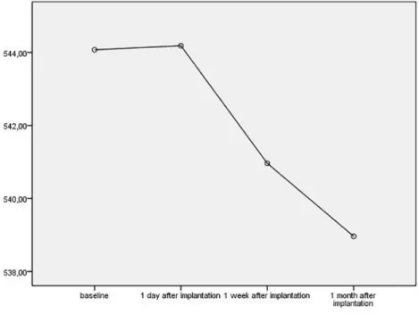
27 eyes of 27 patients were included in the study. The mean age was 58.66 ± 6.59. 15 patients were men, and 12 were women. The mean duration of the diabetic retinopathy was 12.2 ± 2.4 months, and mean HbA1c was 7.2 ± 1.1. Baseline charac-teristics of the patients are shown in Table 1. After IDI, the mean CCT, ECD, and CV showed a statistically significant decrease (p < 0.05; Figures 1 and 2) while the other parameters did not (p > 0.05). These parameters are shown in Table 2.
However, for six patients (19.3%), IOP increased to >24 mmHg. They were all successfully treated with antiglaucoma drops. IOPs did not rise above 28 mmHg, and no patients exhibited corneal edema. No other complications, such as intraop-erative lens injuries, endophthalmitis, retinal detachment, and/or migration of the implant to the anterior chamber, were observed.
Discussion
Diabetic retinopathy is the most frequent retinal vascular disease.6 Several options are available for
DME treatment. These include focal/grid laser, photocoagulation, subthreshold micropulse laser
Table 1. The Baseline Characteristics of the Patients.
Mean age 58.66 ± 6.59
Gender (women/man) 12/15
Mean HbA1c 7.2 ± 1.1
photocoagulation, intravitreal corticosteroids, intravitreal anti-vascular endothelial growth factor (VEGF), and pars plana vitrectomy (PPV).7 In
many studies, the dexamethasone implant was found to be effective in the treatment of DME.8
Many studies have shown the effects of intravitreal anti-VEGF on the cornea.9–11 We wanted to
examine the effects of IDI, one of the effective treatments for DME, on the cornea.
In previous studies, 29.7% of the implanted patients had IOPs >25 mmHg, 66% had cata-ract-related side effects, and most of them required cataract surgery in addition to
Figure 1. The changes in CCT.
presenting vitreous hemorrhage (10%), and injection-related side effects (<2%).12 In our
study, we took pseudophakic patients in order to avoid cataract progression. IOPs were controlled with antiglaucomatous treatment in cases of increased IOP, and none of patients needed glau-coma surgery. Six of the patients had increased IOP. Although, compared to other patients, the patients with increased IOP did not have any additional decrease in ECD, but we did not know whether increased IOP had effects on ECD. There was no significant difference in CCT in previous studies after IDI.3 However, in our
study, we found that CCT decreased signifi-cantly one after injection (p = 0.011). We know that the corneal endothelium contains glucocor-ticoid receptors based on results from previous studies, and it was shown in an in vitro study that IDI can induce high levels of apoptosis. We think that the decrease in CCT may have occurred as a result of steroid-triggering apoptosis.4 Even if we
had found a statistically significant decrease, this decrease will not be reflected in a clinical setting because it is only 6 µm on average. Decreases in CCT which are not clinically significant do not suggest that dexamethasone is unsuitable.
In the literature, there are studies showing that intracameral dexamethasone applied after cataract
surgery and after penetrating keratoplasty is not toxic to the corneal endothelium.5,13 However, in
our study, we observed that ECD showed a statis-tically significant decrease (p < 0.001). ECD was found to be approximately <200 cells/mm² on average. Bourne and colleagues14 reported a
16.1% decrease in ECD after cataract surgery in their large series, which was observed for a period of 1 year. In age-related endothelium changes, the average annual loss was shown to be 0.5%. ECD between the second and eighth decades was shown to change from 3000 to 4000 to 2600 cells/mm².15
As shown in these studies, the corneal endothelial number decreases with age and surgery. The decrease in the number of corneal endothelial cells in our study was less than that caused by age and surgery. In addition to this finding, the decrease in these cells was not reflected in the clinic. Endothelial failure was not observed in any of our patients.
Effects of IDI on corneal changes have been pre-viously studied. Kwak and colleagues16 reported
no toxic effect on cornea, retina, and lens in a rab-bit model following 400-μg IDI injection. Ilhan and colleagues3 reported that 0.7-mg IDI
applica-tion probably had no side effects on the corneal endothelium at 6 months in patients with macular edema caused by RVO. In another study, Güler and colleagues17 show that IDI application had Table 2. Changes of the corneal parameters.
Parameters Baseline 1 day after imp. 1 week after imp. 1 month after imp p value
HVID 11.78 ± 0.38 11.73 ± 0.37 11.7 ± 0.38 11.69 ± 0.37 0.87 CCT 544 ± 36 544 ± 36 540 ± 37 538 ± 27 0.011 ACD 3.69 ± 0.68 3.63 ± 0.72 3.68 ± 0.74 3.75 ± 0.78 0.308 ICA 44.51 ± 9.87 45 ± 9.64 46.44 ± 9.05 46.85 ± 8.75 0.102 K1 43.34 ± 1.67 43.51 ± 1.62 43.49 ± 1.56 43.45 ± 1.56 0.440 K2 44.27 ± 1.56 44.29 ± 1.62 44.29 ± 1.54 44.35 ± 1.53 0.327 CYL −0.93 ± 0.60 −0.77 ± 0.55 −0.8 ± 0.49 −0.91 ± 0.69 0.412 AX 90.7 ± 62.49 83.77 ± 59.99 88.44 ± 57.33 74.14 ± 57 0.530 ECD 2361 ± 330 2288 ± 341 2255 ± 361 2224 ± 386 <0.001 CV 41.22 ± 6.29 39.74 ± 5.09 38.11 ± 5.01 38.44 ± 5.13 0.036
ACD, anterior chamber depth; AX, axis of cylindrical value; CCT, central corneal thickness; CV, coefficient variation; CYL, cornea cylindrical value; ECD, endothelial cell density; HVID, horizontal visible iris diameter; ICA, iridocorneal angle. Statistically signifcant values are shown with bold number.
decreased ECD in RVO patients third month after injections. They did not find any decrease in ECD at first month, while we found decrease at first month. Güler and colleagues studied RVO patients, while we examined the diabetic eyes. It is well known that diabetic patients has lower ECD than healthy subject.18 These rapid decrease
in ECD could be related to combination effects of dexamethasone and DM on ECD.
There are many studies about corneal changes related to post-20-23-25-27-G microincision vitrectomy (especially corneal astigmatism and corneal endothelium).19–21 Many authors have
reported that the corneal contour is signifi-cantly changed after 20-G pars plana vitrec-tomy (PPV), inducing surgically induced astigmatism.22–25 In a recent study in which the
corneal topographic changes following PPV with the 23- and 25-G transconjunctival suture-less vitrectomy (TSV) in addition to the stand-ard 20-G PPV were compared, it was found that 23- and 25-G TSV did not induce changes in corneal topographic parameters following surgery, whereas 20-G PPV was found to have induced transient topographic corneal changes that returned to pre-operative levels at third month post-operatively.22
In this study, IDI had no effects on anterior corneal parameters, as ACD, iridocorneal ICA, sim K1 and K2 keratometry, PD, HVID, and corneal astigmatism. These findings appear to confirm the safety of IDI on the anterior cornea.
The short follow-up period and the small sample size were limitations of our study. However, we believe that our study is valuable because it is the one of the few studies showing the effects of IDI on the cornea in patients with DME. However, it should be supported by long-term, large sample-size studies.
Conflict of interest statement
The authors declare no potential conflicts of interest with respect to the research, authorship, and/or publication of this article.
Ethics statement
All necessary authorizations were obtained from the Institutional Review Board of Okmeydanı Research and Training Hospital, İstanbul, Turkey with number 894 at 08/05/2018. All patients pro-vided a written informed consent.
Funding
The authors received no financial support for the research, authorship, and/or publication of this article.
ORCID iD
Alper Halil Bayat https://orcid.org/0000-0003- 1827-968X
References
1. London NJ, Chiang A and Haller JA. The dexamethasone drug delivery system: indications and evidence. Adv Ther 2011; 28: 351–366. 2. Haller JA, Bandello F, Belfort R Jr, et al.
OZURDEX GENEVA Study Group Randomized, sham-controlled trial of
dexamethasone intravitreal implant in patients with macular edema due to retinal vein occlusion. Ophthalmology 2010; 117: 1134–1146.
3. Ilhan N, Coskun M and Ilhan O. Effect of intravitreal injection of dexamethasone implant on corneal endothelium in macular edema due to retinal vein occlusion. Cutan Ocul Toxicol 2015; 34: 294–297.
4. Chen WL, Lin CT, Yao CC, et al. In-vitro effects of dexamethasone on cellular proliferation, apoptosis, and Na+-K+-ATPase activity of bovine corneal endothelial cells. Ocul Immunol Inflamm 2006; 14: 215–223.
5. Jamil AZ, Ahmed A and Mirza KA. Effect of intracameral use of dexamethasone on corneal endothelial cells. J Coll Physicians Surg Pak 2014; 24: 245–248.
6. Satman I, Yilmaz T, Sengül A, et al. Population-based study of diabetes and risk characteristics in Turkey: results of the Turkish Diabetes Epidemiyology study (TURDEP). Diabetes Care 2002; 25: 1551–1556.
7. Yau JW, Rogers SL, Kawasaki R, et al. Global prevalence and major risk factors of diabetic retinopathy. Diabetes Care 2012; 35: 556–564. 8. Gillies MC, Lim LL, Campain A, et al.
A randomized clinical trial of intravitreal bevacizumab versus intravitreal dexamethasone for diabetic macular edema. Ophthalmology 2014; 121: 2473–2481.
9. Guler M, Capkin M, Simsek A, et al. Short-term effects of intravitreal bevacizumab on cornea and anterior chamber. Curr Eye Res 2014; 39: 989–993.
10. Chiang CC, Chen WL, Lin JM, et al. Effect of bevacizumab on human corneal endothelial cells:
a six-month follow-up study. Am J Ophthalmol 2008; 146: 688–691.
11. Omay E, Elgin U, Sen E, et al. The early effects of intravitreal anti vascular endothelial growth factor agents on intraocular pressure and central corneal thickness. Int Ophthalmol 2016; 36: 665–670.
12. Dugel PU, Bandello F and Loewenstein A. Dexamethasone intravitreal implant in the treatment of diabetic macular edema. Clin Ophthalmol 2015; 9: 1321–1335.
13. Reinhard T and Sundmacher R. Adjunctive intracameral application of corticosteroids in patients with endothelial immune reactions after penetrating keratoplasty: a pilot study. Transpl Int 2002; 15: 81–88.
14. Bourne RRA, Minassian DC, Dart JKG, et al. Effect of cataract surgery on the corneal endothelium: modern phacoemulsification compared to extracapsular cataract surgery. Ophthalmology 2004; 111: 679–685. 15. Tuft SJ and Coster DJ:. The corneal
endothelium. Eye 1990; 4: 389–424. 16. Kwak HW and D’Amico DJ. Evaluation of
the retinal toxicity and pharmacokinetics of dexamethasone after intravitreal injection. Arch Ophthalmol 1992; 110: 259–266.
17. Güler HA, Örnek N, Örnek K, et al. Effect of dexamethasone implant (Ozurdex) on corneal endothelium in retinal vein occlusion patients. BMC Ophthalmol 2018; 18: 235.
18. Sudhir RR, Raman R and Sharma T. Changes in the corneal endothelial cell density and
morphology in patients with type 2 diabetes mellitus: a population-based study, Sankara Nethralaya Diabetic Retinopathy and Molecular Genetics Study (SN-DREAMS, Report 23). Cornea 2012; 31: 1119–1122.
19. Oshima Y, Wakabayashi T, Sato T, et al. A 27-gauge instrument system for transconjunctival sutureless microincision vitrectomy surgery. Ophthalmology 2010; 117: 93–102.
20. Toygar O, Mi CW, Miller DM, et al. Outcomes of transconjunctival sutureless 27-gauge
vitrectomy with silicone oil infusion. Graefes Arch Clin Exp Ophthalmol 2016; 254: 2111–2118. 21. Galway G, Drury B, Cronin BG, et al. A
comparison of induced astigmatism in 20- vs 25-gauge vitrectomy procedures. Eye (Lond) 2010; 24: 315–317.
22. Grandinetti AA, Kniggendorf V, Moreira LB, et al. A comparison study of corneal topographic changes following 20-, 23-, and 25-G pars plana vitrectomy. Arq Bras Oftalmol 2015; 78: 283–285.
23. Slusher MM, Ford JG and Busbee B. Clinically significant corneal astigmatism and pars plana vitrectomy. Ophthalmic Surg Lasers 2002; 33: 5–8.
24. Azar-Arevalo O and Arevalo JF. Corneal topography changes after vitreoretinal surgery. Ophthalmic Surg Lasers 2001; 32: 168–172. 25. Weinberger D, Lichter H, Loya N, et al.
Corneal topographic changes after retinal and vitreous surgery. Ophthalmology 1999; 106: 1521–1524.
Visit SAGE journals online journals.sagepub.com/ home/oed