Prediction of the development of pulmonary arterial hypertension
with Tei Index in congenital heart diseases with left-to-right shunt
Soldan sağa şantlı doğumsal kalp hastalıklarında pulmoner arteriyel hipertansiyon
gelişiminin Tei indeksi ile öngörülmesi
1Department of Pediatrics, Beyhekim State Hospital, Konya, Turkey
2Department of Pediatric Cardiology, Dr. Ali Kemal Belviranlı Obstetrics and Children Hospital, Konya, Turkey 3Department of Pediatrics, Selçuk University Selçuklu Faculty of Medicine, Konya, Turkey
4Department of Pediatric Cardiology, Necmettin Erbakan University Meram Faculty of Medicine, Konya, Turkey Mehmet Yücel, M.D.,1 Hayrullah Alp, M.D.,2 Alaaddin Yorulmaz, M.D.,3
Sevim Karaarslan, M.D.,4 Tamer Baysal, M.D.4
Objective: The aim of this study was to determine the use-fulness of the Tei Index, an echocardiographic parameter, in the early determination of pulmonary artery pressure (PAP) in congenital heart disease (CHD) with a left-to-right shunt.
Methods: Right and left ventricular functions were evalu-ated using Tei Index values determined with tissue Doppler echocardiography. Cardiac catheterization was performed in all cases. The presence of pulmonary arterial hyperten-sion (PAH) was defined as a mean PAP of ≥25 mm Hg and a pulmonary vascular resistance index of >3 WU/m2. Patients with a pulmonary/systemic blood flow ratio of ≥2 were con-sidered candidates for surgery.
Results: The Tei Index values measured from the left ven-tricular posterior wall and the right venven-tricular anterior wall were found to be significantly higher in the patients with PAH (0.68±0.18, 0.67±0.16, respectively) compared with the patients without PAH (0.56±0.16, p=0.027; 0.51±0.12 p=0.001). A significant correlation was detected between the Tei Index value measured from the left ventricular posterior wall and the mean PAP (correlation coefficient: 0.491).
Conclusion: The right ventricular Tei Index values in chil-dren with CHD and a left-to-right shunt can be used as a pa-rameter to follow up on the potential development of PAH, to make a diagnosis in the early period, and to make a timely decision about surgery.
Amaç: Çalışmamızın amacı, soldan sağa şantlı doğumsal kalp hastalıklarında pulmoner arter basıncını belirlemede bir ekokardiyografik parametre olan Tei İndeksi’nin kullanı-labilirliğinin belirlenmesidir.
Yöntemler: İzole soldan sağa şantlı doğumsal kalp hasta-lığı olan ve cerrahi öncesi tanı amaçlı kalp kateterizasyonu yapılan 30 olgu ile 30 sağlıklı çocuk çalışmaya alındı. Grup-ların sağ ve sol ventrikül fonksiyonları Tei İndeksi belirlemek amacıyla doku Doppler ekokardiyografi ile değerlendirildi. Pulmoner ve sistemik kan akımları hesaplandı. Ortalama pulmoner arter basıncı ≥25 mm Hg ve pulmoner vasküler rezistans indeksi >3 WU/m2 olan olgular pulmoner arteriyel hipertansiyon olarak kabul edildi. Pulmoner/sistemik kan akımı oranı ≥2 olanlar cerrahiye uygun olgular olarak de-ğerlendirildi.
Bulgular: Sol ventrikül arka duvar ve sağ ventrikül ön du-vardan ölçülen Tei İndeksi değerleri pulmoner arteriyel hi-pertansiyonu olan olgularda (0.68±0.18, 0.67±0.16) pulmo-ner arteriyel hipertansiyonu olmayan olgulara (0.56±0.16, 0.51±0.12) göre belirgin olarak yüksek tespit edildi (p=0.027, p=0.001). Ayrıca, sol ventrikül arka duvardan elde edilen Tei İndeksi ile ortalama pulmoner arter basıncı arasında belirgin korelasyon olduğu görüldü (korelasyon katsayısı: 0.491).
Sonuç: Soldan sağa şantlı doğumsal kalp hastalığı bulunan çocuklarda özellikle sağ ventrikülün çeşitli bölgelerinden öl-çülen Tei İndeksi değerleri, pulmoner arteriyel hipertansiyon gelişiminin takibinde, bunun erken tanısında ve uygun za-manda cerrahiye karar vermede kullanılabilir.
Received:July 12, 2018 Accepted:February 05, 2019
Correspondence: Dr. Hayrullah Alp. Dr. Ali Kemal Belviranlı Kadın Doğum ve Çocuk Hastalıkları Hastanesi, Çocuk Kardiyoloji Kliniği, 42060 Konya, Turkey.
Tel: +90 332 - 235 42 05 / 2221 e-mail: [email protected]
© 2019 Turkish Society of Cardiology
P
ulmonary arterial hypertension (PAH) is a patho-logical condition that occurs as a result of struc-tural and functional impairment of the pulmonary vascular bed, and often develops secondary to heart and lung disease. PAH in childhood is associated with congenital heart disease (CHD), particularly in patients with a left-to-right shunt. Eisenmenger syn-drome may occur as a complication of a congenital heart defect that allows intracardiac communication, and subsequent closure of the hole is usually con-traindicated.[1,2]Cardiac catheterization is the gold standard to de-termine the pulmonary artery pressure (PAP). The Doppler method, which measures the tricuspid regur-gitation to determine the pressure difference between the 2 spaces and the tricuspid annular plane systolic excursion is another widely used non-invasive method. [1] However, there are various difficulties in determin-ing the PAP usdetermin-ing these non-invasive methods. Studies have shown that right ventricular functions are impor-tant variables and indicators of a natural process in pa-tients with pulmonary hypertension.[2,3]
Tei et al.[4,5] calculated the Tei Index (Myocardial Performance Index) using Doppler parameters that can be easily determined in clinical practice and the results are not affected by changes in pre-afterload, heart rate and blood pressure.[6]
The aim of this study was to examine whether the Tei Index can be used to predict the development of PAP in children with CHD and a left-to-right shunt.
METHODS
This study included 30 children with CHD and an isolated left-to-right shunt who underwent cardiac catheterization for diagnosis before surgery and 30 healthy children of the same age range. Cardiac catheterization was performed due to a suspicion of the development of PAH based on echocardiography results, inadequate weight gain, frequent pulmonary infections or other lower respiratory tract infections, progressive left ventricular dilatation, or suspicion of possible other cardiac lesions observed on echocardio-graphy. Patients who were using PAH-specific therapy, had chronic pulmonary disease, or had a left or right ventricular outflow tract obstruction were excluded. Local hospital ethics committee approval and written informed consent was obtained from the patients.
Echocardiographic evaluation Echocardiographic examinations were performed using a Sonos 5500 (Phillips Health-care, Andover, MA, USA) with a 5.0 MHz transducer in the pediatric cardi-ology echocardio-graphy laboratory.
Echocardiograms were recorded on video tape and were analyzed by a second, blinded pediatric cardi-ologist directly from the stored images. The average result of 3 cycles was used for each parameter. All of the measurements were performed according to the guidelines of the American Society of Echocardiog-raphy.[7,8]
A routine echocardiographic examination was performed on all of the patients and control subjects. Measurements were obtained from 4 locations with the sample volume positioned on the lateral aspect of each atrioventricular valve annulus and the basal portion of the interventricular septal region of each ventricle. Tissue Doppler velocity measurements of peak early diastolic myocardial (Em), peak atrial systolic (Am), and peak systolic myocardial (Sm) ve-locity were performed using the standard technique. Em/Am ratios were also calculated. Time intervals of isovolumetric contraction time (IVCTm), myocardial ejection contraction time (CTm), and isovolumetric relaxation time (IVRTm) were measured in order to calculate the Tei Index values using the formula of the sum of isovolumetric contraction and relaxation times divided by the ejection time.[4,5] A Tei index value of >0.5 was considered high.
Hemodynamic evaluation
Midazolam and/or chloral hydrate was administered to the patients to induce sedation during catheterization. Ketamine was not used since it can affect pressure measurements. Pressure and oxygen measurements were performed in room air using an appropriate-size catheter (fluid-filled). Pulmonary and systemic blood flows and resistance were calculated. A mean PAP of ≥25 mm Hg and a pulmonary vascular resistance
in-Abbreviations:
Am Peak atrial systolic velocity ASD Atrial septal defect AVSD Atrioventricular septal defect CHD Congenital heart disease CTm Myocardial ejection contraction time
Em Peak early diastolic myocardial velocity
IVCTm Isovolumetric contraction time IVRTm Isovolumetric relaxation time PAH Pulmonary arterial hypertension PAP Pulmonary artery pressure PDA Patent ductus arteriosus Sm Peak systolic myocardial velocity VSD Ventricular septal defect
dex of >3 WU/m2 were considered to indicate PAH. [9] Patients with a mean pulmonary artery pressure of <25 mm Hg were classified as the group without PAH. Patients with a pulmonary/systemic blood flow ratio of ≥2 were considered candidates for surgery.
Statistical analysis
SPSS for Windows, Version 13.0 (SPSS Inc., Chicago, IL, USA) software was used to analyze the findings. The descriptive findings were presented as a mean±SD. The conformity to normal distribution was examined and variance analysis was used to compare the groups. The Student’s t-test was applied for binary comparisons and Pearson’s correlation analysis was used to examine the relationship between parameters. The significance level applied was p<0.05. Intraob-server and interobIntraob-server variability were assessed us-ing Pearson’s correlation analysis, coefficient of vari-ance, and Bland-Altman analysis.
RESULTS Patient characteristics
A total of 30 patients who were diagnosed with CHD using echocardiography and who underwent cardiac catheterization for consideration of surgical closure of the defect were included in the study. The diagno-sis and the echocardiographic, clinical, and hemody-namic characteristics of the patients are provided in Table 1. Of our patients, 14 had a ventricular septal defect (VSD), 8 had an atrial septal defect (ASD), and 4 had both defects. Two had an atrioventricular septal defect (AVSD), 1 had a patent ductus arterio-sus (PDA), and 1 had a single (common) atrium. The gender distribution of patients was 19 females and 11 males, and the mean age was 56 months. In all, 11 patients had PAH. There was no significant difference between the patient and control groups in terms of gender and age (p>0.05 for all).
The patients were divided into groups of those with PAH and those without PAH based on the PAP and blood flow ratio measured during cardiac catheterization. The congenital heart defects present in each group are shown in Table 2. In the patients with PAH, 7 (64%) had a VSD, 2 (18%) had an ASD, 1 (9%) had both atrial and ventricular septal defects, and 1 (9%) had an AVSD. In the patients without PAH, 7 (36%) had a VSD, 6 (31%) had an ASD, 3 (17%) had both atrial and ventricular septal defects,
1 (8%) had a single (common) atrium, and 1 (8%) had PDA.
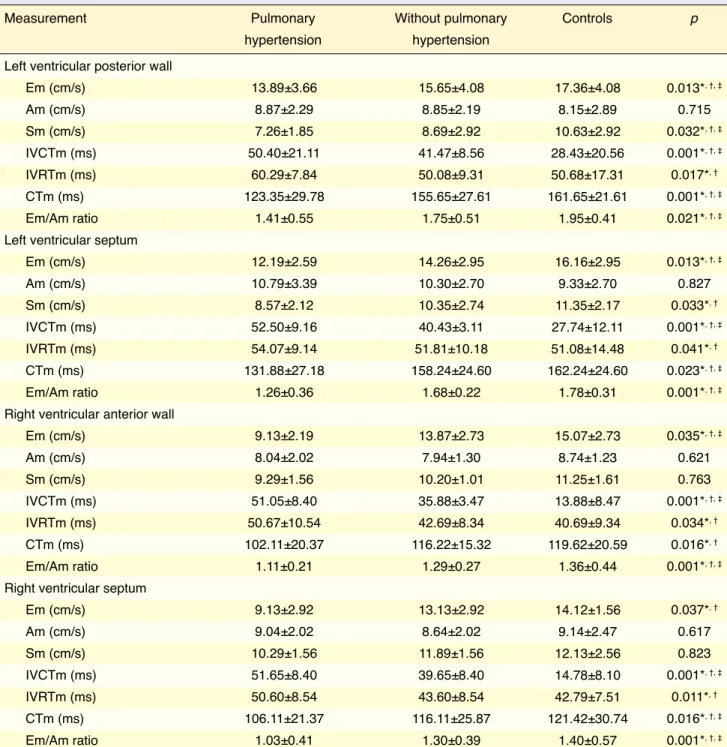
Tissue Doppler echocardiographic measurements
The Em velocity, Sm velocity, CTm, and Em/Am ratio measured at 4 four levels were all significantly lower in patients with PAH (Table 3). The IVCTm and IVRTm intervals measured at 4 levels were signifi-cantly higher in the PAH group.
Tei Index measurement with tissue Doppler echocardiography
Table 4 provides the mean Tei Index values measured from the left ventricular posterior wall, the right ven-tricular anterior wall, and the edges of the septum with tissue Doppler in the patients with PAH, the patients without PAH, and the controls. The Tei Index values measured at these sites were significantly higher in the patient group compared with those of the control group. The left ventricular posterior wall Tei Index measurement of the patients with PAH (0.68±0.18) was statistically higher than that of the patients with-out PAH (0.56±0.16) (p=0.027). There was no sta-tistically significant difference between the patients with PAH (0.57±0.12) and the patients without PAH (0.52±0.09) in the Tei Index value measured from the edges of the septum (p=0.389). The right ventricular anterior wall Tei Index value was significantly higher in the patients with PAH (0.67±0.16) than in the pa-tients without PAH (0.51±0.12) (p=0.001). However, there was no statistically significant difference be-tween the groups in the Tei Index value for the right ventricular edge of the septum (p=0.884).
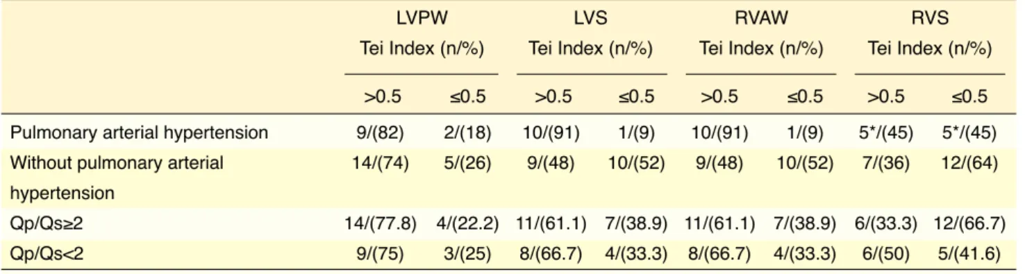
The Tei Index measurement from the left ventri-cular posterior wall was >0.5 in 9 (82%) of 11 patients with PAH and 14 (74%) of 19 patients without PAH. The distribution of the pathological values of the left ventricular Tei Index measured with tissue Doppler echocardiography according to PAH status and pul-monary/systemic blood flow ratio is presented in Table 5. The right ventricular anterior wall Tei Index measurement was >0.5 in 10 (91%) of 11 patients with PAH and 9 (48%) of 19 patients without PAH. A Tei Index value >0.5 for the right ventricular anterior wall demonstrated a sensitivity and specificity for deter-mining patients with PAH of 91% and 93.3%, respec-tively. The sensitivity and specificity of left ventricu-lar posterior wall Tei Index results of >0.5 in patients with PAH was 81.8% and 96.6%, respectively.
The left ventricular posterior wall Tei Index value was >0.5 in 14 (77.8%) of 18 patients with a pul-monary/systemic blood flow ratio ≥2. The sensitivity and specificity of the Tei Index result in determining a pulmonary/systemic blood flow ratio of ≥2 was 77.7% and 96.6%, respectively. The Tei Index mea-surement from the right ventricle anterior wall was >0.5 in 11 (61.1%) of 18 patients with a pulmonary/ systemic blood flow ratio of ≥2. The sensitivity and specificity of a Tei Index value of >0.5 for the right The Tei Index value measured from the left
ven-tricular edge of the septum was >0.5 in 7 (64%) pa-tients with PAH and 13 (68%) papa-tients without PAH. The sensitivity and specificity in identifying patients with PAH was 70% and 96.6%, respectively. The Tei Index measured from the right ventricular edge of the septum was >0.5 in 5 (45%) patients with PAH and 7 (36%) patients without PAH, which demonstrated a sensitivity and specificity in distinguishing patients with PAH of 50% and 93.3%, respectively.
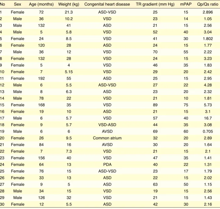
Table 1. Demographic features of the study population, echocardiographic diagnoses, tricuspid regurgitation gradient on echocardiography, and Qp/Qs ratio determined with cardiac angiography
No Sex Age (months) Weight (kg) Congenital heart disease TR gradient (mm Hg) mPAP Qp/Qs ratio
1 Female 72 21.3 ASD-VSD 25 15 2.896 2 Male 36 10.2 VSD 23 14 1.05 3 Male 132 41 ASD 21 15 2.56 4 Male 5 5.8 VSD 52 40 3.04 5 Female 24 8.5 VSD 41 30 1.802 6 Female 120 28 ASD 24 15 1.77 7 Male 36 12 VSD 70 55 2.22 8 Female 132 28 VSD 24 15 3.23 9 Female 5 4 VSD 46 35 1.83 10 Female 7 5.15 VSD 29 20 2.42 11 Female 192 55 ASD 25 15 2.95 12 Male 6 5.5 ASD-VSD 27 22 4.28 13 Male 8 6.3 ASD 23 20 2.32 14 Male 78 22 VSD 21 10 1.81 15 Female 168 35 VSD 89 75 5.73 16 Female 19 10 ASD 21 15 3.1 17 Male 6 5.7 VSD 57 40 16.7 18 Female 9 5.7 VSD-ASD 44 35 3.08 19 Male 6 6 AVSD 69 60 0.705
20 Female 26 9.5 Common atrium 32 20 2.89
21 Female 84 16 AVSD 30 20 1.64 22 Female 7 7.3 VSD 21 15 2.1 23 Female 156 40 VSD 47 35 1.41 24 Female 64 13 PDA 40 22 1.31 25 Female 76 15 ASD-VSD 23 17 1.79 26 Female 33 13 ASD 22 15 2.02 27 Female 9 5 ASD 63 50 1.15 28 Male 34 15 VSD 19 15 2.56 29 Male 126 32 VSD 21 15 1.43 30 Female 12 5.5 ASD 42 30 2.16
ASD: Atrial septal defect; VSD: Ventricular septal defect; AVSD: Atrioventricular septal defect; PDA: Patent ductus arteriosus; TR: Tricuspid regurgitation; mPAP: Mean pulmonary artery pressure; Qp/Qs: Pulmonary/systemic blood flow ratio.
ventricle anterior wall in isolating patients with a pul-monary/systemic blood flow ratio of ≥2 was 61% and 93.3%, respectively.
The left ventricular edge of the septum Tei Index value was >0.5 in 13 (72.2%) of 18 patients with a pulmonary/systemic blood flow ratio of ≥2, indicating a sensitivity and specificity of 72% and 96.6%, re-spectively. The Tei Index measurement performed from the right ventricular edge of the septum was >0.5 in 6 (33.3%) of 18 patients with a pulmonary/ systemic blood flow ratio ≥2, which demonstrated a sensitivity and specificity in detecting patients with a pulmonary/systemic blood flow ratio ≥2 of 33.3% and 93.3%, respectively.
A significant correlation was detected between the Tei Index value measured from the left ventricular posterior wall and the mean PAP (correlation coeffi-cient: 0.491). Similarly, a significant correlation was detected between the Tei Index result measured from the left ventricular edge of the septum and the pul-monary/systemic blood flow ratio (correlation coef-ficient: 0.431). While there was a significant correla-tion between Tei Index measurement from the right ventricular anterior wall and the pulmonary/systemic blood flow ratio (correlation coefficient: 0.495), no correlation was detected between the Tei Index value measured from the right ventricular anterior wall and the mean PAP.
The interobserver variability of the left ventricle Em and Am velocities, right ventricular IVCTm and CTm measurements was less than 5.1% (0.2%–7.3%). The intraobserver variability of right ventricular Em, Am, and Sm velocities was also low (coefficient of variation: 4%–8%).
DISCUSSION
The Tei Index is a non-invasive tool used to mea-sure the right and left ventricular systolic and dias-tolic functions.[3,10–17] The index is equivalent to the sum of isovolumetric contraction time and isovolu-metric relaxation time divided by ejection time.[5] These time intervals can be measured easily during routine Doppler echocardiographic examination.[1,18] This index is unaffected by the geometric shape of the ventricles and heart rate.[5,19] The PAP value and the pulmonary/systemic blood flow ratio are impor-tant in planning the treatment of CHD patients with a left-to-right shunt. On Doppler echocardiography, after systolic and diastolic gradients are determined from the tricuspid valve and pulmonary valve, respec-tively, the systolic and mean PAP can be measured using a simplified Bernoulli equation.[20] Pulmonary artery systolic pressure=4x(tricuspid regurgitation peak velocity)2+right atrial pressure[21–26] and mean PAP=4(pulmonary regurgitation peak velocity)2.[27] However, the lack of adequate regurgitation to mea-sure peak velocity makes these methods impossible to apply in some PAH cases.
Eidem et al.[16] reported that the Tei Index quan-titatively demonstrated right ventricular performance and is relatively independent of changes in preload and afterload. It has also been demonstrated that right ventricular Tei Index results were not different in pe-diatric patients with ASD compared with normal chil-dren while values were significantly higher in adult patients with ASD in contrast to healthy adults.[1] On the other hand, Baysal et al.[17] found increased Tei Index values in both the left and right ventricles in Table 2. Congenital heart diseases in groups with and without pulmonary arterial hypertension
Congenital heart disease Pulmonary arterial hypertension Without pulmonary arterial hypertension n
(mPAP ≥25 mm Hg) (mPAP <25 mm Hg) VSD 7 7 14 ASD 2 6 8 ASD+VSD 1 3 4 AVSD 1 1 2 Common atrium – 1 1 PDA – 1 1 Total 11 19 30
ficantly prolonged, the ejection time was significantly shortened, and that the Tei Index results were signifi-cantly greater in patients with primary pulmonary hy-pertension compared with controls. In a recent study performed in 12 children with idiopathic pulmonary hypertension using pulsed-wave Doppler, the mean patients with left-to-right cardiac shunt lesions.
Addi-tionally, in a study performed on animals by Grignola et al.,[28] it was reported that the Tei index was a sensi-tive indicator to identify right ventricular dysfunction occurring in acute PAH. Dujardin et al.[29] found that the right ventricular IVCTm and IVRTm were
signi-Table 3. Tissue Doppler echocardiographic measurements of the patients and controls
Measurement Pulmonary Without pulmonary Controls p
hypertension hypertension
Left ventricular posterior wall Em (cm/s) Am (cm/s) Sm (cm/s) IVCTm (ms) IVRTm (ms) CTm (ms) Em/Am ratio Left ventricular septum Em (cm/s) Am (cm/s) Sm (cm/s) IVCTm (ms) IVRTm (ms) CTm (ms) Em/Am ratio
Right ventricular anterior wall Em (cm/s) Am (cm/s) Sm (cm/s) IVCTm (ms) IVRTm (ms) CTm (ms) Em/Am ratio
Right ventricular septum Em (cm/s) Am (cm/s) Sm (cm/s) IVCTm (ms) IVRTm (ms) CTm (ms) Em/Am ratio
Em: Peak early diastolic myocardial velocity; Am: Peak atrial systolic velocity; Sm: Peak systolic myocardial velocity; CTm: Contraction time; IVCTm: Izovol-umetric contraction time; IVRTm: IzovolIzovol-umetric relaxation time. *Pulmonary hypertension group vs. control subjects; †Non-pulmonary hypertension group vs. control subjects; ‡Pulmonary hypertension group vs. non-pulmonary hypertension group.
13.89±3.66 8.87±2.29 7.26±1.85 50.40±21.11 60.29±7.84 123.35±29.78 1.41±0.55 12.19±2.59 10.79±3.39 8.57±2.12 52.50±9.16 54.07±9.14 131.88±27.18 1.26±0.36 9.13±2.19 8.04±2.02 9.29±1.56 51.05±8.40 50.67±10.54 102.11±20.37 1.11±0.21 9.13±2.92 9.04±2.02 10.29±1.56 51.65±8.40 50.60±8.54 106.11±21.37 1.03±0.41 15.65±4.08 8.85±2.19 8.69±2.92 41.47±8.56 50.08±9.31 155.65±27.61 1.75±0.51 14.26±2.95 10.30±2.70 10.35±2.74 40.43±3.11 51.81±10.18 158.24±24.60 1.68±0.22 13.87±2.73 7.94±1.30 10.20±1.01 35.88±3.47 42.69±8.34 116.22±15.32 1.29±0.27 13.13±2.92 8.64±2.02 11.89±1.56 39.65±8.40 43.60±8.54 116.11±25.87 1.30±0.39 17.36±4.08 8.15±2.89 10.63±2.92 28.43±20.56 50.68±17.31 161.65±21.61 1.95±0.41 16.16±2.95 9.33±2.70 11.35±2.17 27.74±12.11 51.08±14.48 162.24±24.60 1.78±0.31 15.07±2.73 8.74±1.23 11.25±1.61 13.88±8.47 40.69±9.34 119.62±20.59 1.36±0.44 14.12±1.56 9.14±2.47 12.13±2.56 14.78±8.10 42.79±7.51 121.42±30.74 1.40±0.57 0.013*, †, ‡ 0.715 0.032*, †, ‡ 0.001*, †, ‡ 0.017*, † 0.001*, †, ‡ 0.021*, †, ‡ 0.013*, †, ‡ 0.827 0.033*, † 0.001*, †, ‡ 0.041*, † 0.023*, †, ‡ 0.001*, †, ‡ 0.035*, †, ‡ 0.621 0.763 0.001*, †, ‡ 0.034*, † 0.016*, † 0.001*, †, ‡ 0.037*, † 0.617 0.823 0.001*, †, ‡ 0.011*, † 0.016*, †, ‡ 0.001*, †, ‡
regurgitation.[27] Cheung et al.[31] reported that a sud-den increase in ventricular volume caused a sudsud-den increase in Tei Index values.
In our study, the Tei Index values were measured in 4 sites using tissue Doppler: the right ventricular anterior wall, the left ventricular posterior wall, and the right and left ventricular edges of the septum. A systolic PAP of >35 mm Hg or mean PAP of ≥25 mm Hg at rest and >30 mm Hg during exercise or pul-monary vascular resistance index of >3 WU/m2 are the most commonly used criteria for the diagnosis of Tei Index value was 0.64±0.30 in the right ventricle
of the patients with PAH and 0.28±0.03 in the con-trols (p<0.01). The left ventricular Tei Index result of patients with PAH and controls was 0.44±0.15 and 0.34±0.03, respectively (p<0.05).[30] In the same study, the authors reported that the Tei Index mea-surement from the right ventricle was associated with invasively measured PAP and that the Tei values were useful in the clinical follow-up of these patients.[30] The Tei Index and mean PAP have also been accepted as criteria for PAH and have been noted as more reli-able than systolic PAP measured from tricuspid valve
Table 5. Comparison of right and left ventricular Tei Index values according to pulmonary arterial hypertension and pulmonary to systemic flow rate ratio
LVPW LVS RVAW RVS Tei Index (n/%) Tei Index (n/%) Tei Index (n/%) Tei Index (n/%)
>0.5 ≤0.5 >0.5 ≤0.5 >0.5 ≤0.5 >0.5 ≤0.5
Pulmonary arterial hypertension 9/(82) 2/(18) 10/(91) 1/(9) 10/(91) 1/(9) 5*/(45) 5*/(45)
Without pulmonary arterial 14/(74) 5/(26) 9/(48) 10/(52) 9/(48) 10/(52) 7/(36) 12/(64)
hypertension
Qp/Qs≥2 14/(77.8) 4/(22.2) 11/(61.1) 7/(38.9) 11/(61.1) 7/(38.9) 6/(33.3) 12/(66.7)
Qp/Qs<2 9/(75) 3/(25) 8/(66.7) 4/(33.3) 8/(66.7) 4/(33.3) 6/(50) 5/(41.6)
LVPW: Left ventricular posterior wall; LWS: Left ventricular septum; RVAW: Right ventricular anterior wall; RVS: Right ventricular septum; Qp/Qs: Pulmonary/ systemic blood flow ratio. *Tei Index value could not be measured in 1 patient with an atrioventricular septal defect.
Table 4. Right and left ventricular Tei Index values of the study population measured using tissue Doppler echocardiography
Pulmonary arterial Without pulmonary arterial Control group p
hypertension (n=11) hypertension (n=19) (n=30) (<0.05) LVPW 0.68±0.18 0.56±0.16 0.41±0.05 0.001* Tei Index 0.001† 0.027‡ LVS 0.57±0.12 0.52±0.09 0.39±0.05 0.001* Tei Index 0.001† 0.389‡ RVAW 0.67±0.16 0.51±0.12 0.41±0.057 0.001* Tei Index 0.006† 0.001‡ RVS 0.5±0.08 0.48±0.12 0.4±0.07 0.022* Tei Index 0.02† 0.884‡
*Pulmonary hypertension group vs. control subjects; †Non-pulmonary hypertension group vs. control subjects; ‡Pulmonary hypertension group vs. non-pulmonary hypertension group. LVPW: Left ventricular posterior wall; LWS: Left ventricular septum; RVAW: Right ventricular anterior wall; RVS: Right ventricular septum.
for the right ventricular anterior wall and the left ven-tricular edge of the septum (correlation coefficient: 0.495 and 0.431, respectively). Additionally, the Tei Index values obtained from the left ventricular pos-terior wall and the left ventricular edge of the sep-tum were more often >0.5 compared with the right ventricular anterior wall and the right ventricular edge of the septum. Our results suggested that as the pul-monary/systemic blood flow ratio increased, the num-ber of patients with a Tei Index value >0.5 in the left ventricular posterior wall increased.
Limitations of the study
Our patient group consisted of children with CHD, who may experience heart rate variability. The patient group was not homogenous in terms of the type of heart defect and some patients had both increased vol-ume and pressure load. The various defects caused an increase in volume in different ventricles and it was not possible to determine the effects of volume and pressure overload on the Tei Index values.
Conclusion
Our study results indicated that the Tei Index values increased substantially in patients with very large defects, which can affect the ventricles hemodynam-ically. The Tei Index measurements from various regions of the right ventricle in children with CHD with a left-to-right shunt can be used as a parameter to follow up on the development of PAH, to diagnose it in the early period, and to make a timely decision about surgery. Finally, higher Tei Index values mea-sured with tissue Doppler can be used as a criterion in directing the treatment of patients with a left-to-right shunt. Additional studies with larger groups are needed.
Peer-review: Externally peer-reviewed. Conflict-of-interest: None.
Funding resources: This research received no specific
grant from any funding agency, commercial or not-for-profit sectors.
Authorship contributions: Concept: M.Y., H.A., A.Y.;
Design: M.Y., H.A., A.Y.; Supervision: M.Y., H.A., A.Y.; Materials: M.Y., H.A., S.K., T.B.; Data: M.Y., H.A., S.K., T.B.; Analysis: M.Y., H.A., S.K., T.B.; Literature search: H.A., A.Y., S.K.; Writing: H.A., A.Y., S.K.; Critical revi-sion: H.A., A.Y., S.K.
pulmonary hypertension.[9,32,33] The Tei Index mea-surements measured from the right ventricular ante-rior wall, the left ventricular posteante-rior wall and the left and right ventricular edges of the septum with tissue Doppler were found to be significantly higher in the patient group than the control group. There was no significant difference in the Tei Index values mea-sured from the left and right ventricular edges of the septum between the patients with and without PAH. Also, there was a significant correlation determined between the Tei Index value measured from the left ventricular posterior wall and the mean PAP (correla-tion coefficient: 0.491).
Roberson et al.[34] reported that the upper limit for right ventricular Tei Index measurement was 0.44 in infants and 0.54 in adolescents. The upper limit for the right ventricle has also been reported as0.44 for infants and 0.49 for 18-year-olds.[35] Our study yielded Tei Index values of the right ventricular ante-rior wall and left ventricular posteante-rior wall that were >0.5 in 91% and 82%, respectively, of the patients with PAH. The sensitivity and specificity of a Tei in-dex value >0.5 in detecting patients with pulmonary hypertension was 91% and 93.3%, respectively, for the right ventricular anterior wall, 81.8% and 96.6% for the left ventricular posterior wall, 50% and 93.3% for the right ventricular edge of the septum, and 70% and 96.6% for the left ventricular edge of the septum. In patients with PAH, the sensitivity was higher on the right ventricular anterior wall compared with the other sites.
In this study, the patients were divided into 2 groups using a cutoff of a pulmonary/systemic blood flow ratio <2. In the patients with a pulmonary/sys-temic blood flow ratio ≥2, the Tei Index values for the left ventricular posterior wall, right ventricular ante-rior wall, left ventricular edge of the septum, and right ventricular edge of the septum were >0.5 in 77.8%, 61.1%, 72.2%, and 33.3% of the patients, respec-tively. The sensitivity and specificity of a Tei Index value >0.5 in detecting patients with a pulmonary/sys-temic blood flow ratio ≥2 was 61% and 93.3% for the right ventricular anterior wall, 77.7% and 96.6% for the left ventricular posterior wall, 33.3% and 93.3% for the right ventricular edge of the septum, and 72% and 96.6% for the left ventricular edge of the septum,. There was a significant correlation between the Tei Index and the pulmonary/systemic blood flow ratio
13. Frieadman D, Buyon J, Kim M, Glickstein JS. Cardiac func-tion assessed by Myocardial Performance Index. Ultrasound Obstet Gynecol 2003;21:33−6. [CrossRef]
14. Gonzalez RR, Dyar D, De Lange M. The use of MPI on pedi-atric heart transplant patients. JDMS 1998;14:51−3. [CrossRef] 15. Duranjin KS, Tei C, Yeo TC, Hodge DO, Rossi A, Seward JB.
Prognostic value of a Doppler index combining systolic and diastolic performance in idiopathic-dilated cardiomyopathy. Am J Cardiol 1998;82:1071−6. [CrossRef]
16. Eidem BW, O’Leary PW, Tei C, Seward JB. Usefulness of the myocardial performance index for assessing right ven-tricular function in congenital heart disease. Am J Cardiol 2000;86:654−8. [CrossRef]
17. Baysal T, Oran B, Doğan M, Cimen D, Karaaslan S. The my-ocardial performance index in children with isolated left-to-right shunt lesions. Anadolu Kardiyol Derg 2005;5:108−11. 18. Chen J, Xie L, Dai L, Yu L, Liu L, Zhou Y, et al. Right Heart
Function of Fetuses and Infants with Large Ventricular Septal Defect: A Longitudinal Case-Control Study. Pediatr Cardiol 2016;37:1488−97. [CrossRef]
19. Eidem BW, Tei C, O’Leary PW, Cetta F, Seward JB. Nonge-ometric quantitative assessment of right and left ventricular function: myocardial performance index in normal children and patients with Ebstein anomaly. J Am Soc Echocardiogr 1998;11:849−56. [CrossRef]
20. Ensing G, Seward J, Darragh R, Caldwell R. Feasibility of generating hemodynamic pressure curves from non-inva-sive Doppler echocardiographic signals. J Am Coll Cardiol 1994;23:434−42. [CrossRef]
21. Hatle L, Angelsen BA, Tromsdal A. Non-invasive estimation of pulmonary artery systolic pressure with Doppler ultra-sound. Br Heart J 1981;45:157−65. [CrossRef]
22. Zimbarra Cabrita I, Ruísanchez C, Grapsa J, Dawson D, North B, Pinto FJ, et al. Validation of the isovolumetric re-laxation time for the estimation of pulmonary systolic arterial blood pressure in chronic pulmonary hypertension. Eur Heart J Cardiovasc Imaging 2013;14:51−5. [CrossRef]
23. Beghetti M. Echocardiographic evaluation of pulmonary pressures and right ventricular function after pediatric cardiac surgery: A Simple approach for the intensivist. Front Pediatr 2017;5:184. [CrossRef]
24. Amaki M, Nakatani S, Kanzaki H, Kyotani S, Nakanishi N, Shigemasa C, et al. Usefulness of three-dimensional echocar-diography in assessing right ventricular function in pa-tients with primary pulmonary hypertension. Hypertens Res 2009;32:419−22. [CrossRef]
25. Xie Y, Burke BM, Kopelnik A, Auger W, Daniels LB, Madani MM, et al. Echocardiographic estimation of pulmonary vas-cular resistance in chronic thromboembolic pulmonary hyper-tension: utility of right heart Doppler measurements. Echocar-diography 2014;31:29−33. [CrossRef]
26. Kushwaha SP, Zhao QH, Liu QQ, Wu WH, Wang L, Yuan P, et al. Shape of the pulmonary artery Doppler-Flow profile
pre-REFERENCES
1. Koestenberger M, Friedberg MK, Nestaas E, Michel-Behnke I, Hansmann G. Transthoracic echocardiography in the eval-uation of pediatric pulmonary hypertension and ventricular dysfunction. Pulm Circ 2016;6:15−29. [CrossRef]
2. Jiang L, Wiegers SE, Weyman AE. Right ventricle. In: Wey-man AE, editor. Principles and practice of echocardiography. 2nd ed. Philadelphia: Lea & Febiger; 1994.p.901−21. 3. Feigenbaum H, Armstrong WF, Ryan T. Pulmonary
hyperten-sion. Feigenbaum’s Echocardiography. London: Lippincott Williams and Wilkins; 2005.
4. Tei C. New non-invasive index for combined systolic and di-astolic ventricular function. J. Cardiol 1995;26:135−6. 5. Tei C, Ling LH, Hodge DO, Bailey KR, Oh JK, Rodeheffer
RJ, et al. New index of combined systolic and diastolic my-ocardial performance: a simple and reproducible measure of cardiac function-a study in normals and dilated cardiomyopa-thy. J Cardiol 1995;26:357−66. [CrossRef]
6. Cahill JM, Horan M, Quigley P, Maurer BJ, McDonald K. Doppler echocardiographic indices of diastolic function in heart failure admissions with preserved left ventricular sys-tolic function. Eur J Heart Failure 2002;4:473−8. [CrossRef] 7. Lopez L, Colan SD, Frommelt PC, Ensing GJ, Kendall K,
Younoszai AK, et al. Recommendations for quantification methods during the performance of a pediatric echocardio-gram: a report from the Pediatric Measurements Writing Group of the American Society of Echocardiography Pe-diatric and Congenital Heart Disease Council. J Am Soc Echocardiogr 2010;23:465−95.
8. Lang RM, Bierig M, Devereux RB, Flachskampf FA, Foster E, Pellikka PA, et al. Recommendations for chamber quantifi-cation: a report from the American Society of Echocardiogra-phy’s Guidelines and Standards Committee and the Chamber Quantification Writing Group, developed in conjunction with the European Association of Echocardiography, a branch of the European Society of Cardiology. J Am Soc Echocardiogr 2005;18:1440−63. [CrossRef]
9. Abman SH, Hansmann G, Archer SL, Ivy DD, Adatia I, Chung WK, et al. Pediatric Guidelines from the American Heart Association and American Thoracic Society. Circula-tion 2015;132:2037−99. [CrossRef]
10. Weyman AE. Interatrial and interventricular septa. Principles and practice of echocardiography. 2nd ed. Philedelphia: PLea & Febiger; 1994.
11. Eidem BW, McMahon CJ, Cohen RR, Wu J, Finkelshteyn I, Kovalchin JP, et al. Impact of cardiac growth on doppler tis-sue imaging velocities: A study in healthy children. J Am Soc Echocardiogr 2004;17:212−21. [CrossRef]
12. Broberg CS, Pantely GA, Barber BJ, Mack GK, Lee K, Thig-pen T, et al. Validation of myocardial performance index by echo in mice.a noninvasive measure of left ventricular func-tion. J Am Soc Echocardiogr 2003;16:814−23. [CrossRef]
terial hypertension. The key role of echocardiography. Chest 2005;127:1836−43. [CrossRef]
33. Galiè N, Torbicki A, Barst R, Dartevelle P, Haworth S, Hi-genbottam T, et al. Guidelines on diagnosis and treatment of pulmonary arterial hypertension. The Task Force on Di-agnosis and Treatment of Pulmonary Arterial Hyperten-sion of the European Society of Cardiology. Eur Heart J 2004;25:2243−78.
34. Roberson DA, Cui W. Right ventricular Tei index in children: effect of method, age, body surface area, and heart rate. J Am Soc Echocardiogr 2007;20:764−70. [CrossRef]
35. Harada K, Tamura M, Toyono M, Yasuoka K. Comparison of the right ventricular Tei index by tissue Doppler imaging to that obtained by pulsed Doppler in children without heart dis-ease. Am J Cardiol 2002;90:566−9. [CrossRef]
dicts the hemodynamics of pulmonary hypertension caused by left-sided heart disease. Clin Cardiol 2016;39:150−6. 27. Rich S. Primary pulmonary hypertension: executive summary
from the World Symposium-Primary Pulmonary Hyperten-sion, 1998. Geneva (Switzerland): WHO.
28. Grignola JC, Ginès F, Guzzo D. Comparison of the Tei index with invasive measurements of right ventricular function. Int J Cardiol 2006;113:25−33. [CrossRef]
29. Tei C, Duranjin KS, Hodge DO, Bailey KR, McGoon MD, Tajik AJ, et al. Doppler echocardiographic index for assess-ment of global right ventricular function. J Am Soc Echocar-diogr 1996;9:838−47. [CrossRef]
30. Dyer KL, Pauliks LB, Das B, Shandas R, Ivy D, Shaffer EM, et al. Use of myocardial performance index in pediatric pa-tients with idiopathic pulmonary arterial hypertension. J Am Soc Echocardiogr 2006;19:21−7. [CrossRef]
31. Cheung MMH, Smallhorn JF, Redington AN, Vogel M. The effects of changes in loading conditions on the myocardial performance index: comparison with conductance catheter measurements. Eur Heart J 2004;25:2238−42. [CrossRef] 32. Bossone E, Bodini BD, Mazza A, Allegra L. Pulmonary
ar-Keywords: Children; Doppler echocardiography; pulmonary arterial
hypertension; Tei Index
Anahtar sözcükler: Çocuklar; Dopler ekokardiyografi; pulmoner