LET T ER T O T HE EDIT O R
Open Access
Ultrasound-guided modified thoracolumbar
interfascial plane block is effective for pain
management following multi-level lumbar
spinal fusion surgery
Mursel Ekinci
1, Bahadir Ciftci
1*and Yunus Oktay Atalay
2Keywords: Modified thoracolumbar interfascial plane (mTLIP) block, Multi-level lumbar spinal fusion surgery, Postoperative analgesia
To the Editor,
The number of spinal surgeries that are performed has been increasing because of the rise in the incidences of spinal diseases. Surgery for lumbar spinal fusion causes severe pain postoperatively, and the mobilization and rehabilitation of patients are very important after surgery (Kim et al.2016). However, rehabilitation processes may be negatively affected by postoperative pain (Pınar et al.2017). Therefore, postoperative pain management is an im-portant issue following multi-level spinal fusion sur-gery. The postoperative pain can be managed with a variety of regional anesthesia techniques. One of these techniques is ultrasound-guided thoracolumbar inter-fascial plane (TLIP) block. TLIP block targets the dorsal rami of the thoracolumbar nerves, and there are in-creasing reports about its efficacy in pain management following spinal surgeries (Hand et al. 2015; Ueshima et al. 2017; Ueshima and Otake 2017). We report here on a case involving successful pain management using modified TLIP (mTLIP) block following multi-level lumbar spinal fusion surgery. Written informed consent was obtained from the patient for the publication of this case report and accompanying images.
A 60-year-old man, who weighed 80 kg and had an American Society of Anesthesiologists (ASA) physical sta-tus of 2 (arterial hypertension), underwent lumbar spinal fusion surgery at three levels (L1–4 vertebrae levels)
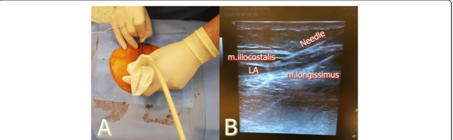
(Fig. 1a, b). After applying standard ASA monitoring, anesthesia was induced and orotracheal intubation was performed using an 8.0-mm tracheal tube. The patient was placed in the prone position. Before beginning the surgery, we performed a mTLIP block bilaterally. The block was performed under aseptic conditions at the level of the L3 vertebrae using the GE Vivid Q® ultrasound
device (Fig. 2a). A 12-MHz linear ultrasound probe was covered with a sterile sheath and placed in a vertical orien-tation. After visualizing the hyperechoic shadow of the spinous process as an anatomical guide point, the probe was moved forward to the lateral to visualize the longissi-mus and iliocostal longissi-muscles. Between these longissi-muscles, a 22-gauge, 80-mm block needle was inserted in a medial-to-lateral direction in the interfascial plane. Once the needle tip had been placed within the interfacial plane and after careful aspiration to rule out intravascular needle place-ment, 2 mL of saline was injected to confirm the proper injection site, and then a dose of 0.25% bupivacaine 20 mL was injected in each side (total 40 mL) (Fig.2b). A dose of 800 mg of ibuprofen IV was administrated intraoperatively 30 min before the end of the surgery for multimodal post-operative analgesia. The operation was uneventful, and there was no complication during the surgery. The patient was extubated and transferred to the post-anesthesia care unit (PACU). At the PACU, the patient’s visual analog score (VAS) was 3; therefore, no analgesic was adminis-tered. After a modified Aldrete score of ≥ 9, the patient was discharged from the PACU. A dose of 400 mg ibupro-fen was administrated routinely, every 8 h. The maximum VAS score that the patient experienced was 3 at rest, and he achieved mobilization within 24 h. The patient was
© The Author(s). 2019 Open Access This article is distributed under the terms of the Creative Commons Attribution 4.0 International License (http://creativecommons.org/licenses/by/4.0/), which permits unrestricted use, distribution, and reproduction in any medium, provided you give appropriate credit to the original author(s) and the source, provide a link to the Creative Commons license, and indicate if changes were made.
* Correspondence:[email protected];[email protected] 1Department of Anesthesiology and Reanimation, Istanbul Medipol
University, School of Medicine, Mega Medipol University Hospital, Bagcilar, 34040 Istanbul, Turkey
Full list of author information is available at the end of the article
Ain-Shams Journal
of Anesthesiology
Ekinci et al. Ain-Shams Journal of Anesthesiology (2019) 11:24mobilized very easily, and no additional analgesic was ad-ministrated during the postoperative period.
Discussion
The anatomy of the lumbar region has been understood clearly for anesthesia practice in the past few years. From medial to lateral respectively, the multifidus, long-issimus, and iliocostal muscles form the muscle layer of the lumbar paravertebral area. The dorsal nerve over-spreads here above the transverse process of vertebrae. The paravertebral area is innervated by the dorsal ramus of the spinal nerve. TLIP block was first defined by Hand et al. in 2015. They injected local anesthetic be-tween the multifidus and longissimus muscles at the third lumbar vertebral level by positioning the block needle at a 30° angle from the skin, and then they ad-vanced the needle from lateral to medial (classic tech-nique) (Hand et al. 2015). Ahiskalioglu et al. asserted that advancing the needle from lateral to medial might have a risk of inadvertent neuroaxial injection. Also, sonographic imaging of the longissimus and iliocostalis muscles can be easier than imaging the multifidus and longissimus muscles. Therefore, Ahiskalioglu et al. de-vised and described a modified TLIP block in which a local anesthetic is injected between the longissimus and
iliocostalis muscles with a needle angled at 15° and the needle is advanced medial to lateral (lateral technique) (Ahiskalioglu et al.2017). The efficacy of the TLIP block for lumbar laminoplasty has been retrospectively investi-gated, and Ueshima et al. found that the TLIP block with the classic approach provides 24 h of effective analgesia following single-level spinal surgery (Ueshima et al.2017). However, although this is effective for single-level surgery, the effectiveness of the TLIP block for multi-level spinal surgery is unclear. Ohgoshi et al. described two cases of multi-level lumbar surgery in which they performed TLIP block using the classic approach (Ohgoshi et al. 2017). The authors noted that this block is also effective for multi-level spinal surgery. With respect to a mTLIP block, Ahiskalioglu et al. suggested that a lateral approach may be useful for two- or three-level spinal surgeries as well (Ahiskalioglu et al.2017). In our case, we also performed a TLIP block using a lateral approach and found this tech-nique to be effective for analgesia after three-level spinal fusion surgery. While Ohgoshi et al. reported the effective-ness of TLIP block for multi-level laminectomy, the surgery in our case involved a multi-level spinal fusion, which is associated with the possibility of more pain after surgery. It is useful to note that although Ohgoshi et al. mentioned that the TLIP block is effective only for short
Fig. 1 a Surgery of multi-level spinal fusion. b X-ray photograph of multi-level spinal fusion
Fig. 2 a Ultrasound and patient setup for block preparation. b Sonographic anatomy and spread of local anesthetic
periods of time, they did not mention any specific amount of time in their report (Ohgoshi et al.2017). In our case, the effectiveness of the mTLIP block was 24 h, which is similar to the classical approach TLIP block that Ueshima et al. performed for single-level spinal surgery (Ueshima and Otake2017).
There is no research that shows results of comparing the classic and modified approaches yet. For this, large randomized studies are needed. In conclusion, we be-lieve that the mTLIP block may be a good option for managing pain in multi-level spinal fusion surgery.
Abbreviations
ASA:American Society of Anesthesiologists; mTLIP: Modified thoracolumbar interfascial plane; PACU: Post-anesthesia care unit; TLIP: Thoracolumbar interfascial plane; VAS: Visual analog score
Acknowledgements Not applicable Authors’ contributions
BC and ME contributed to the writing, literature scanning, and block performing. YOA contributed to the writing, literature scanning, block performing, and review. All authors read and approved the final manuscript. Funding
The authors received no financial support for the research and/or authorship of this article.
Availability of data and materials Not applicable
Ethics approval and consent to participate Not applicable.
Consent for publication
Written informed consent was obtained from the patient for the publication of this case report and accompanying images.
Competing interests
The authors declare that they have no competing interests. Author details
1Department of Anesthesiology and Reanimation, Istanbul Medipol
University, School of Medicine, Mega Medipol University Hospital, Bagcilar, 34040 Istanbul, Turkey.2Department of Anesthesiology and Reanimation, Istanbul Medipol University, International School of Medicine, Mega Medipol University Hospital, Istanbul, Turkey.
Received: 19 June 2019 Accepted: 22 October 2019
References
Ahiskalioglu A, Alici HA, Selvitopi K, Yayik AM (2017) Ultrasonography-guided modified thoracolumbar interfascial plane block: a new approach. Can J Anesth 64(7):775–776
Hand WR et al (2015) Thoracolumbar interfascial plane (TLIP) block: a pilot study in volunteers. Can J Anesth 62:1196–1200
Kim SI, Ha KY, Oh IS (2016) Preemptive multimodal analgesia for postoperative pain management after lumbar fusion surgery: a randomized controlled trial. Eur Spine J 25(5):1614–1619
Ohgoshi Y, Namiki R, Kori S, Matsukawa M (2017) The use of ultrasound-guided thoracolumbar interfascial plane block for multi-level lumbar spinal surgery. J Clin Anesth 37:162
Pınar HU, Karaca Ö, Karakoç F, Doğan R (2017) Effects of addition of preoperative intravenous ibuprofen to pregabalin on postoperative pain in posterior lumbar interbody fusion surgery. Pain Res Manag.https://doi.org/10.1155/ 2017/1030491
Ueshima H, Otake H (2017) Clinical experiences of ultrasound-guided lateral thoracolumbar Interfascial plane (TLIP) block. J Clin Anesth 39:145 Ueshima H et al (2017) Efficacy of the thoracolumbar interfascial plane block for
lumbar laminoplasty: a retrospective study. Asian Spine J 11(5):722–725
Publisher’s Note
Springer Nature remains neutral with regard to jurisdictional claims in published maps and institutional affiliations.