AHAlYSiS О? THS FUNCTЮ^^ CH THE ^:TClEvчR I:.
ASSOCIATED PROTEIN TC
»Гл ♦ 4. i,iím w V il·« ■··' < · » * /’*' ч С î · · í' ί·, '7 J / r : : f : :
Q »
6 ô 3
■ N B Z
B S 5/З З Э
ANALYSIS OF THE FUNCTION OF THE NUCLEAR
MATRIX-ASSOCIATED PROTEIN CID
A THESIS SUBMITTED TO
THE DEPARTMENT OF MOLECULAR BIOLOGY AND GENETICS
AND
THE INSTITUTE OF ENGINEERING AND SCIENCE
OF BILKENT UNIVERSITY
IN PARTIAL FULFILMENT OF THE REQUIREMENTS
FOR THE DEGREE OF MASTER OF SCIENCE
By
BILADA BILICAN
ß l \ \ Q H Ó03 ' K83
■ІЭЗЭ
6
о VI certify that I read this thesis and that in my opinion it is fully adequate, in
scope and in quality, as thesis for the degree of Master of Science.
I certify that I read this thesis and that in my opinion it is fully adequate, in
scope and in quality, as thesis for the degree of Master of Science.
. Ugur Yavuzer
I certify that I read this thesis and that in my opinion it is fully adequate, in
scope and in quality, as thesis for the degree of Master of Science.
Approved for Institute of Engineering and Science.
ABSTRACT
ANALYSIS OF THE FUNCTION OF THE NUCLEAR MATRIX- ASSOCIATED PROTEIN C ID
BILADA BILİCAN
M.Sc. in Molecular Biology and Genetics Supervisor: Dr. Uğur Yavuzer
July 1999, 86 pages
DNA Double-strand breaks (DSBs) are generated as intermediate stnictures during V(D)J recombination, as a consequence o f oxidative metabolism, or can be induced by exogenous factors such as gamma irradiation and radiomimetic drugs. Mutational studies identified the serine/threonine kinase, DNA-PK, as an essential component o f DNA DSB repair machinery. The activation o f the multi-component DNA-PK complex requires either free DNA ends or an association with the nuclear-matrix associated protein C ID , which facilitates the activation o f DNA-PK in a DNA end-independent fashion. The activation o f DNA-PK through its interaction with C ID , joins an increasing body o f evidence which suggests a role for higher order nuclear organisation in the orchestration o f complex cellular processes such as transcription, RNA splicing, nucleotide excision repair, replication and double-strand DNA break repair.
In this study, the yeast two hybrid system was employed to screen a B-cell cDNA library to identify the interacting proteins with C ID , and the interactions determined were further characterised. It was found that, C ID interacts specifically with the recombinational hotspot binding protein Translin and Translin associated factor X, TRAX, both
in vitro
andin vivo,
providing evidence that C 1D may play a critical role in DNA repair and recombination. Interestingly, an interaction between TRAX and DNA- PKcs has also been identified underin vivo
conditions. Tlie interaction o f TRAX with DNA-PKcs and C ID indicates a connection between DNA double-strand break repair, recombination, and dynamic nuclear architecture.ÖZET
NÜKLEER MATRİKS PROTEİNİ C ID ’NİN FONKSİYONUNUN İNCELENMESİ
BILADA BILİCAN
Yüksek Lisans Tezi, Moleküler Biyoloji ve Genetik Bölümü Tez Yöneticisi: Dr. Uğur Yavuzer
Temmuz 1999, 86 sayfa
DNA çift sarmal kırıkları V(D)J rekombinasyonu sırasında ve oksidatif metabolizma nedeniyle oluşabildiği gibi, gama radyasyonu ve kanser terapisinde kullanılan ilaçlar sonucunda da ortaya çıkmaktadır . Mutasyon çalışmaları, bir serine/threonine kinaz olan DNA-PK’nın DNA çift sarmal kırıklarının onanm ında önemli bir rol oynadığını göstermiştir. DNA-PK enzim kompleksinin aktivasyonu, serbest DNA ucu veya bir nükleer matriks proteini olan C İD ile etkileşimi gerektirmektedir. DNA-PK’nın C ID ile aktivasyonu, serbest DNA ucuna gereksinim duymaması yönünden diğer aktivasyon mekanizmalarından aynimaktadır. DNA-PK’nın C İD ile olan etkileşimi nükleer matriks yapılanmasının transkripsiyon, replikasyon, DNA onarımı ve RNA işlenmesi gibi kompleks hücresel işlevlerin eş güdümü ve organisazyonunda önemli rol alabileceğini göstermektedir.
Bu çalışmada maya ikili hibrid sistemi kullanılarak bir B-lıücresi kütüphanesi taranmış, C ID proteini ile etkileşime giren proteinler belirlenmiş, ve bu etkileşimler karakterize edilmiştir. C ID ’nin
in vitro
vein vivo
koşullarda rekombinasyon noktalarına bağlanan Translin ve Translin Associated Factor X, TRAX, proteinlerine spesifik olarak bağlandığı gösterilmiştir. Ayrıca, TRA X ’ınin vivo
şartlarda DNA-PKcs ile de etkileşime girdiği belirlenmiştir. TRAX proteininin DNA-PKcs ve C ID ile olan bu etkileşimi DNA çift sarmal kınk onanm ı, rekombinasyon, ve dinamik nükleer matriksACKNOWLEDGEMENTS
Above all, I would like to express my gratitude to my thesis supervisor,
Dr. Uğur
Yavuze/%
my mentor and guru, for her talent in creating a memorable experience o f life out from a scientific “sacrifice” with an elegant touch o f humour, wisdom and scientific knowledge. I am indebted her for providing the foresight and courage 1 needed to chase after my fate.1 would like to thank to
Prof. Dr. Mehmet Öztürk
for providing the opportunity to carry my plans to real life, and sharing his vision which influenced my scientific view and broadened my future perspectives.I would like to name and thank to
S. Hollenberg
for providing the yeast strains,S.EIIedge
for cDNA library and yeast plasmids.Dr. C. Yakicier
for cDNAs and being so kind both scientifically and personally, and C.R. Coding
for restriction enzymes, consumables, and valuable discussions.Very special thanks to
Ash Öztan,
my somewhat simultaneous reincarnation, for being a part o f my conscious and unconscious;Gülayşe İnce,
95% Dr. Jeykll, 5% Mr. Hyde, for the great laughter and her intimate friendship;Tuba Dinçer,
the ever-known living legend o f humour to me, for being an ineplaceable lab partner with endless support and hopefully friend for life;Hilal Özdağ,
the earth mother put up to test with a Ph.D., for her lovely chat and sincere interest; and Enise Peker, the queen mother, for always being the last resort to shelter.1 also would like to thank to Çağla, Esra, Reşat, Buket, Temre, Tolga, Arzu, Cemaliye, Hani, Oytun, Burta and Onur for having the time to share and the joy they brought, and Lütfiye Mesci, Sevim Baran, Birsen Cevher and Füsun Elvan for their helps.
TABLE OF CONTENTS
SIGNATURE P A G E ...II A B STR A C T... I ll Ö ZET... r v A C K N O W L E D G E M E N T S...V TABLE O F C O N T E N T S...VILIST OF FIGURES... VIII
A BBR EV A TIO N S... IX
IN TR O D U C TIO N ... 1
CHAPTER I; DNA D A M A G E ...2
1.1 DNA DOUBLE STRAND B R E A K S... 3
1.2 NON-HOMOLOGOUS DNA END JOINING...3
1.3 ROLE or DNA-PK .vnd ASSOCIATED FACTORS IN DNA NHEJ... 5
1.4 COMPONENTS OF DNA-PK 6 1.5 DNA-PK .AND C I D ... 11
1.6 IDENTIFIC.ATION OF THI· Fl NCTION OF THE C 1D PROTEIN... 12
CH APTER II: THE NUCLEAR M A TR IX ... 14
2.1 The Nu c l e u s...14
2.2 Organisationofthe Nucleus .and Nuclear Matrix... 14
2.3 Functionsof Nucle.ar Ma t r ix...16
CH APTER III: M ATERIALS AND M E T H O D S ...20
3.1 Ye.ast Two Hybrid System...21
3.1.1 Constructs:...22
3.1.2 Yeast St rat ns...23
3.1.3 Transformation...23
3.1.4 f-g a l Filter A ssay...26
3.1.5 DN.4 Isolation from Yeast Colonies...26
3.2 Recombin.w tD N A M .\nipulation Techniques... 29
3.2.1 Preparation o f Competent Cells for Transformation and Transformation with Plasmid DMA -CaCl: Method.... *... !...29
3.2.2 Preparation o f Competent Cells for Transformation and Transformation with Plasmid DNA -Simple and Efficient Method...30
3.2.3 Plasmid DNA Purification and Quantitation...31
3.2.4 Restriction Enzyme Digestion o f DNA...33
3.2.5 DNA Ligation... 33
3.2.6 Polymerase Chain Reaction...33
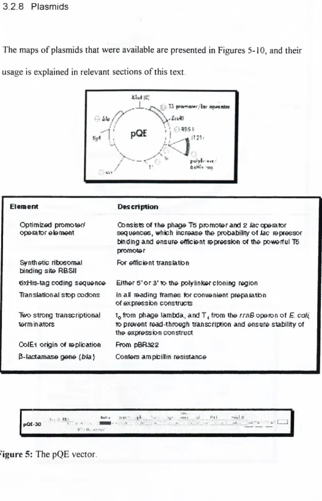
3.2.7 Bacterial Strains...34 3.2.8 Plasmids...35 3.3 Pr im er s... 39 3.4 Constructs... 40 3.5 Sequencing... 41 3.6 Gel Electrophoresis... 41
3.6.1 Agarose Gel Electrophoresis o f DNA Fragments...41
3.6.2 SDS Polyacrylamide Gel Electrophoresis...41
3.7 Biochemic.alTechniqlt-s... 42
3.7.7 Cell Growth and Induction...42
3.7.2 Cell Harx^est and Lysis...43
3.7.3 Protein Purification o f Glutathione-S-Transferase Tagged CID Protein...44
3.7.4 GST Pulldown Assay Using Immobilised GST-CID...45
3.7.5 Protein Purification by Immobilised Metal Ion Affinity Chromatography and in vitro Interaction 46 3.7.6 Protein Refolding...47
3.7.7 Pull-down ...47
3.7.8 Immunological Detection o f Immobilised Proteins (Western Blotting)...48
3.7.9 In vitro Transcription and Translation...49
3.8 Restriction Endonucleases. DNA a n dRNA Modifying Enzymes... 49
3.9 R.ADIOCHEMIC.ALS...50
3.10 Chemic.als. Kits .and Speclal. Consum ables...50
3.11 Equipmen’F... 50
3.12 Stand.ard Solitions .a n dBuffers... 51
CHAPTER 4: R E SU L T S... 52
4.1 Introduction...53
4.2 Ye.ast Two Hybrid Screening...54
4.3 tVlignmentof TRiAX cD N A s with Full Length T R A X ...57
4.4 Amplificationofthe Full Length ORF of TRAX and TranslinfromcD N A ...58
4.5 Cloningofthe PCR Pi^m^cTs of TRAX and Translinintothe Yeastand Bacterial Expression Vectors...60
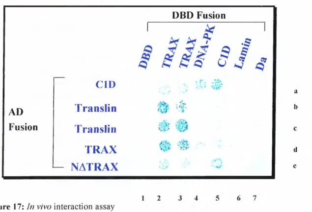
4.6 Invivo Inter.action Ass.a y s... 61
4.7 IN VITRO Interaction Assays... 63
4. 7.1 Purification o f GST-CID Protein Under Nondenaturing Conditions...63
4.7.2 Small Scale Induction o f the His Tagged CID Protein...64
4.7.3 Purification o f His Tagged CID Protein Under Denaturing Conditions...66
4.7.4 In vitro Transcription and Translation...68
4.7.5 Ni-NIA Pull-down A ssa y ...69
CH APTER 5: D ISC U SSIO N ... 72
5.1 PERSPECTIVES...79
R EFER EN C ES...80
LIST OF FIGURES
Figure 1
Mechanism o f V(D)J recombination 5Figure 2
DNA-PKcs structure 7Figure 3
Model o f possible interactions o f DNA-PK with DNA andother proteins 9
Figure 4
Sequence o f operations 19Figure 5
The pQE vector 35Figure 6
The pACT vector 36Figure 7
The pACT2 vector 36Figure 8
The pBTM l 16 poly-stop 37Figure 9
Multiple cloning site o f the T7 pLink vector 37Figure 10
The pGEX TK2 vector 38Figure 11
Flowchart o f yeast two hybrid screening 56Figure 12
Summary o f the yeast two hybrid screen 57Figure 13
Alignment o f clone41 against full length TRAX 58Figure 14
First round PCR o f TRAX and Translin 59Figure 15
Second round PCR o f TRAX 59Figure 16
Sample digest result 60Figure 17
In vivo
interaction assay 62Figure 18
Purification o f G ST-C ID protein 64Figure 19
Small scale induction o f His-CID protein 65Figure 20
Ni-NTA immobilised C ID 67Figure 21
Ni-NTA immobilised C ID western blot result 67Figure 22
In vitro transcription and translation o f TRAX and Translin 68Figure 23
Ni-NTA pull-down 70ABBREVATIONS
APS Ammonium persulfate
bp base pairs
BSA Bovine Serum Albumin
cDNA Complementary DNA
C-terminus Carboxyl-terminus
ddH20 deionised distilled water
DNA Deoxyribonucleic Acid
DNA-PK DNA dependent protein kinase
DNA-PKcs DNA dependent protein kinase catalytic subunit
dNTP deoxynucleotide triphosphate
ds double strand
DSB Double Strand Break
DTT dithiothretiol
EDTA diaminoethane tetra-acetic acid
EtBr Ethidium Bromide
g gram
His Histidine
HRP Horse Radish Peroxidase
Ig Immunoglobulin
IPTG Isopropylthio-P-D-galactoside
IR Ionising Radiation k kilo kb(s) kilobase(s) kD kilo Dalton LB Luria-Bertoni media Leu Leucine Lys Lysine M molar Ug microgram ml milliliter microliter mM millimolar jiM micromolar
N-terminus Amino terminus
OD Optical Density
PAGE Polyacrylamide Gel Electrophoresis
PBS Phosphate Buffered Saline
PBST Phosphate Buffered Saline with Tween
PCR Polymerase Chain Reaction
PI Phosphatidyleinositol
RNA Ribonucleic Acid
RNase ribonuclease
rpm revolutions per minute
SDS Sodium Dodecyl Sulfate
ss single strand
TBE tris-borate-EDTA solution
TCR T-cell Receptor
TE tris-EDTA solution
TEMED N,N,N,N-tetramethyl-1,2 diaminoethane
TRAX Translin associated factor X
Tris T ris-methylamine
Trp Tryptophan
Ura Uracil
UV Ultraviolet
X-gal 5-bromo-4chloro-3-indolyl P-D-galactopyranoside
XRCC X-ray cross complementing
CHAPTER I: DNA DAMAGE
Damage to genomic DNA occurs spontaneously and can be fiirther enhanced by environmental mutagens. We can define “damage” to DNA as o f any change introducing a deviation from the usual double-helical structure. In this context DNA damage can be classified broadly into tw o as:
• Single base changes; Alterations that affect the sequence but not the overall structure o f DNA. They do not affect transcription or replication when the strands o f the DNA duplex are separated.
• Structural distortions; Alterations that may physically hinder replication or transcription.
Chemical or physical mutagens induce a variety o f lesions in DNA including base modifications, intra-strand and inter-strand crosslinking and strand breaks. If left unrepaired, base modifications can cause mutations and crosslinkings, and strand breaks that may interfere with transcription, DNA replication and chromosome segregation, resulting in the loss o f viability. To ensure survival, cells must be equipped with mechanisms to repair these DNA lesions. Proliferating cells also need to delay cell-cycle progression to avoid the replication or the segregation o f damaged DNA. As a safeguard, cells o f multicellular organisms have the option o f activating programmed cell death in response to DNA damage (Wang 1998). There are three categories o f DNA repair. First one is excision repair, which includes both base and nucleotide excision repair that uses the information on one DNA strand to repair the damaged one. Second, mismatch repair, which occurs immediately after replicative DNA synthesis in S phase and relies on the parental strand to correct a misincorporation in the newly synthesised one. The third category is double-strand break repair. In this last system, three different ways are employed in joining the DNA ends: The first one is homologous recombination, in which the double-strand break on one chromosome is repaired using the information on the homologue. The second is single-strand annealing, and the third one is non-homologous or illegitimate end joining. Mammalian cells rely mostly on non-homologous end-joining activity^
and appear to rejoin the majority o f their breaks by a mechanism(s) that does not require extensive homology (Lewin 1997).
1.1
DNA DOUBLE STRAND BREAKS
A
specific type o f DNA damage, DNA Double Strand Breaks (DSBs), pose a major threat to the successful propagation and integrity o f the genome. If left unrepaired, they can cause cell death and, in multicellular organisms can lead to cancer. A variety o f exogenous agents, including ionising radiation (IR) and a number o f anti-cancer drugs, cause DSBs, as do endogenous free radicals, the by products o f oxidative metabolism. Furthermore, DNA DSBs occur normally as intermediates in V(D)J recombination, the process that helps to generate the vast range o f antigen-binding sites o f antibody and T-cell receptor proteins during mammalian lymphoid development (Gellert 1992). Thus, double-strand break repair is both essential for maintaining genomic integrity and for the normal development o f immune system.1.2
NON’HOMOLOGOUS DNA END JOINING
DNA end-joining is probably the most demanding form o f DNA repair because the two ends are initially physically unlinked and must be brought together into some form o f synapsis. If the break leads to the loss o f several nucleotides, the two ends might begin to diffuse apart; this type o f damage, therefore, begins with a variable distance between the two ends. While the two ends are maintained in synapsis, the ends must be reconfigured biochemically into a ligatable form. Small blocks o f terminal homology are frequently observed to be the points o f alignment o f the two ends. Following the alignment step, excess single-stranded DNA on one or
necessary enzymatic component. Polymerases are then needed to fill in gaps, and ligases are required to seal the nicks (Lees-Miller 1996).
Genetic and biochemical complementation studies have revealed that double strand break repair and V(D)J recombination (Figure 1) are reliant on the DNA- dependent protein kinase (DNA-PK). DNA-PK is an abundant nuclear protein serine/threonine kinase that is activated
in vitro
by DNA double-strand breaks and certain other discontinuities in the DNA double helix. DNA-PK is a multimeric complex with a catalytic subunit named DNA-PKcs and two non-specific DNA end binding proteins, Ku70 and Ku 80 (Jackson and Jeggo 1995).F igure 1: M echanism o f V(D)J recombination. A V -D joining event is shown. The V (D )J recombination apparatus recognises recombination signal sequences (triangles) and induces D N A double-strand breaks. At their jiuiction w idi coding sequences (rectangles). Tlie recombination- signal-sequence reaction intermediates are blunt D S B s, whereas tliose for coding segm ents are D N A hairpins. Ligation is mediated b y die D S B repair apparatus.
1.3
ROLE of DNA-PK and ASSOCIA TED FACTORS in DNA NHEJ
The involvement o f DNA-PK in DSB repair became evident from analysis’ involving a specific series o f mutant rodent cell lines. Early studies to these cells found them to be hypersensitive to IR and radiomimetic agents with little or no cross sensitivity to other types o f DNA damaging agents, showed them to be defective in the repair o f chromosomal DNA DSBs. Subsequent cell fusion studies allowed these cells to be placed in three distinct complementation groups, termed IR4, IRS, and
and Jeggo 1995). Later it was demonstrated that cells o f IRS and IR7 harboured inactivating mutations in the genes o f Ku80 and DNA-PKcs, respectively, and revealed that inactivation o f Ku80 leads to a dramatic destabilisation o f both itself and Ku70 (Peterson et al., 1995). It was therefore concluded that mutations in Ku80 and DNA-PKcs lead to IR sensitivity, that
XRCC5
andXRCC7
encode Ku80 and DNA- PKcs respectively, and that DNA-PK is a crucial component o f the mammalian DNA DSB repair apparatus (Priestley et al., 1998).Targeted disruption o f the gene for Ku70 in mouse cells confirmed that these cells have a similar phenotype to IR4-7. Hence, such cells defective in Ku70 were designated to be IR6 and the gene for Ku70 designated to be
XRCC6
(Gu et al.,1997) . It was also noted that cells defective in IR4, 5, 6, and 7 were defective in coding joint formation, whereas defects in IR4, 5, and 6 affected signal joint formation only ( see Figure 1).
In another work, the XRCC4 gene product was cloned, and IR4 cells were shown to be deleted for this gene (Li et al., 1995). Subsequent studies have shown that this protein forms a tight and specific association with DNA ligase IV, a protein which itself has been revealed recently to function in DNA NHEJ (Grawunder et al.,
1998) .
1.4
Components of DNA-PK
Heterodimeric Ku protein, the DNA-targeting component o f DNA-PK, binds without sequence specificity to double strand (ds) DNA ends, and also recognises ds to single strand (ss) transitions in DNA including gapped and hairpin molecules. Ku represents the major ds DNA end binding protein, and can be easily identified by band shift experiments (Blier et al., 1993). The assembly o f the heterodimer is required for DNA binding activity and it can translocate along DNA molecules forming a multimeric protein-DNA complex. A putative nuclear localisation signal has been identified in Ku70, but not in Ku80, suggesting that the transport to the nucleus might take place after assembly. Although Ku has been reported to have ATPase and ATP dependent helicase activity, convincing ATP binding domains have not been identified
in either o f the subunit. Ku is autophosphorylated
in vitro,
but lacks an identifiable DNA-PK phosphorylation site. Also, its autophosphorylation does affect DNA-PK activity (Lees-Miller 1996).DNA-PKcs is a 460-kD protein, which is stable in the absence o f Ku, but the activation o f the kinase is dependent upon the presence o f DNA ends as well as Ku proteins, indicating that a conformational change in DNA-PKcs takes place when Ku becomes DNA bound. DNA-PKcs has one o f the largest cDNAs known (14 kb), and is a member o f phosphatidylinositol (PI) 3-kinase superfamily (Figure 2). However, to date no lipid phosphorylation activity has been detected (Hartley et al., 1995). DNA-PKcs also possesses a leucine-zipper motif, which has been found to be important for interaction with other proteins ( Yaneva et al., 1989; Yavuzer et al., 1998), and an autophosphorylation site. Autophosphorylation o f DNA-PKcs is involved in regulation o f DNA-PK activity by affecting dissociation o f DNA-PKcs from the Ku-DNA complex (Chan and Lees-Miller 1996).
PI-3 Kinase Homology Domain
SLD LSCK Q LASG LLELAFAFG G LCERLVSLLLNPAVLS...
Figure 2:
DNA-PKcs structure. PI-3 kinase homology domain and the putative leucine zipper regions are indicated.The ability o f DNA-PK to recognise ds DNA ends and to initiate a signal transduction pathway by phosphorylation o f relevant substrates thereby alerting the cell to the presence o f a DSB is a candidate mechanism by which a cell might respond
RNA pol I transcription by phosphorylation, suggesting that one role o f DNA-PK can be to inhibit transcription in the vicinity o f the break, thereby allowing DNA repair machinery to perform its function.
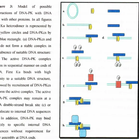
Currently there are numerous speculations about the precise function o f DNA- PK in DNA DSB repair and V(D)J recombination. For example, it has been suggested that DNA-PK may align the broken DNA ends or interact directly with the other components o f the repair machinery to target them to sites o f DNA damage. Alternatively, or in addition, DNA-PK might function by modifying the activity o f DNA repair factors by phosphorylation. It could also counteract the action o f transcription factors or chromatin, which might otherwise interfere with the assembly o f the DNA repair complex. In addition to these, consistent with the fact that DNA- PKcs is related to several factors involved in DNA damage-induced cell cycle checkpoint control processes, DNA-PK activation could trigger DNA damage signalling cascades. Figure 3 summarises the possible modes o f action. (Lees-Miller
Figure 3: Model of possible interactions o f DNA-PK with DNA and with other proteins. In all figures the Ku heterodimer is represented by the yellow circles and DNA-PKcs by the blue rectangle, (a) DNA-PKcs and Ku do not form a stable complex in the absence o f suitable DNA staicture: (b) The active DNA-PK complex forms in sequential manner on ends of DNA. First Ku binds with high affinity to a suitable DN A structure, followed by recruitment o f DNA-PKcs to form the active complex. The active DNA-PK complex may remain at a DNA double-strand break site (c) or translocate to internal D NA sequences (d) . In addition, DNA-PK may bind directly to specific internal DNA sequences without requirement for prior assembly at DNA ends.
(e) Autophosphorylation occurs on DNA-PKcs, Ku70, and Ku80 and results in dissociation of DNA-PKcs from DNA bound heterodimer, (f) DNA-PK can phosphorylate either soluble or DNA-bound substrates (orange boxes) in vitro, (g) DNA-PK may assemble at DNA double strand break and tether the ends o f D NA together, (h) The DNA-PK complex may act as a scaffold to which other proteins are recruited (Lees-Miller 1996).
Recently, it has been reported that DNA-PK activates p53 in response to DNA damage so that p53 can bind to specific DNA sequences. As phosphorylation o f p53 (at serines 15 and 37) by DNA-PK induced by ionising radiation prevents MDM2 from inhibiting p53-dependent transactivation, the action o f DNA-PK on p53 serves tv^o purposes: It activates p53 DNA-binding while blocking its inactivation by
Interestingly, DNA-PK is present at relatively high levels (1% o f HeLa cell nuclear protein) and its levels do not appear to be regulated throughout the cell cycle and despite its activity is induced upon DNA damaging agents, its levels do not show major alterations in response to DNA damaging agents. These observations led to the idea that DNA-PK may act as a damage sensor rather than as an inducible downstream effector (Lee et al., 1997).
Although DNA-PK levels are constant during cell cycle, its activity is regulated in a cell-cycle dependent manner, with peaks o f activity found at the Gi/early S phase and again at the G2 phase in wild type cells. The period o f the G]/early S phase DNA-PK activity correlates with an increased hypersensitivity to gamma-irradiation and a DNA DSB repair deficit in synchronised
scid
pre-B cells. However no difference is observed in the capacity o f the repair o f DSBs at G2 o fscid
cells when compared with wild-type cells, and
scid
cells remain permanently arrested in G2. These findings suggest that DNA-PK may provide an activity, distinct from its activity required for DNA DSB repair, that is necessary for DNA-damaged cells to traverse a G2 checkpoint (Lee et al., 1997).The most interesting mode o f DNA-PK regulation is its inactivation during programmed cell death and upon viral infection. During apoptosis, DNA-PKcs is specifically cleaved by caspase-3 or a caspase-3 like protease with subsequent loss o f its kinase activity. This DNA-PK inactivation is probably to prevent signalling from or repair o f the degraded genomic DNA that is formed during the latter steps o f the apoptotic pathway (Song et al., 1996). In a different study, it has been demonstrated that DNA-PKcs is degraded upon herpes simplex virus type infection o f mammalian cells. Thus suggests that inhibiting DNA-PK function aids virus replication and/or packaging. (Lees-Miller et al., 1996)
1.5
DNA-PK and C1D
Elucidation o f the exact role o f the DNA-PK in these processes require the exact DNA species essential for the activation o f DNA-PK, the identification o f other
proteins that interact with the DNA-PK complex at the DNA break site, and identification o f true physiological substrates o f DNA-PK. Likely substrates and/or interacting proteins may be other DNA end-binding proteins, or helicases, nucleases, or ligases that may be involved in the repair or recombination processes. Some other questions on the activation o f DNA-PK can be stated as whether it always needs free DNA ends or if an accessory protein is enough to recruit DNA-PK to DNA? Though Ku helps to recruit DNA-PKcs to DNA
in vitro
and likely to be required for physiological activation o f DNA-PK at damage sites, there is evidence that DNA- PKcs can bind to DNA and exhibit kinase activity without Ku.To gain more insight into the functions o f DNA-PK, the yeast-two hybrid system was applied using the 1501-1602 amino acid region o f DNA-PKcs that contains the putative leucine zipper m otif and a human B-lymphocyte cDNA library was screened (Yavuzer et al., 1998). The putative leucine zipper region was chosen as bait as leucine zippers have well documented roles in mediating protein-protein interactions. The screen revealed 17 clones containing overlapping sequences derived form the same cDNA. This protein which is termed C ID is a ubiquitously expressed nuclear protein with no significant homology with any known functional protein domains.
in this same study, it has been demonstrated that C ID interacts specifically with the leucine zipper region o f DNA-PKcs both
in vitro
andin vivo,
and is also an efficient substrate for DNA-PKin vitro,
with phosphorylation being o f similar efficiency to the well-characterised DNA-PK substrate p53. Moreover, C ID is capable o f activating DNA-PK in the absence o f free DNA ends. The importance o f this last finding lies in the fact that till then it was believed that DNA-PK interacts with free DNA ends, and therefore, is activated strongly by linear DNA but only weakly, if at all, by closed-circular plasmid DNA. Interestingly, C ID that is bound to supercoiled plasmid DNA not only activates DNA-PK to phosphorylate C ID itself but other substrates like p53 as well.1.6
Identification of the Function of the C1D Protein
The fact that interphase chromosomes and individual genes occupy distinct territories and sites in the interphase nucleus makes the analysis o f DNA-polypeptide complexes potentially involved in the three-dimensional genome organisation a necessity. In this context, residual polypeptides which are most resistant during DNA isolations are o f interest with respect to their potential involvement in the topological organisation o f genomic DNA.
To address this problem, monoclonal antibodies to such polypeptides were prepared by
in vitro
immunisation and used to screen a murine cDNA expression library. A positive cDNA (M C ID ) was detected encoding a polypeptide o f the size 16 kD exhibited by one o f the most resistant polypeptides detectable in phenol- extracted DNA by '^’iodine labelling and SDS-polyacrylamide gel electrophoresis. Later, a homologous human cDNA (H C ID ) was identified in the EST-sequence library by sequence comparison and by complete sequencing. This cDNA encodes a 16 kD polypeptide whose cellular counterpart is able to form phenol-stable complexes with DNA. This suggestion is based on the fact that phenol-extracted DNA contains a residual antigen o f 16 kD and that the antibodies prepared against the residual antigens could detect a cDNA which encodes a 16 kD nuclear polypeptide with DNA affinity. Immunostaining o f subnuclear structures by the antiserum to the recombinant mouse C ID protein showed that in human fibroblast cells only the cell nucleus shows a fibrogranular staining pattern, and cytoplasmic localisation o f the antigen is generally nonsignificant (Nehls et al., 1998).C ID was also identified independently by another group as a nuclear receptor co-represor which is expressed at high levels in adult tissues. In this study, it was demonstrated that C ID potentiates transcriptional repression by thyroid hormone receptor RevErb
itt vivo,
represses transcription when fused to a heterologous DNA binding domain, and interacts with RevErb as well as thyroid hormone receptorin
vitro
(Zamir et al., 1997).In another study, C ID is determined to be interacting with Rac3, which is a small GTPase o f the Rho family, members o f which have been implicated in
tumorigenesis, cell growth/death, and organisation o f the actin cytoskeleton. Many Rac interacting proteins or effectors identified to date have a role in cytoskeletal organisation. In yeast cells, C ID binds to constitutively activated but not GDP-bound Rac3, When coexpressed with Rac3 in COS cells, C ID complexed with constitutively active Rac3 but not with wild-type Rac3, demonstrating that C ID - Rac3 interactions take place />;
vivo
in mammalian cells and that C ID appears to be an effector o f Rac3 (Haataja et al., 1998).Recently, a new study demonstrated that apoptosis is induced in various tumour cell lines by vector dependent over expression o f the C ID protein. C ID protein should have a basic cellular function as it is physiologically expressed in 50 human tissues. Cells transfected with ClD-expressing constructs show terminal deoxynucléotidyl transferase-mediated dUTP nick end-labelling o f DNA ends along with morphologic changes typical for apoptotic cell death, e g. cytoplasmic vacuolation, membrane blebbing, and nuclear disintegration. Moreover, it is observed that C 1 D-dependent apoptosis is not induced in cells with no functional p53 in agreement with the function o f DNA-PK as an upstream activator o f p53, end- independent activation o f DNA-PK by C ID and ability o f DNA-PK activated by C ID to phosphorylate p53 (Rothbarth et al., 1999).
CHAPTER II: THE NUCLEAR MATRIX
2.1
The Nucleus
The eukaryotic nucleus, the largest organelle, is surrounded by two membranes, each one a phospholipid bilayer containing many different types o f proteins. The inner nuclear membrane defines the nucleus itself In many cells, the outer membrane is continuous with the rough endoplasmic reticulum, and the space between the inner and outer nuclear membranes is continuous with the lumen o f the rough endoplasmic reticulum (Lewin 1997).
The major physiological function o f the nucleus is to direct the synthesis o f RNA. In a growing or differentiating cell, the nucleus is the site o f vigorous metabolic activity. In other cells, such as resting mast cells, the nucleus is inactive or dormant and minimal synthesis o f DNA and RNA takes place. Still, less is known about the large scale organisation o f the nucleus.
2.2
Organisation of the Nucieus and Nuciear Matrix
Mammalian cell nucleus is a three-dimensional mosaic complex o f condensed chromatin, interchromatinic regions, nucleolar compartments, and a surrounding double-membraned nuclear envelope containing nuclear pore complexes. Interchromatinic region can be divided into euchromatin, which is actively transcribed portion o f the chromatin, and the non-chromatin nuclear matrix, which is a dynamic fibrogranular matrix that radiates from the nuclear interior to the nuclear pore complexes. The nuclear matrix is a structure that can be isolated from interphase cells or metaphase chromosomes after removal o f soluble proteins and histones, and digestion o f majority o f DNA (Berezney and Coffey 1974). The nuclear matrix has a tripartite structure consisting o f pore complex lamina; a peripheral lamina that forms a continuous structure surrounding the periphery o f the nuclear sphere with residual nuclear pore complexes embedded in, residual nucleoli, and fibrogranular internal matrix which is network made up o f protein and RNA (Nelson et al., 1986).
Nuclear matrix contains 10-25% o f the total nuclear protein and tightly bound DNA and RNA, Nuclear matrix proteins are the nonhistone proteins which compromise the nuclear matrix following the nuclease, salt, and detergent extraction o f isolated cell nuclei. These nonhistone proteins have a heterogeneous profile and include cell type and differentiation state-specific proteins as well as common proteins termed the nuclear matrins. Nuclear lamina has a simpler profile with lamins A, B, and C (60-70 kD) predominating. (Berezney et al., 1996). In contrast to the nuclear lamins, little is known about the nature and function o f internal nuclear matrix proteins.
The presence o f two meters o f DNA and approximately three times as much protein mass in the ~500pm ' volume o f a typical mammalian cell nucleus generates a crowded macromolecular environment, hence mammalian nucleus requires a structural order to conduct various processes like DNA packaging, replication and transcription. The nature o f this organisation must be such that:
• It must permit diversity o f function, DNA must be topographically and topologically partitioned into independent functional units or domains
• It must carry out complex biosynthetic processes involving nucleic acids using multienzyme systems, eukaryotic cells appear to favour massive biosynthetic enzyme complexes, must be strategically positioned with respect to DNA domains • It must direct dramatic spatial and temporal rearrangements such as the
segregation o f genetic material during mitosis, nuclear structural elements must be dynamic (Nelson et al., 1986).
Over the past several years evidence has accumulated that the nuclear matrix is not simply a static structure, but is rather a dynamic scaffolding system that is intimately associated with such fundamental nuclear processes as DNA organisation, DNA replication, heterogeneous nuclear RNA synthesis and processing, and hormone action. These findings suggest that many important nuclear events occur not in solution but rather in association with relatively insoluble structural components that are firmly bound to the nuclear matrix (Nelson et al., 1986).
For instance, it has been proposed that eukaryotic nuclear DNA is organised into supercoiled loops anchored at their bases to the nuclear matrix, and during DNA replication these loops are reeled loop by loop through fixed replication sites on the nuclear matrix (Vogelstein et al., 1980).
Also, fixed transcriptional complexes that synthesise RNA have been identified on the nuclear matrix. Some studies suggest that over 95% o f newly synthesised hnRNA is associated with the nuclear matrix (Nelson et al., 1986).
In a relevant study, it has been demonstrated that o f 11 genomic sequences examined, only actively transcribed genes were quantitatively bound to the nuclear matrix. Nontranscribed sequences are localised in the unattached loop structures and can be released by restriction enzyme treatment. Also, the association o f the ovalbumin gene with the nuclear matrix is reversible when hormone is withdrawn and transcription ceased (Ciejek et al., 1983). Also, precursor mRNAs are preferentially associated with the nuclear matrix compared to mature mRNAs (Gallinaro et al.,
1983).
The spaces between the chromatin contain two types o f ribonucleoprotein elements, called perichromatin fibrils and interchromatin granule clusters, that have subsequently been functionally connected to sites o f pre-mRNA transcription and processing (Spector 1993). Ribunucleoproteins, constituting after all abundant elements o f the nuclear mass, are likely to be functionally integrated elements o f nuclear architecture. The presence o f pre-mRNA splicing activity and splicing
co-2.3
Functions of Nuclear Matrix
factor proteins were demonstrated in nuclear matrix preparations (Blencowe et al., 1994)
Two recent studies relating to RNA polymerase II have provided strong clues on dynamic nuclear structure functionally related to gene expression. Mortillaro
ei al.
(1996) reported that hyperphoshorylated form o f the largest subunit o f RNA polymerase II is associated with nuclear sites at which pre-mRNA splicing factors are concentrated, and was selectively retained in extracted nuclear matrix preparations (Mortillaro et al., 1996). Moreover, Vincent
el al.
(1996) demonstrated that the largest subunit o f RNA polymerase II is a component o f ribonucleoprotein depleted nuclear matrix preparations, and observed that this protein is only present in these preparations in hyperphoshorylated form. Hyperphoshorylation o f the largest subunit o f RNA polymerase II is a transient modification, functionally linked to the most active phase o f this enzyme (Dahmus 1996), and hyperphosphorylation- dephosphorylation cycles are known to occur for numerous other nuclear proteins involved in DNA replication, transcription, and RNA processing.Newly transcribed extranucleolar RNA is preferentially localised along the borders and within the interchromatinic regions. Once released from the chromatin template, the newly transcribed RNA migrates through the interchromatinic region as RNP particles or fibrils toward the nuclear periphery. This suggests that nuclear matrix contains a network o f RNP particles and/or fibrils extending from the sites o f transcription to final release through the nuclear pore complexes. The strands or channels o f nuclear matrix which lead from the interior to the nuclear pore complexes may, therefore, represent dynamic assembly lines for the co-ordinate transcription, splicing, processing, and transport o f RNA.
A study employing fluorescent nuclear proteins demonstrated a non chromatin, internal nuclear structure in living cells. Images taken in living mammalian cells reveal that a green fluorescent protein-tagged splicing factor (SF2/ASF) in many instances leaves its nuclear sites o f high accumulation as discrete packages moving
100 protein changes have been observed as a result o f this aggregation. Protein aggregation with the nuclear matrix is associated with the disruption o f nuclear matrix-dependent DNA replication, DNA transcription, hnRNA processing, and DNA repair. Disruptions o f these processes lead to cell death. Nuclear matrix protein changes affect these processes by inhibiting DNA supercoiling ability and inhibiting the access to matrix-associated DNA. The nuclear matrix appears to be a target for the detrimental effects o f heat shock, and certain heat shock proteins serve to protect against these defects (Roti Roti et al., 1997).
Despite the vast amount o f evidences indicating the importance o f nuclear matrix in several cellular processes, little is known about the molecular mechanisms underlying these events. As being a nuclear matrix protein, C ID should play a very important role in regulation o f at least some o f these important biological processes.
The available information suggests that the interaction between C ID and DNA-PKcs may serve as a mechanism to target DNA-PKcs to a DNA molecule. Once DNA-PK is DNA bound and activated, it may then phosphorylate and regulate proteins that are directly involved in DNA repair. Alternatively, or in addition, C ID might target DNA-PK to the nuclear matrix constitutively.
As being a nuclear matrix protein C 1D should also interact with other proteins apart from DNA-PK. Identification o f such proteins will not only provide valuable clues about the function o f the C ID protein, but function o f the nuclear matrix as well. It is possible that C ID may act as an accessory protein to recruit DNA and several proteins to certain regions o f nuclear matrix.
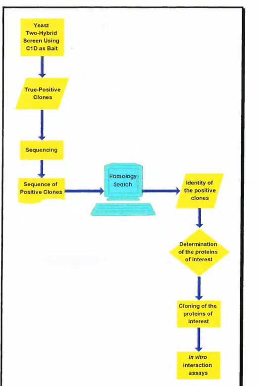
Therefore, the aim o f the project is the identification o f the interacting proteins with C ID by yeast-two-hybrid system, and further characterisation o f these interactions with potential significance o f providing clues about DNA-PK, C 1D, and nuclear matrix function (Figure 4).
Yeast Two-Hybrid Screen Using C1 D a s Bait
I4
True-Positive ClonesI
1
Sequencing1
i IHomologyjSequence of ; i Search j Identity of
ii|^ the positive clones
1
Determination of the proteins of interest Cloning of the proteins of interest in vitro interaction assaysMATERIALS AND METHODS
CHAPTER III: MATERIALS AND METHODS
3.1
Yeast Two Hybrid System
The yeast two-hybrid system provides a relatively straight forward approach to understanding protein interactions. The basic protocol was developed by Stan Fields (Fields and Song 1989) and several different two-hybrid systems have been developed to study protein function since then. The garden-variety application is to learn about the function o f a given protein by isolating proteins that interact with it, usually by screening a cDNA library. To conduct such an interactor hunt, a protein is expressed in yeast as a fusion to the DNA-binding domain o f a transcription factor lacking a transcription activation domain. The DNA-binding fusion protein is generally called the bait . The yeast strain also contains one or more reporter genes with binding sites for the DNA-binding domain. To identify proteins that interact with the bait, a plasmid library that expresses cDNA-encoded proteins fused to a transcription activation domain is introduced into the strain. Interaction o f a cDNA- encoded protein with the bait results in activation o f the reporter genes, allowing cells containing the interactors to be identified.
DNA Binding Domain Fusion Construct:
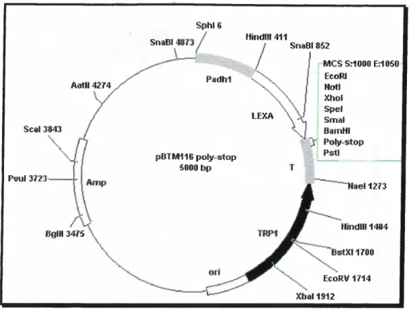
This construct was generated by U. Yavuzer and the full length open reading frame o f cDNA encoding C ID protein (nucleotides between 118-543) was cloned into the Xhol site o f the multiple cloning site o f vector pBTM l 16 polystop. The bait vector, pBTM116 poly-stop, was also modified by U. Yavuzer from the original plasmid constructed by S. Fields, carries the TR Pl gene and has a polylinker downstream o f LexA coding sequence. The DNA-BD is provided by the entire prokaryotic LexA protein, which normally functions as a repressor o f SOS genes in
E.coli
when it binds to LexA operators.The cDNA Library:
The cDNA library was cloned into the Xhol site located in the multiple cloning site o f the pACT vector (The library was kindly provided by S. Elledge). pACT vector carries the LEU2 gene and contains NLS-Gal4-linker unit driven by the ADH promoter. The library was generated by random-primed cDNA synthesis from human B-lymphocyte polyA+ RNA. Following second strand synthesis with RNAse H, pol
I,
andE.coli
DNA ligase, the cDNA was repaired with T4 DNA polymeraseand linkered appropriately for cloning in to the vector. Fragments were size selected to have insert sizes greater than 600 bp. The library was originally received in bacteriophage, lysed, transformed into HBlOl
E.coli
strain, amplified, titrated andpurified.
3.1.1 Constructs;
L40:
M A T a , his3A 2 00 , trp I-9 0 1 , k u 2 - 2 J 1 2 , a Je2 , L Y S2::(L exA op)4-H lS3, U R A 3 ::(L e xA o p )s-L a c Z G A L 4. L40 can detect weak LexA activators as histidine prototrophs without the use o f 3-aminotriazoIe. The expression o f the LacZ and HI S3 coding sequences are driven, respectively by minimal GALİ and HI S3 promoters fiised to multimerised LexA binding sites. The reporters, LacZ and HIS3, are integrated to the genes URA3 and LYS2 respectively o f the yeast genome.\ M K 1 0 : M A T a . his3, lys2, tr p i, leu2, U R A 3 ::(L e x A o p )s -L a c Z G A IJ .
3.1.2 Yeast Strains
3.1.3 Transformation
Small Scale Transformation:
A single colony o f L40 was inoculated in 10 ml YPAD (10 g yeast extract, 20 g peptone, 0.1 g adenine, 900 ml ddH20, autoclaved, cooled and 100 ml 20% glucose added) and grown overnight at 30“C with shaking at 200 rpm. 50 ml YPAD medium was then inoculated with overnight grown culture to produce an OD(,oo between 0.2- 0.3 and incubated further till the ООбоо reaches 0.6-0.75. All subsequent steps were carried at room temperature. Yeast cells were pelleted at 2500 rpm for 10 minutes. After resuspending the pellet in 40 ml IxTE, cells were repelleted, resuspended in 2 ml LiAc/TE (100 mM LiAc in 0.5xTE), and incubated at room temperature for 10 minutes. Meanwhile 0 .1 pg o f DNA was dissolved in 8 pi ddH20 and 10 pi o f salmon sperm DNA was added. 150 pi competent yeast cells was dispensed to DNA
DMSO was added, followed by a heat shock at 42"C for 7 minutes. Cells were then pelleted, supernatant aspirated, and pellet resuspended in 1.0 ml TE. Cells were pelleted once more as above, resuspended in 100 pi TE, and was plated on appropriate selective medium.
Large Scale Transformation:
The LexA-ClD fusion plasmid was first introduced into L40 by selecting growth on trpytophane(-) plates after a small scale transformation. The resulting strain was used for growing a 2 ml overnight culture in YC - Ura, Trp (1.2 g yeast nitrogen base without amino acids, 5 g ammonium sulfate, 10 g succinic acid, 6 g NaOH, 900 ml ddH20, autoclaved, 100 ml 20% glucose and all amino acids other than stated were added). This 2 ml culture was then used to inoculate 100 ml o f the same medium and incubated for another 18 hours. At the end o f this period, 1.0 1 YPAD (pre-warmed to 30‘C ) was inoculated with the 100 ml overnight culture to obtain an optical density ООбоо o f 0.3, and grown at 30°C 200 rpm for three hours. Cells were pelleted at 2500 rpm for 5 minutes at room temperature and pellet was resuspended in 500 ml TE. The suspension was respun and pellet was resuspended in 20 ml 100 mM LiAc/0.5xTE, and the DNA mixture was added (1.0 ml o f 10 mg/ml denatured salmon sperm DNA and 500 pg o f library plasmid DNA). After mixing well, 140 ml o f lOOmM LiAc/40% PEG/lxTE was added and incubated at 30"C for 30 minutes. The mixture was then transferred to a sterile two litre beaker and covered with a foil. 17.6 ml o f DMSO was added, mixed by swirling, and heat shocked at 42“C in a water bath for 6 minutes with occasional swirling to facilitate heat transfer. After the heat shock, the mixture was immediately diluted with 400 ml
YPA ( YPAD without glucose) and rapidly cooled down to room temperature in a water bath. Cells were pelleted at 2500 rpm for 5 minutes at room temperature. Pellet was washed with 500 ml YPA, resuspended in 1.0 1 pre-warmed YPAD, and incubated at 30°C for one hour with gentle shaking. 1.0 ml o f the cells was removed, pelleted, supernatant removed, and pellet resuspended in 1.0 ml YC without uracil, tryptophane, and lysine. 10 and 1 pi o f these cells were plated on YC -Ura,Trp,Leu plate. This measures the primary transformation efficiency which should be between 10-100x10^ transformants. The 1.0 1 o f cells was pelleted at 2500 rpm for 5 minutes at room temperature, pellet resuspended in 500 ml YC without uracil, tryptophane, and leucine, respun and then resuspended again in 1.0 L prewarmed YC without uracil, tryptophane, and leucine and incubated at 30°C for 4-6 hours. The plating efficiency increases during this period. At the end o f the incubation period, the cells were pelleted at 2500 rpm for 5 minutes at room temperature, washed twice with YC -Trp,His,Ura,Leu,Lys, and final pellet was resuspended in 10 ml YC without Trp,His,Ura,Leu,Lys. Aliquots o f 200 pi were plated on 144 mm plates containing YC without Trp,His,Ura,Leu,Lys. His+ colonies were then taken to a grid for p-gal analysis.
A dry nitrocellulose filter was laid onto yeast colonies paying attention not to trap air bubbles. The filter was marked for orientation determination, removed and placed colony side up on a pre-cooled aluminium boat floating on liquid nitrogen. After 30 seconds, the boat was immersed in liquid nitrogen for 10 seconds. Then, the filter was removed, and left at room temperature colony side up till it was thawed. In the lid o f a petri dish, 1.5 ml Z buffer containing 15 |il o f 50 mg/ml X-gal was placed (Z buffer: 60 mM Na2HP04, 40 mM NaH2P04, 10 mM KCl, 1 mM MgS04, pH 7.0 ). One #1 Whatman filter circle was placed in the lid avoiding any air bubbles. Then, the nitrocellulose filter was placed on top o f Whatman filter colony side facing up. The lid was covered with the bottom o f the dish and was incubated at 30‘’C till it yields a detectable colour.
3.1.4 (î-gal Filter Assay
3.1.5 DNA Isolation from Yeast Colonies
The yeast plasmid DNA was extracted and transferred to
E.coli
to facilitatefurther analysis. The procedure is modified from Ward A C. (Ward 1990). 3 ml o f culture was grown overnight at 30"C 200 rpm with appropriate selection. 1.5 ml o f the culture was pelleted by centrifuging at 2500 rpm for 5 minutes at room temperature. The pellet was resuspended in 120 pi Lysis buffer ( 2% Triton-X, 1% SDS, 100 mM NaCl, 10 mM Tris pH,:8.0, 1 mM EDTA). 150 pi acid washed glass beads and 300 pi phenol/chloroform were added, and the mixture was vortexed vigorously for 1 minute. Then, it was centrifuged at 13,000 rpm for 2 minutes and the supernatant was transferred to a clean tube. In cases where supernatant was not clear enough, equal volume o f phenol/chloroform was added and previous step was2(^
repeated. 2.5 volumes o f cold (-20“C) absolute ethanol was added to the supernatant, centrifuged at 13,000 rpm for 10 minutes. The supernatant was removed, and the pellet was washed with 70% o f cold (-20‘*C) ethanol, dried and resuspended in 5 pi o f sterile ddH20. The DNA tubes were stored at -80°C till usage.
3.1.6 Isolation of cDNA Clones
To verify the specificity o f interactions, cDNA clones have to be rescued and put back into yeast to check for the repetition o f interaction. Also specificity should be confirmed by using heterologous LexA-fusion proteins. Plasmids purified from yeast contain both LexA -C lD fusion plasmid and the cDNA library plasmid. To obtain specifically the cDNA clone, the plasmid isolated from L40 yeast strain was transformed into
E.coli
strain H B lO l which is leucine deficient, and therefore, theLEU2 gene from yeast complements for the leucine deficiency in bacteria.
Overnight grown HBlO l culture was diluted in 100 ml to obtain an OD6oo o f
0.2-0.3 and incubated at 37°C, 200 rpm till OD6oo was in between 0.6-0.75. Culture was chilled on ice for 15 minutes and centrifuged at 2500 G for 15 minutes at 4“C. The pellet was resuspended in 50 ml o f cold sterile 10% glycerol and centrifuged at 4000 rpm at 4"C for 15 minutes. The pellet was resuspended in the remaining few drops and kept on ice. The electroporation cuvettes were chilled prior to use, and 40 pi cell suspension and 2 pi DNA (from plasmid isolates o f yeast colonies) were mixed in chilled eppendorfs. The mixture was transferred to electroporation cuvettes on ice
added immediately. The cell suspension was then transferred to a 15 ml falcon tube with a sterile pasteur pipette, and incubated at 37“C for 1 hour. Samples were then centrifuged at 2500 rpm for 5 minutes, pellet was washed with 1 ml M9 [0.025 g proline, 7.5 g bacto-agar, 390 ml ddH20, autoclaved, 100 ml 5x M9 salt solution (30 g Na2HP04, 15 g KH2PO4, 5 g NH4CI, 2.5 g NaCl, 1 1 dd H2O, autoclaved) and 10 ml 20% glucose added.] medium, and re-centrifuged. Final pellet was dissolved in
100 μ1 M9, spread on ampicilline (50 pg/ml) M9 plates, and incubated at 37‘*C overnight. The plasmid DNA was isolated from the colonies as described in section J .2.J
3
.
1.7Mating Assay
The plasmid isolated from H BlO l strain was then transformed back to L40 yeast strain and grown on trp(+) leu(-) medium. The plasmids expressing the LexA fusion proteins were transformed into AMR70 (M ata). The basic mating protocol is as follows. Single colonies from primary positive clones rescued in L40 trp(+) leu(-) and AMR70/bait test strains leu(+) trp(-) are mixed separately in 5 μ1 sterile ddH20. Then, 2 μ1 from each mixture is mixed, dotted on a YPAD plate and incubated at 30“€ for 4 hours. The colony was transferred to YC -Ura, Trp, Leu plate and grown at 30"C for two days. This step selects for diploid growth. At the end o f incubation period, the plates were tested for β-gal activity as described in section 3.1.4. After elimination o f the false-positives, the real clones were amplified and sequenced.
The sequences o f the real positive cDNAs were first translated to protein sequence using Expasy Molecular Biology Server (http://expasy.nhri.org.tw) and Translation Machine program at http://www2.ebi.ac.uk/translate. The translations were performed in three reading frames and were submitted to BLAST2 (http://www.ncbi.nlm.nih.gov/BLAST/) for homology search using basic search parameters, and the match with the highest meaningfial score was taken into consideration. The full length nucleotide sequence o f the clones were then retrieved from Entrez server (http://www.ncbi.nlm.nih.gov/Entrez/), and subjected to restriction enzyme analysis using W ebcutter program, which was again running online on WWW (http://www.firstmarket.com/cutter/cut2.html).
3.1.8 Database Search
3 .2
Recombinant DNA Manipulation Techniques
3.2.1 Preparation of Competent Cells for Transformation and
Transformation with Plasmid DNA - CaCb Method
50-200 ml o f LB was inoculated with 0.5 ml o f overnight culture o f the appropriate strain o f
E.coli
and incubated until the culture reached an OD600 o f 0.6 0.7. The cells were pelleted by centrifugation at 3000 rpm 4“C for 10 minutes, resuspended in 0.4 volume o f ice cold sterile 100 mM CaCb and incubated on ice for 1 hour. The cells were pelleted as above and resuspended in 0.5-2 ml ice cold sterileDNA (less than 100 ng) was added to 150 |il competent cells, and incubated on ice for 30 minutes. The cells were then exposed to a 90 second heat shock at 42°C, chilled on ice for 1-2 minutes, 800 pi Luria-Bertoni (LB) (1.0% tryptone, 0.5% yeast extract, 1.0% NaCl) was added and incubation continued at 37°C for one hour. The culture was then pelleted, resuspended in 100 pi LB, and was spread on L-agar plates supplemented with 50 pg/ml ampicillin and/or 25 pg/ml kanamaycin, inverted and incubated overnight at 37‘'C.
3.2.2 Preparation of Competent Cells for Transformation and
Transformation with Plasmid DNA - Simple and Efficient Method
250 ml o f SOB (0.5% bacto tryptone, 0.5% yeast extract, lOmM NaCl, 2.5mM KCl, lOmM MgCl2, lOmM MgSOa, sterilised by autoclaving) was inoculated with appropriate
E.coli
strain, grown to an ODeoo o f 0.6 at 37"C shaker at 200 rpm.The culture was chilled on ice for 10 minutes, centrifuged at 2500 g for 10 minutes at 4“C. The pellet was then resuspended in 80 ml o f ice-cold TB (10 mM Pipes, 55 mM MnCL, 15 mM
CaCh,
250 mM KCl, pH 6.7, sterilised by filtration through 0.45 pmfilter, stored at 4"C), and incubated on ice for 10 minutes. The mixture was pelleted as above, resuspended gently in 20 ml TB. DMSO was added to 7%, mixed gently and incubated on ice for 10 minutes. Aliquots o f this mixture were then immediately chilled in liquid nitrogen, and stored in liquid nitrogen.
For transformation with supercompetent cells, an aliquot was thawed at room temperature. 200 pi o f the cells was mixed with plasmid DNA (less than 100 ng) in a 15 ml tube, and incubated on ice for 30 minutes. The cells were then exposed to a 30 second heat shock at 42°C, chilled on ice for 1-2 minutes, 800 pi SOC (SOB with 20
mM glucose) was added and incubation continued at 37"C for one hour with vigorous shaking at 250 rpm. The culture was then pelleted, resuspended in 100 pi SOC, and was spread on L-agar plates supplemented with 50 pg/ml ampicillin and/or 25 pg/ml kanamaycin, inverted and incubated overnight at 37°C (Inoue et al., 1990).
3.2.3 Plasmid DNA Purification and Quantitation
Small Scale Isolation
Small scale preparation o f plasmid DNA was performed by standard methods based on NaOH/SDS cell lysis and potassium acetate precipitation o f cellular debris (Maniatis et al., 1982).
Cells were harvested by centrifugation (13,000 rpm, 3 minutes, at room temperature) from a 1.5 ml overnight culture o f bacteria carrying the plasmid o f interest. Following resuspension o f the bacterial pellet in 100 pi solution I (50 mM glucose, 25 mM Tris-Cl [pH 8.0], 10 mM EDTA), 200 pi solution II (200 mM NaOH, 1% SDS) and 150 pi solution III (3M potassium acetate, 11.5% acetic acid [glacial] ) was added, mixed and then DNA was extracted with an equal volume o f phenol.chloroform 1:1. The phases were separated by centrifugation (13,000 rpm, 5 minutes, room temperature), and the DNA was precipitated from the aqueous phase by addition o f 1 ml cold absolute ethanol. After washing once with 70% ethanol, the pellet was dried and resuspended in 30 pi ddH20 with lOpg/ml DNase-free RNase A to destroy the RNA in the product.
Medium Scale Isolation
Cells were grown in 50-100 ml LB (containing appropriate antibiotic) overnight to saturation and plasmid DNA was isolated by using QIAfilter Plasmid Midi Kit (Cat No: 10 12145, Qiagen Inc.), according to the manufacturer’s instructions.
DNA Puriflcation from Agarose Gel
The desired DNA bands were cut from the agarose gel under UV illumination and DNA was extracted from agarose gel slices by QIAquick Gel Extraction Kit (Cat No; 28106, Qiagen Inc.), according to the manufacturer’s instructions.
Quantification of DNA
Concentrations and purity o f nucleic acids were determined by measuring absorbency at 260nm and 280nm in a spectrophotometer (Beckmann Instruments Inc., CA, USA). The ratio between absorbance values at OD260 and OD280 was taken. Nucleic acid samples displaying OD260 and OD280 values in the range o f 1.8 to 2.0 are regarded as highly pure. A value o f OD26o=l 0 corresponds to a concentration o f approximately 50 pg/^tl double stranded DNA, and 20 pg/pl for oligonucleotides (Maniatis.et al 1982.).
3.2.4 Restriction Enzyme Digestion of DNA
Restriction enzyme digests o f DNA were carried out in a total volume o f 20 |il with 5-10 units o f restriction enzyme, volume o f which was always smaller than 1/10*'’ o f the total reaction volume. Buffers and incubation conditions were used for each digest as recommended by the manufacturer.
3.2.5 DNA Ligation
DNA fragments were ligated into plasmid vectors as described in Current Protocols (Ausubel 1987). Prior to ligation, vector and insert concentrations were checked by agarose gel electrophoresis, and 1:4 vector;insert ratio maintained in the ligation reactions. Ligations were performed in 15 pi reaction volumes containing 0.5 pg plasmid DNA and molar excess o f insert in the presence o f 1-4 Weiss units o f T4 DNA ligase and standard ligation buffer supplied by the manufacturer at either room temperature for 4 hours or at 16"C overnight.
3.2.6 Polymerase Chain Reaction
Cyclical amplification o f target DNA sequences between a pair o f oligonucleotide primers is possible using the thermostable DNA Polymerase o f