E-mail: [email protected] doi:10.3906/sag-0803-10
Can [F-18] fluorodeoxyglucose positron emission tomography
replace sentinel lymph node biopsy for the detection of axillary
metastases in patients with early-stage breast cancer?*
Semih GÖRGÜLÜ1, Mehmet Fatih CAN1, Oğuz HANÇERLİOĞULLARI1, Nuri ARSLAN2,
Erkan ÖZTÜRK1, Emel ÖZTÜRK3, Müjdat BALKAN1, Turgut TUFAN1
Aim:To investigate the value of fluorodeoxyglucose positron emission tomography (FDG-PET) in detecting axillary involvement, and to compare its accuracy with sentinel lymph node biopsy (SLNB) in patients with clinically early-stage breast cancer.
Materials and methods:Twenty-eight female patients with histologically-confirmed T1-2 breast cancer who were scheduled to have SLNB were included in the study. FDG-PET images were obtained 1-7 days prior to surgery with an intravenous injection of 370 MBq of FDG, while plasma glucose levels were maintained below 120 mg/dL. All the images were interpreted by 2 independent nuclear medicine specialists, who were blinded to the histological diagnoses. SLNB was performed in standard fashion with peri-tumoral injection of isosulphan blue dye. In all cases, a level I-II axillary dissection was performed following SLNB. Sentinel nodes were processed after formalin fixation; no frozen sections were used.
Results:Thirteen (46%) patients were found to have axillary involvement. SLNB (an average of 2.3 LNs removed per patient) demonstrated metastases in all 13 patients. The diagnostic accuracy of FDG-PET was as follows: true-positive in 4 out of 13 patients (overall sensitivity = 31%), false-negative in 1 patient with metastasis (overall specificity = 94%), positive predictive value = 80%, negative predictive value = 63%, and accuracy = 68%.
Conclusion:FDG-PET appears to be significantly less accurate than SLNB at detecting axillary metastases. In patients with an axillary-positive PET scan, however, axillary lymph node dissection may be performed without prior SLNB.
Key words:Breast cancer, sentinel lymph node biopsy, positron emission tomography, axillary metastasis
[F-18] Deoksiglukoz pozitron emisyon tomografisi erken evre meme kanserli
hastaların aksiller metastazlarının tespitinde sentinel lenf nodu biyopsisinin yerini
alabilir mi?
Amaç:Florodeoksiglukoz positron emisyon tomografisinin (FDG-PET) erken evre meme kanserli hastalarda aksiller tutulumu saptamadaki değerini araştırmak ve doğruluğunu sentinel lenf nodu biyopsisi (SLNB) ile karşılaştırmak.
Yöntem ve gereç:Histolojik olarak meme kanseri tanısı almış ve SLNB planlanan yirmi sekiz kadın hasta çalışmaya dahil edildi. FDG-PET görüntüleri cerrahiden 1-7 gün önce, kan glukoz düzeyleri 120 mg/dL. nin altında tutularak 370 MBq FDG’nin intravenöz enjeksiyonu ile elde edildi. Görüntüler birbirinden bağımsız ve histolojik tanılardan habersiz iki nükleer tıp uzmanınca yorumlandı. SLNB standart şekilde izosülfan mavisinin peritümöral enjeksiyonu ile
Received: 06.03.2008 – Accepted: 13.05.2009
1Department of General Surgery, Gülhane Military Medical Academy, 06018 Etlik, Ankara - TURKEY 2Department of Nuclear Medicine, Gülhane Military Medical Academy 06018 Etlik, Ankara - TURKEY 3Department of Nuclear Medicine, Faculty of Medicine, Ufuk University, 06520 Balgat, Ankara - TURKEY
Correspondence: Semih GÖRGÜLÜ, Department of Surgery, Gülhane Military Medical Academy, 06018 Etlik, Ankara - TURKEY
E-mail: [email protected]
Introduction
Breast cancer is the most common malignancy in women (1), and the second most lethal cancer in the US, despite the fact that 80% of all patients present with stage I or II disease (2,3). The status of the axillary lymph node is the single most powerful independent predictor of prognosis, both for disease recurrence and survival in breast cancer. No imaging technique currently is available for the accurate evaluation of axillary lymph node involvement. Therefore, the best procedure for examining axillary lymph nodes remains axillary lymph node dissection (ALND) followed by pathological examination. As a secondary function, axillary lymph node dissection also helps to control local axillary disease. However, only 30% of women with an invasive breast tumor of diameter of ≤2 cm have axillary lymph node metastases (4). Axillary lymph node dissection confers no survival advantage when the lymph nodes are not involved. Axillary dissection also can result in significant short- and long-term sequelae and tends to delay discharge from hospital (5). The morbidity of ALND has led to the increased use of sentinel lymph node biopsy (SLNB) as a less invasive alternative. This technique has become the new standard of care in patients with early-stage breast cancer in whom the nodes clinically appear uninvolved (6). Nonetheless, any technique that could identify positive lymph nodes before surgery would have several advantages. Imaging by positron emission tomography (PET) using the radio-labeled glucose analogue 2-(fluorine-18)-fluoro-2-deoxy-D-glucose (F-18 FDG) allows for visualization of a wide variety of tumors, because of the enhanced tracer uptake by malignant tissue, when compared with surrounding benign tissues (7-9). The available literature comparing the utility of PET
scanning versus SLNB while staging the axilla in early stage breast cancer is limited. The general aim of this study was to determine the appropriateness of PET as an alternative to SLNB and ALND in early-stage breast cancer patients.
Materials and methods
Twenty-eight women, mean age 54.4 ± 13.0 years, with histologically proven breast cancer and scheduled to have SLNB and ALND as part of their surgical therapy, constituted the study population. The study was conducted between May 2005 and October 2006. The preoperative diagnosis was confirmed by fine needle aspiration with cytology. Males and patients with clinical stage III or IV disease, uncontrolled diabetes mellitus, prior SLNB or ALND, inflammatory breast cancer, or pregnancy were excluded. The protocol of the study was approved by the Institutional Ethics Committee, and written informed consent was obtained from all patients.
PET imaging
PET studies, using an Ecat Exact scanner (Siemens/CTI Knoxville, TN, USA), were performed in patients 1-7 days before surgery. To standardize blood glucose levels, patients were required to fast for at least 4 h before scanning. Plasma glucose levels were determined before FDG injection, and PET imaging performed with plasma glucose levels below 120 mg/dL; 370 MBq (or 10 mCi) of FDG was injected into an antecubital vein contralateral to the tumor. Patients remained in the supine position with their arms raised while in the imager. Positron emission imaging commenced 45 min after the administration of FDG. Before FDG injection, 2 contiguous bed position transmission scans (10 min
gerçekleştirildi. Tüm olgularda SLNB işlemini level I-II aksilla disksiyonu izledi. Sentinel nodlar formalin fiksasyonu sonrası rutin takiple değerlendirildi.
Bulgular:13 (% 46) hastada aksiller tutulum saptandı. SLNB (ortalama 2.3 lenf nodu/hasta) tüm hastalardaki metastazları gösterdi. FDG-PET’in tanısal doğruluğu aşağıdaki şekildeydi: 13 hastanın 4’ünde gerçek-pozitif (duyarlılık =% 31), hastaların 1’inde yalancı-negatif (özgüllük =% 94), pozitif prediktif değer =% 80, negatif prediktif değer =% 63, ve doğruluk =% 68.
Sonuç:FDG-PET’in aksiller metastazları saptamadaki doğruluğu SLNB’den belirgin şekilde düşük bulunmuştur. Ancak PET incelemesinin aksillayı pozitif gösterdiği olgularda SLNB uygulanmaksızın aksiller diseksiyona gidilebilir.
each) were acquired with a rotating 68Ge rod source for correction of attenuation of the mammary and axillary regions. Two 20-min static emission studies were acquired in the transmission positions, 45-60 min after FDG injection.
All images were interpreted separately by 2 experienced nuclear medicine physicians. PET interpretations were blinded to the histopathological findings. The images were read as positive if localized FDG-uptake was increased relative to the surrounding tissue. Quantitative measurement of the single-pixel maximal standardized uptake value (SUV), normalized to body weight, was performed for any suspicious lesion. PET results were evaluated for sensitivity, specificity, accuracy, and positive and negative predictive values relative to the histopathological diagnosis.
Surgery
Definitive surgery was performed within 7 days of the PET scan. Since SLNB was not a routine operation at the time of the study, complete ALND was performed as part of SLNB validation and implementation studies. SLNB was carried out and followed by standard ALND (level I and II) and breast conserving resection or modified radical mastectomy. SLNB was performed using a blue dye technique, with peri-tumoral injection (1 mL into each 4 side) of isosulphan blue dye (Lymphazurin, 1%, Autosuture, USA) using a 25-gauge needle. Approximately 10 min after the dye injection, a small transverse incision was made, slightly anterior to and just below the hair-bearing area in the axilla. The dissection then was extended down to the superficial fascia covering the axillary fat pad. Meticulous blunt dissection was performed to identify the dye-filled lymphatic tract or blue stained SLN. When a dye-filled lymphatic tract was detected first, it was followed proximally and distally until the blue stained SN was identified. In cases in which the SN was found first, the proximal lymphatic channel was followed until it reached the breast parenchyma. If a secondary lymph node was stained blue, it also was dissected.
Histopathological examination
All SLNs were processed following formalin fixation; no frozen sections were performed. SLNs were cut into 2 mm thin slices and each slide was embedded in separate paraffin blocks. For each block,
2 × 2 sections were cut at a distance of 100 μm. Half of the sections were stained with hematoxylin-eosin, and the rest underwent immunohistochemistry using pancytokeratin staining. The specimen from ALND was formalin fixed and paraffin embedded, and each mold was sectioned twice for hematoxylin-eosin staining. In invasive carcinoma cases, immunohistochemistry using pancytokeratin staining also was performed. Size, grade, margins, and estrogen and progesterone status were determined.
Statistical analysis
Sensitivity, specificity, positive and negative predictive values, and the accuracy of the PET imaging technique were calculated using the following formulas:
Sensitivity = TP/(TP + FN) Specificity = TN/(TN + FP)
Positive predictive value = TP/(TP + FP) Negative predictive value = TN/(TN + FN) Accuracy = (TP + TN)/(TP + FP + FN + TN) where TP is true positive, TN is true negative, FP is false positive, and FN is false negative.
Results
The clinical characteristics of all 28 patients are summarized in Table 1. Of these 28 patients, 13 (46%) were noted to have at least one axillary metastasis. An average of 2.3 lymph nodes were removed per patient and labeled as SLN. The SLNs were positive for metastasis in all 13 patients. The FDG-PET was true-positive in 4 of 13 patients with an axillary metastasis, and the overall sensitivity of FDG-PET was 31%. There was one false-positive result with FDG-PET, and the overall specificity of PET scanning for the detection of axillary lymph node metastases was 94%. The positive and negative predictive value and accuracy of FDG-PET for detection of axillary metastases were 80%, 63%, and 68% respectively.
FDG-PET was false-negative for axillary lymph node metastasis in 9 of 13 metastases. The smallest metastatic lymph node that was visible using PET was 11 mm in diameter. The mean size of the lymph node was 8.8 mm in false-negative cases, while it was 14.7 mm in patients with true positive PET scans.
Discussion
Axillary lymph node status is the single most important prognostic factor and best predictor of recurrence and survival in primary breast cancer (4,10,11). Surgical removal and histopathological examination of nodes still remains the standard way to assess their involvement (12). However, axillary dissection causes significant morbidity, including wound infection, seroma formation, lymphedema, restriction of arm and shoulder movement, and numbness involving upper arm skin. These sequelae are a major cause of emotional distress, functional impairment, and additional cost of treatment. Over recent decades, the mean size of presenting primary tumors and, correspondingly, the percentage of
women with axillary lymph node involvement have decreased (13). These factors have led many authors to advocate more restricted use of ALND in patients with early breast cancer, and the development of less invasive and noninvasive means to stage axilla (14,15).
There is no imaging technique currently available permitting the accurate determination of axillary involvement. The development of sentinel lymph node biopsy has enabled precise axillary staging through a minimally invasive and less morbid approach (16,17), but it still involves a surgical procedure. A noninvasive imaging test that could accurately stage the axilla in early stage breast cancer clearly would be ideal (14).
Sentinel lymph node mapping and dissection is a highly sensitive and accurate technique for nodal evaluation. It has been applied to the staging of axillary lymph nodes in patients with breast cancer, providing prognostic information, yet producing less surgical morbidity than ALND. When performed by an experienced surgeon and analyzed by an experienced pathologist with serial sectioning and immunohistochemical evaluation, SLNB is the most accurate detection tool used in the staging of breast cancer (18). In the present study, no false-negative SLNB was observed. Nonetheless, SLNB has certain limitations, in both locally advanced and clinically early-stage breast cancer patients, including a false-negative rate up to 17% (19). The common causes of false-negative SLNB are blockage of lymphatic vessels by tumor cells, improper injection techniques, removal of only a single lymph node, and inadequate surgical experience (18).
The clinical use of FDG-PET for staging is being investigated for many human tumors, including breast cancer (20-23). Since the first reported visualization of lymph node metastases with FDG-PET in a preclinical animal study in 1990 (24), a number of studies have compared the accuracy of PET scanning and ALND, with conflicting results. Some investigators have concluded that FDG-PET is capable of accurately assessing the nodal status of breast cancer, and has relatively high sensitivity and specificity (18). Greco et al. reported a sensitivity of 94% and specificity of 86% in 167 patients undergoing ALND, but more than 40% of their patients had T2 tumors, and 43% of the patients had positive nodes,
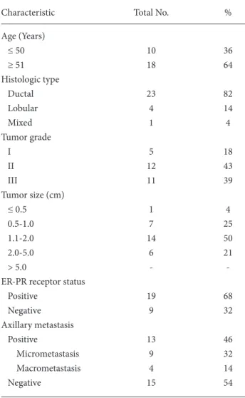
Table 1. Clinical characteristics of the 28 patients.
Characteristic Total No. %
Age (Years) ≤ 50 10 36 ≥ 51 18 64 Histologic type Ductal 23 82 Lobular 4 14 Mixed 1 4 Tumor grade I 5 18 II 12 43 III 11 39 Tumor size (cm) ≤ 0.5 1 4 0.5-1.0 7 25 1.1-2.0 14 50 2.0-5.0 6 21 > 5.0 -
-ER-PR receptor status
Positive 19 68 Negative 9 32 Axillary metastasis Positive 13 46 Micrometastasis 9 32 Macrometastasis 4 14 Negative 15 54
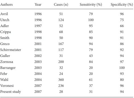
suggesting that the subjects in this study generally had more advanced disease (25). In a study by Schirrmeister et al. sensitivity and specificity were 79% and 92%, respectively, with a false-negative rate of 20%, when compared with ALND. However, only 89 of 117 patients were found to have breast cancer (26). Based upon their series of 200 patients, Zornosa et al. reported 84% sensitivity and 97% specificity for FDG-PET at detecting axillary metastases (27). In other investigations with smaller patient numbers, researchers have reported similar findings, with the sensitivity of preoperative FDG-PET ranging from 85% to 100% (9,21,25-31) (Table 2).
Despite the optimistic conclusions of initial reports, recent studies have yielded conflicting results, wherein the accuracy of FDG-PET has been compared to the accuracy of SLN biopsy and ALND (32-35). For instance, Barranger et al. reported a sensitivity of 20% and specificity of 100% for the detection of nodal metastases by means of FDG-PET (32). In patients qualifying for sentinel node biopsy, FDG-PET seems to perform less well. FDG-PET missed 8 of 14 positive axillary metastases evaluated by SLNB in a study by Guller et al., 8 of 10 patients in a study by Fehr et al., and 13 of 36 in a study by Kumar
et al. (18,20,34). Similarly, Veronesi et al. compared FDG-PET with SLNB for the identification of occult metastases, and reported that the sensitivity of FDG-PET for the detection of axillary metastases was 37%, while the specificity and positive predictive values were 96% and 88%, respectively (35).
In our study, the sensitivity and specificity of FDG-PET for detecting axillary metastatic lymph nodes were 27% and 100%, respectively. Despite the limited sample size of our study, the results were enhanced by blinded PET interpretation and the use of 2 PET examiners. The present study demonstrates the inability of PET to visualize axillary metastases. Our results are similar to those published by Wahl et al. and Veronesi et al. from larger series (33,35). The limited spatial resolution of commercially available PET devices and the presence of fewer metabolically hyperactive cells in patients with micro-metastases certainly appear to be the primary explanations for the low sensitivity of PET at detecting microscopic lymph node metastases.
Contrary to the limited sensitivity of PDG-PET at detecting axillary metastases, we found PET scanning to have high specificity and positive predictive value, as reported in previous studies (20,27,32,35). There
Table 2. Sensitivity and specificity of FDG-PET scanning in detection of sentinel lymph node and axillary metastasis in breast cancer patients.
Authors Year Cases (n) Sensitivity (%) Specificity (%)
Avril 1996 51 79 96 Utech 1996 124 100 75 Adler 1997 52 95 66 Crippa 1998 68 85 91 Smith 1998 50 90 91 Greco 2001 167 94 86 Schirrmeister 2001 117 79 92 Guller 2002 31 43 94 Zornosa 2003 200 84 97 Barranger 2003 32 20 100 Fehr 2004 24 20 93 Wahl 2004 360 61 80 Veronesi 2007 236 37 96 Present study 2007 28 31 94
was only one false positive scan in our study, and the specificity and positive predictive values of FDG-PET were 94% and 80%, respectively, indicating that a positive PET scan is a highly accurate diagnostic test, when histopathological examination is used as the gold standard. Therefore, a positive PET scan may be useful for identifying patients for whom the axillary approach could proceed directly to the ALND, without prior SLNB.
Previous studies have found that FDG-PET is able to provide additional information when assessing patients with internal mammary or supraclavicular metastatic nodes or distant metastasis (9,31). However, it currently is premature to recommend a preoperative PET scan for all patients as a routine diagnostic tool (33).
In conclusion, SLNB remains the procedure of choice for evaluating the histological status of axillary lymph nodes in patients with early-stage breast carcinoma. Given its current spatial resolution, PET imaging appears insufficiently sensitive for localizing microscopic sentinel node metastases. Based upon our results and data from the literature, we conclude that FDG-PET cannot replace SLNB in the detection of axillary metastases. On the other hand, the high specificity of FDG-PET imaging may be useful to identify patients who should undergo ALND rather than SLNB for axillary staging. Better imaging devices with higher resolution than current PET scanners, better tracers, and novel technical developments in PET technology may improve the detection rate of axillary metastases in the near future.
References
1. Ergul E, Sazci A. Molecular genetics of breast cancer. Turk J Med Sci 2001; 31: 1-14.
2. Greenlee RT, Hill-Harmon MB, Murray T, Thun M. Cancer statistics, 2001. CA Cancer J Clin 2001; 51: 15-36.
3. Chung A, Liou D, Karlan S, Waxman A, Fujimoto K, Hagiike M et al. Preoperative FDG-PET for axillary metastases in patients with breast cancer. Arch Surg 2006; 141: 783-8. 4. Carter CL, Allen C, Henson DE. Relation of tumor size, lymph
node status, and survival in 24,740 breast cancer cases. Cancer 1989; 63: 181-7.
5. Warmuth MA, Bowen G, Prosnitz LR, Chu L, Broadwater G, Peterson B et al. Complications of axillary lymph node dissection for carcinoma of the breast: a report based on a patient survey. Cancer 1998; 83: 1362-8.
6. Lyman GH, Giuliano AE, Somerfield MR, Benson AB 3rd, Bodurka DC, Burstein HJ et al. American Society of Clinical Oncology guideline recommendations for sentinel lymph node biopsy in early-stage breast cancer. J Clin Oncol 2005; 23: 7703-20.
7. Strauss LG, Conti PS. The applications of PET in clinical oncology. J Nucl Med 1991; 32: 623-48.
8. Hawkins RA, Hoh CK. PET FDG studies in oncology. Nucl Med Biol 1994; 21: 739-47.
9. Avril N, Dose J, Jänicke F, Ziegler S, Römer W, Weber W et al. Assessment of axillary lymph node involvement in breast cancer patients with positron emission tomography using radiolabeled 2-(fluorine-18)-fluoro-2-deoxy-D-glucose. J Natl Cancer Inst 1996; 88: 1204-9.
10. Soni NK, Spillane AJ. Experience of sentinel node biopsy alone in early breast cancer without further axillary dissection in patients with negative sentinel node. ANZ J Surg 2005; 75: 292-9.
11. Fisher ER, Anderson S, Redmond C, Fisher B. Pathologic findings from the National Surgical Adjuvant Breast Project. Discriminants for 15-year survival. National Surgical Adjuvant Breast and Bowel Project Investigators. Cancer 1993; 71(6 Suppl): 2141-50.
12. Spillane AJ, Sacks NP. Role of axillary surgery in early breast cancer: review of the current evidence. Aust N Z J Surg 2000; 70: 515-24.
13. Cady B, Stone MD, Schuler JG, Thakur R, Wanner MA, Lavin PT. The new era in breast cancer: invasion, size, and nodal involvement dramatically decreasing as a result of mammographic screening. Arch Surg 1996; 131: 301-8. 14. Lovrics PJ, Chen V, Coates G, Cornacchi SD, Goldsmith CH,
Law C et al. A prospective evaluation of positron emission tomography scanning, sentinel lymph node biopsy, and standard axillary dissection for axillary staging in patients with early stage breast cancer. Ann Surg Oncol 2004; 11: 846-53. 15. Haffty BG, Ward B, Pathare P, Salem R, McKhann C, Beinfield
M et al. Reappraisal of the role of axillary lymph node dissection in the conservative treatment of breast cancer. J Clin Oncol 1997; 15: 691-700.
16. Roumen RM, Kuijt GP, Liem IH, van Beek MW. Treatment of 100 patients with sentinel node-negative breast cancer without further axillary dissection. Br J Surg 2001; 88: 1639-43.
17. Schrenk P, Rieger R, Shamiyeh A, Wayand W. Morbidity following sentinel lymph node biopsy versus axillary lymph node dissection for patients with breast carcinoma. Cancer 2000; 88: 608-14.
18. Kumar R, Zhuang H, Schnall M, Conant E, Damia S, Weinstein S et al. FDG PET positive lymph nodes are highly predictive of metastasis in breast cancer. Nucl Med Commun 2006; 27: 231-6.
19. Zavagno G, De Salvo GL, Scalco G, Boza F, Barutta L, Del Bianco P et al. A Randomized Clinical Trial on Sentinel Lymph Node Biopsy Versus Axillary Lymph Node Dissection in Breast Cancer: Results of the Sentinella/GIVOM Trial. Ann Surg 2008; 247: 207-213.
20. Fehr MK, Hornung R, Varga Z, Burger D, Hess T, Haller U et al. Axillary staging using positron emission tomography in breast cancer patients qualifying for sentinel lymph node biopsy. Breast J 2004; 10: 89-93.
21. Adler LP, Crowe JP, al-Kaisi NK, Sunshine JL. Evaluation of breast masses and axillary lymph nodes with [F-18] 2-deoxy-2-fluoro-D-glucose PET. Radiology 1993; 187: 743-50.
22. Pietrzyk U, Scheidhauer K, Scharl A, Schuster A, Schicha H. Presurgical visualization of primary breast carcinoma with PET emission and transmission imaging. J Nucl Med 1995; 36: 1882-4.
23. Scheidhauer K, Scharl A, Pietrzyk U, Wagner R, Göhring UJ, Schomäcker K et al. Qualitative [18F]FDG positron emission tomography in primary breast cancer: clinical relevance and practicability. Eur J Nucl Med 1996; 23: 618-23.
24. Wahl RL, Kaminski MS, Ethier SP, Hutchins GD. The potential of 2-deoxy-2[18F]fluoro-D-glucose (FDG) for the detection of tumor involvement in lymph nodes. J Nucl Med 1990; 31: 1831-5.
25. Greco M, Crippa F, Agresti R, Seregni E, Gerali A, Giovanazzi R et al. Axillary lymph node staging in breast cancer by 2-fluoro-2-deoxy-D-glucose-positron emission tomography: clinical evaluation and alternative management. J Natl Cancer Inst 2001; 93: 630-5.
26. Schirrmeister H, Kühn T, Guhlmann A, Santjohanser C, Hörster T, Nüssle K et al. Fluorine-18 2-deoxy-2-fluoro-D-glucose PET in the preoperative staging of breast cancer: comparison with the standard staging procedures. Eur J Nucl Med 2001; 28: 351-8.
27. Zornoza G, Garcia-Velloso MJ, Sola J, Regueira FM, Pina L, Beorlegui C. 18F-FDG PET complemented with sentinel lymph node biopsy in the detection of axillary involvement in breast cancer. Eur J Surg Oncol 2004; 30: 15-9.
28. Utech CI, Young CS, Winter PF. Prospective evaluation of fluorine-18 fluorodeoxyclucose positron emission tomography in breast cancer for staging of the axilla related to surgery and immunocytochemistry. Eur J Nucl Med 1996; 23: 1588-93. 29. Adler LP, Faulhaber PF, Schnur KC, Al-Kasi NL, Shenk RR.
Axillary lymph node metastases: screening with [F-18]2-deoxy-2-fluoro-D-glucose (FDG) PET. Radiology 1997; 203: 323-7. 30. Crippa F, Agresti R, Seregni E, Greco M, Pascali C, Bogni A et
al. Prospective evaluation of fluorine-18-FDG PET in presurgical staging of the axilla in breast cancer. J Nucl Med 1998; 39: 4-8.
31. Smith IC, Ogston KN, Whitford P, Smith FW, Sharp P, Norton M et al. Staging of the axilla in breast cancer: accurate in vivo assessment using positron emission tomography with 2-(fluorine-18)-fluoro-2-deoxy-D-glucose. Ann Surg 1998; 228: 220-7.
32. Barranger E, Grahek D, Antoine M, Montravers F, Talbot JN, Uzan S. Evaluation of fluorodeoxyglucose positron emission tomography in the detection of axillary lymph node metastases in patients with early-stage breast cancer. Ann Surg Oncol 2003; 10: 622-7.
33. Wahl RL, Siegel BA, Coleman RE, Gatsonis CG; PET Study Group. Prospective multicenter study of axillary nodal staging by positron emission tomography in breast cancer: a report of the staging breast cancer with PET Study Group. J Clin Oncol 2004; 22: 277-85.
34. Guller U, Nitzsche EU, Schirp U, Viehl CT, Torhorst J, Moch H et al. Selective axillary surgery in breast cancer patients based on positron emission tomography with 18F-fluoro-2-deoxy-D-glucose: not yet! Breast Cancer Res Treat 2002; 71: 171-3. 35. Veronesi U, De Cicco C, Galimberti VE, Fernandez JR,
Rotmensz N, Viale G et al. A comparative study on the value of FDG-PET and sentinel node biopsy to identify occult axillary metastases. Ann Oncol 2007; 18: 473-8.